
Biomarkers are extensively used to evaluate the early adverse effects of a toxicant exposure in an organism. Freshwater is exposed to many anthropogenic compounds that adversely affect aquatic life especially, fish population. Effect of acute exposure of low concentrations of BPA (10, 100, 1000 µg/l) on liver cyp1a1 and heat shock proteins mRNA level was evaluated using real time qPCR. Results revealed that exposure to bisphenol A caused decrease expression of cyp1a1 in a concentration dependent manner. Exposure to graded concentrations of bisphenol-A resulted in significant increase in mRNA expression of heat shock proteins (hsp70 and hsp90). In the light of present results, expression of cyp1a1 or heat shock proteins may be used as biomarkers to assess the presence of BPA in the environment.
Short Communication · Volume 52, Issue 1, 8 January 2020 · Open Access
Heat Shock Proteins and Cytochrome P450 1a1 Expression as Biomarkers of Exposure to Bisphenol A in Catla catla
· · ·
Affiliations & Notes ⌄
- 1Department of Zoology, Government College University, Lahore 2Department of Physiology and Cell Biology, University of Health Sciences, Lahore , * Corresponding author: mehwishfaheem@gcu.edu.pk
Article Info ⌄
- Journal:
- Pakistan Journal of Zoology
- Published:
- 8 January 2020

From the article
- In the present study, effect of BPA was studied on mRNA expression of cyp1a1 in liver of Catla catla which is endemic to Pakistan and commercially important species.
- Graded concentration of BPA (10,100,1000µg/l) for 14 days resulted in marked decrease in cyp1a1 transcript level in a concentration dependent manner.
- Exposure of 10µg/l BPA for 14 days caused 3 folds’ significant increase in mRNA level of HSP 70 while 2.4-fold increase was recorded in fish exposed to 1000µg/l BPA for 14 days (Fig. 2B).
- Exposure of 10,100 and 1000µg/l BPA resulted in significantly higher expression of HSP 90 in liver of Catla catla.
Selected from the article's own text.
Abstract
Table I. Primer sequences amplicon lengths and accession numbers of genes studied.
| Gene name | Primer sequence (5’-3’) | Product size | Accession number |
| Cytochrome p450 1a1 | TGTGTCTGAGAGCCTTGTGG GGCTCAGATGTGGGTTGTTT | 164bp | JX480500.2 |
| Heat shock protein 70 | TGAGATCGACTCGCTGTACG CCACCAACCAACACAATGTC | 170bp | KC599207.1 |
| Heat shock protein 90 | GAACTCATCCCCGATCTGAA CCGAATTGACCGATCATAGAA | 171bp | KC800801.1 |
| Tata box binding protein (tbp) | AACAGCTTGTCCCTCCTGGA CAGGAGTGATGGGGGTCATA | 213bp | KX371090.1 |
| gapdh | ATCACAGCCACGCAGAAGACC CAGGAATGACTTTGCCCACAGC | 126bp | JX480499.1 |
been used as biomarker of exposure in various fish species and other aquatic organisms ; ; . The results of these studies showed that heat shock proteins are sensitive markers of chemical exposure and use of molecular approaches towards this end can be very promising.
Gene expression studies are now increasingly used to determine protein and mRNA levels and can be good prognostic parameters for predicting protein metabolism . Moreover, measuring mRNA level is sensitive method and provide mechanistic approach to evaluate toxic endpoints. Therefore, in the present study, cytochrome P4501A1 and heat shock protein 70 and 90 expression was studied as potential biomarkers of exposure to BPA in a commercial edible fish, Catla catla.
Materials and methods
A stock solution of BPA was prepared in ethanol (2mg/ml), exposure concentrations (10, 100, 1000µg/l) were prepared by dissolving stock solution in aquarium water. Female C. ctala were purchased from a commercial fish farm. After acclimatization for two weeks, fish were divided into four groups (n=3) in replicate and exposed to various concentrations (10, 100 and 1000 µg/l) of BPA for 14 days. Toxicant solution was replaced every other day. At the end of exposure period, fish were anesthetized with clove oil and length and weight were measured. Fish were humanely dissected and liver was removed, snap frozen in liquid nitrogen and stored at -80oC until further analysis.
RNA was isolated from frozen tissues using Trizol reagent, cDNA was prepared with 1µg of total RNA using Reverse aid MMLV cDNA synthesis kit (Thermo Fischer Scientific). Geometric mean of tata box binding protein and glyceraldehyde-3-phosphate was used as reference control following Faheem et al. (2018). Gene sequence was obtained from National Center for Biotechnology Information (NCBI) and primers for all genes were designed using primer 3plus software. Primer sequence, gene accession number and product size is listed in Table I. Real time q-PCR was performed on CFX 96 (Bio-Rad, Hercules, CA, USA) with Syber green fluorescent label. A melt curve analysis was performed at the end of reaction to ensure amplification of the single product and PCR product was run on 1.5 % agarose gel. Ct values generated by software were converted to fold change relative to control using 2-∆∆Ct method as described by Livak and Schmittgen (2001).
Gene expression analysis is expressed as mean±S.E.M. Data was checked for homogeneityof varience through Shapiro-Wilk test. To evaluate significant difference among groups (p<0.05), one way analysis of variance was used followed by Tukey’s post hoc test. All statistical analysis was performed using IBM SPSS (Version: 20).
Results and discussion
Primers of genes were specific in binding and yielded a single band of desired product size (Fig. 1). Expression of cytochrome P450 1a1 gene relative to control is shown in Figure 2. The cytochrome P450 enzymes metabolize toxic compounds and CYP1A1 is most important as it is involved in metabolism of estrogens . In the present study, effect of BPA was studied on mRNA expression of cyp1a1 in liver of Catla catla which is endemic to Pakistan and commercially important species. Graded concentration of BPA (10,100,1000µg/l) for 14 days resulted in marked decrease in cyp1a1 transcript level in a concentration dependent manner. Similar decreases in cyp1a1 level were reported in Atlantic cod exposed to 50 µg/l of BPA for 21 days . Similarly, a dietary exposure of bisphenol A (5 and 50 mg/kg bw) for 21 days in Sparus aurata juveniles resulted in significant down regulation of cyp1a1 level. In present study, down-regulation of cyp1a1 recorded in all treatment groups (Fig. 2A) is in agreement with available data on the cross-talk between the reproductive system and the detoxification pathway and is supported by studies showing an inverse relationship between plasma E2 levels and EROD activity during the reproductive cycle . Faheem et al. (2017) recently reported that exposure of same concentrations of BPA to Catla catla females resulted in increased plasma estradiol concentration. The decrease in cyp1a1 transcript level may be the cause of increased E2 levels reported by Faheem et al. (2017), as elevated E2 concentrations are known to be associated with decreases in CYP1A1 activity . These studies and results of present study confirmed that estrogenic compounds like BPA inhibits the detoxification process and therefore, mRNA level of key gene (cyp1A1) can be used as potential biomarker to confirm presence of estrogenic compounds in water bodies.
Heat shock proteins (HSPs) are highly conserved cellular proteins present in all organisms studied to date and are known to function as stress proteins or molecular chaperones ; . HSP levels are mostly studied to assess the toxicity in organism both in laboratory conditions and at polluted environmental sites and therefore used as biomarkers to evaluate toxicity and overall ecological status of a water body . In the present study, mRNA level of HSP 70 and HSP 90 was evaluated after 14 days’ exposure to gradient concentrations of BPA. Expression of heat shock protein 70 and 90 (Fig. 2B). Exposure of 10µg/l BPA for 14 days caused 3 folds’ significant increase in mRNA level of HSP 70 while 2.4-fold increase was recorded in fish exposed to 1000µg/l BPA for 14 days (Fig. 2B). Exposure of 10,100 and 1000µg/l BPA resulted in significantly higher expression of HSP 90 in liver of Catla catla. Pufferfish (Takifugu obscurus) exposed to 600 µg/l of bisphenol-A and 300 µg/l of 4-tert-octylphenol for 96 h resulted in significant upregulation of hsp 70 and hsp 90 after 24 h till 96 h . Sparus aurata juveniles fed with octylphenol for 21 days resulted in significant increased HSP70 mRNA expression compared to control . Male and female black goby injected with nonylphenol and estradiol resulted in significantly increased expression of hsp70 mRNA after 72 hours . Many earlier studies also confirmed the elevated expression of HSP mRNA in fish exposed to pollutants hence, it can be used as a biomarker of exposure in fish .
Conclusion
In conclusion, the inhibition of cyp1a1 and elevation of hsp transcript result in decreased detoxification ability which may lead to compromised health of organisms living in areas contaminated with environmental estrogens such as BPA. Our results offer baseline information which can be used for further research related to the EDCs and use of native carps like Catla catla as a bio-indicator for monitoring environmental health status.
Statement of conflict of Interest
The authors have declared no conflict of interest.
Figures
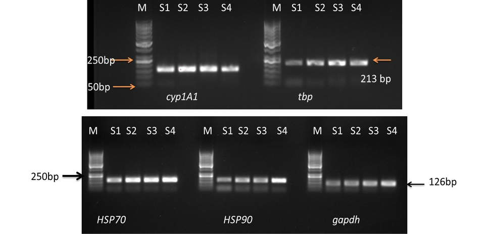
Liver heat shock proteins (70 and 90), cyp1A1 expression of adult female Catla catla exposed to graded concentration of BPA for 14 days. Gapdh, tbp used as reference control; M, marker; S1, control; S2, 10 μg/I BPA; S3, 100 μg/I BPA; S4, 1000 μg/I BPA.
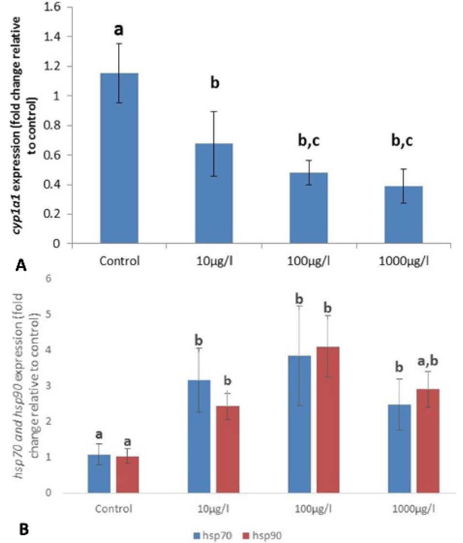
Relative gene expression of the cytochrome p450 1a1 gene normalized tbp and gapdh (A) and heat shock protein 70 and 90 normalized to mean of three selected gene (gapdh, tbp) (B) in liver of Catla catla (n=3) after 14 days of exposure to graded concentration of BPA. Data expressed as mean fold change ±S.E.M. Different letters indicate significant differences among groups. P<0.05.
References
- Ait-Aissa, S., Porcher, J.M., Arrigo, A.P. and Lambre, C., 2000. Toxicology, 145: 147–157. https://doi.org/10.1016/S0300-483X(00)00145-1[CrossRef]
- Akdogan, H.A. and Sen, A., 2010. Vet. Med., 55: 463-471. https://doi.org/10.17221/2982-VETMED[CrossRef]
- Arukwe, A. and Goksøyr, A., 1997. J. exp. Zool. 277: 313–325. https://doi.org/10.1002/(SICI)1097-010X(19970301)277:4<313::AID-JEZ5>3.3.CO;2-#[CrossRef]
- Bierkens, J.A., 2000. Toxicology, 153: 61–72. https://doi.org/10.1016/S0300-483X(00)00304-8[CrossRef]
- Chen, C., Zhou, Q., Liu, S. and Xiu, Z., 2011. Chemosphere, 83: 1147–1154. https://doi.org/10.1016/j.chemosphere.2011.01.006[CrossRef]
- Cheng, X. and Klaassen, C.D., 2008. Toxicol. Sci., 106: 29–36. https://doi.org/10.1093/toxsci/kfn147[CrossRef]
- Dang, W., Xu, N., Zhang, W., Gao, J., Fan, H. and Lu, H., 2018. Pakistan J. Zool., 50: 1043-1051
- de Pomerai, D., 1996. Hum. exp. Toxicol., 15: 279–285. https://doi.org/10.1177/096032719601500401[CrossRef]
- Deane, E.E. and Woo, N.Y.S., 2011. Rev. Fish. Biol. Fish. 21: 153–185. https://doi.org/10.1007/s11160-010-9164-8[CrossRef]
- Faheem, M., Jahan, N., Khaliq, S. and Lone, K.P., 2018. Rendiconti. Lincei., 29: 13–22. https://doi.org/10.1007/s12210-017-0653-8[CrossRef]
- Faheem, M., Khaliq, S., Ahmad, H.U. and Lone, K.P., 2017. Pak. Vet. J., 37: 326-330.
- Gao, K., Brandt, I., Goldstone, J.V. and Jönsson, M.E., 2011. Comp. Biochem. Physiol. C., 154: 42–55. https://doi.org/10.1016/j.cbpc.2011.02.005[CrossRef]
- Gupta, S.C., Sharma, A., Mishra, M. and Chowdhuri, D.K., 2010. Life Sci., 86: 377–384. https://doi.org/10.1016/j.lfs.2009.12.015[CrossRef]
- Handy, R.D., Galloway, T.S. and Depledge, M.H., 2003. Ecotoxicology, 12: 331-43. https://doi.org/10.1023/A:1022527432252[CrossRef]
- Hook, S.E., Gallagher, E.P. and Batley, G.E., 2014. Integr. Environ. Assess. Manage., 10: 327-341. https://doi.org/10.1002/ieam.1530[CrossRef]
- Jeong, H.G., Kim, J.Y., Choi, C.Y., You, H.J. and Hahm, K.S., 2001. Cancer Lett., 165: 95–101. https://doi.org/10.1016/S0304-3835(01)00407-4[CrossRef]
- Jin, Y., Zhang, X., Shu, L., Chen, L., Sun, L., Qian, H., Liu, W. and Fu, Z., 2010. Chemosphere, 78: 846–852. https://doi.org/10.1016/j.chemosphere.2009.11.044[CrossRef]
- Kim, J.H., Jeon, H.J., Baek, J.M., Han, K.N. and Dahms, H.U., 2013. Aquacul. Res., 44: 985–994. https://doi.org/10.1111/j.1365-2109.2012.03104.x[CrossRef]
- Kroon, F., Streten, C. and Harries, S., 2017. PLoS One, 12: e0174762. https://doi.org/10.1371/journal.pone.0174762[CrossRef]
- Liao, C. and Kannan, K., 2014. Fd. Addit. Contam., 31: 319-329. https://doi.org/10.1080/19440049.2013.868611[CrossRef]
- Lin, P., Hu, S.W. and Chang, T.H., 2003. Toxicol. Sci., 71: 20–26. https://doi.org/10.1023/A:1021281217589[CrossRef]
- Livak, K.J. and Schmittgen, T.D., 2001. Methods, 25: 402–408. https://doi.org/10.1006/meth.2001.1262[CrossRef]
- Maradonna, F. and Carnevali, O., 2007. Biomarkers, 12: 240-255. https://doi.org/10.1080/13547500601070859[CrossRef]
- Mendum, T., Stoler, E., VanBenschoten, H. and Warner, J.C., 2011. Green Chem. Lett. Rev., 4: 81–86. https://doi.org/10.1080/17518253.2010.502908[CrossRef]
- Nelson, D.R., 2011. BBA Proteins Proteom., 1814: 14–18. https://doi.org/10.1016/j.bbapap.2010.08.008[CrossRef]
- Olsvik, P.A., Lie, K.K., Sturve, J., Hasselberg, L. and Andersen, O.K., 2009. Chemosphere, 75: 360- 367. https://doi.org/10.1016/j.chemosphere.2008.12.039[CrossRef]
- Rajeshkumar, S., Mini, J. and Munuswamy, N., 2013. Ecotoxicol. Environ. Safe., 98: 8–18. https://doi.org/10.1016/j.ecoenv.2013.07.029[CrossRef]
- Silver, J.T. and Noble, E.G., 2012. Cell Stress Chaperones, 17: 1–9. https://doi.org/10.1007/s12192-011-0290-6[CrossRef]
- Staples, C.A., Woodburn, K., Caspers, N., Hall, A.T. and Klecka, G.M., 2002. Hum. Ecol. Risk. Assess., 8: 1083-1105. https://doi.org/10.1080/1080-700291905837[CrossRef]
- Sung, Y.Y., MacRae, T.H., Sorgeloos, P. and Bossier, P., 2011. Aquaculture, 3: 120-137. https://doi.org/10.1111/j.1753-5131.2011.01049.x[CrossRef]
- Traversi, I., Gioacchini, G., Scorolli, A., Mita, D.G., Carnevali, O. and Mandich, A., 2014. Gen. Comp. Endocrinol., 205: 185-196. https://doi.org/10.1016/j.ygcen.2014.06.015[CrossRef]
- van der Oost, R., Beyer, J. and Vermeulen, N.P.E., 2003. Environ. Toxicol. Pharmacol., 13: 57-149. https://doi.org/10.1016/S1382-6689(02)00126-6[CrossRef]
- Vijayan, M.M., Pereira, C., Kruzynski, G. and Iwama, G.K., 1998. Aquat. Toxicol., 40: 101–108. https://doi.org/10.1016/S0166-445X(97)00057-X[CrossRef]
- Whyte, J.J., Jung, R., Schmitt, C.J. and Tillitt, D.E., 2000. Crit. Rev. Toxicol., 30: 347-569. https://doi.org/10.1080/10408440091159239[CrossRef]
- Wong, C.K.C., Yeung, H.Y., Cheung, R.Y.H., Yung, K.K.L. and Wong, M.H., 2000. Arch. environ. Contam. Toxicol., 38: 468-483.
Services
Article usage statistics
Loading…