Research Article · Volume 13, Issue 2, 28 May 2025 · Open Access
Micro-Nanoplastics Pollution and Its Oxidative Stress-Induced Effects on Fish Physiology in Coastal Waters of Surabaya
· · · · · · · ·
Affiliations & Notes ⌄
1Department of Biology, Faculty of Science and Technology, University of Airlangga, Indonesia
2Department of Biology, Faculty of Science and Data Analitics Institut Teknologi Sepuluh Nopember, Indonesia
3Aquatic Resources Management, Department of Marine Affairs and Fisheries, Faculty of Agriculture, Trunojoyo University, Madura, Indonesia
4Department of Toxicology, Advanced Medical and Dental Institute Universiti Sains Malaysia, Malaysia. *Correspondence | Alfiah Hayati, Department of Biology, Faculty of Science and Technology, University of Airlangga, Indonesia
PSA analysis results indicate that all examined fish are exposed to MNP.
This study confirms that MNP pollution in the waters of East Surabaya, Indonesia, has contaminated fish bodies.
MNP found in seawater and fish tissues (gastrointestinal, liver and meat) with varying distribution and sizes in each fish tissue.
The accumulation of MNP in fish tissues induces health disorders mediated by ROS, as evidenced by the examination of indicators such as MDA, endogenous antioxidants such as SOD, cytokine expression (IL-12, TNF-α, IFN-γ), and apoptotic enzymes (Caspase-9 and Caspase-3), affecting the vitality and life of the fish.
Selected from the article's own text.
Abstract
This study investigates the impact of micro-nanoplastics (MNPs) as water pollutants on the East Coast of Surabaya, Indonesia, and examines their biochemical effects on the blood serum of exposed fish. The physiological condition of fish serves as a bioindicator of environmental quality. The objective of this research was to assess the enzymatic biochemical responses of eight fish species (86 specimens in total) commonly caught by local fishermen. Observed parameters include species identification, MNP distribution and particle size in seawater and fish tissues (gastrointestinal tract, liver, and muscle), as well as biochemical markers indicating oxidative stress, immune response, and apoptosis. The identified fish species include Mugil cephalus, Johnius trachycephalus, Plotosus canius, Harpadon nehereus (H. sagor), Sillago verrucosa, Dasyatis sp., Megalops nigriceps, and Galeocerdo cuvier. Micro-nanoplastics were detected in seawater and within fish tissues, with varying distribution and particle sizes across different organs. Histopathological analysis revealed MNP adhesion on gill lamellae surfaces and lesions in intestinal villi. Biochemical assays showed malondialdehyde (MDA) levels ranging from 2.24 to 4.30 nmol/mL, while superoxide dismutase (SOD) levels ranged from 13.83 to 24.75 ng/mL. The highest levels of interleukin-12 (IL-12), tumor necrosis factor-alpha (TNF-α), and interferon-gamma (IFN-γ) were observed in S. verrucosa, Dasyatis sp., and G. cuvier, respectively. Additionally, elevated levels of Caspase-9 and Caspase-3 indicated increased pro-apoptotic activity. These findings confirm that MNP contamination is present in the waters of the East Coast of Surabaya and has bioaccumulated in fish tissues. The accumulation induces physiological disturbances mediated by reactive oxygen species (ROS), as reflected in elevated oxidative stress markers, altered immune cytokine expression, and increased apoptosis, ultimately affecting the health, vitality, and survival of the fish.
Plastics are synthetic polymers composed of repeating chains of carbon atoms, often bonded with elements such as oxygen, nitrogen, chlorine, or sulfur. These long-chain organic compounds have high molecular weight and structural stability. Since their development in the early twentieth century, plastics have evolved rapidly, resulting in a significant increase in global production . Due to their exceptional durability and resistance to environmental degradation, plastics persist and accumulate across various ecosystems. Over time, the degradation of plastics through physical, chemical, and biological processes leads to the formation of smaller particles known as microplastics (MPs) and nanoplastics (NPs) ; .
A substantial proportion of this waste originates from the widespread use of single-use plastic items, such as bags, bottles, gloves, and face masks ; . Human activities associated with aquatic environments, particularly aquaculture, are also notable contributors to marine plastic pollution. In aquaculture, plastics are used extensively for items such as floats, cages, nets, fishing lines, trawls, and traps . Abandoned, lost, or improperly discarded fishing gear significantly exacerbates oceanic plastic contamination (FAO, 2019; ; , 2023). The widespread distribution of plastic materials in surface waters, sediments, and deep-sea environments has resulted in their ingestion by marine organisms, leading to bioaccumulation and biomagnification throughout aquatic food webs ; .
MPs, defined as plastic particles smaller than 5 mm, are either intentionally manufactured for industrial and consumer products or result from the fragmentation of larger plastic items. Even smaller are NPs, typically ranging from a few nanometers to several micrometers in size, with most commonly reported dimensions below 100 nm. The fragmentation of plastics into MPs and NPs has become a global concern, contributing an estimated 4.8 to 12.7 million tons of plastic waste to the oceans annually. Indonesia alone is responsible for approximately 1.29 million tons of this input .
The structural diversity of MNPs, shaped by their sources and forms, has facilitated their pervasive presence across ecosystems globally . The problem is intensified by poor plastic waste management, high consumption rates, and increasing leakage into water bodies ; . Climate-related factors such as heavy rainfall and strong wind patterns further enhance the transport of MNPs into aquatic systems . These pollutants have been shown to exert adverse effects on aquatic organisms, including delayed growth, oxidative stress, and neurotoxicity .
MNPs enter aquatic organisms through ingestion, inhalation, and dermal contact, and can accumulate in their tissues and cells ; ; ; . Their toxicity stems from both their intrinsic physical and chemical characteristics such as small size and reactive surface groups, and from their ability to adsorb environmental pollutants, including hydrophobic toxicants ; ; . Particularly in coastal and marine ecosystems, high MNP concentrations have been associated with disruptions to ecological function and organismal health, notably in fish ; . Prolonged exposure to MNPs in fish has been linked to impaired immune function, hormonal imbalance, and structural alterations in reproductive organs .
Plastic particles may enter aquatic organisms through both gill respiration and gastrointestinal ingestion . Particles adhering to gill lamellae can, in the case of smaller sizes, penetrate the epithelial membrane and enter the circulatory or lymphatic systems, thereby impairing nutrient uptake and growth. Alternatively, ingested plastics may accumulate throughout the digestive tract, from the stomach to the intestines, where they provoke inflammation and reduce digestive efficiency, ultimately causing feeding impairment and malnutrition ; . This condition can lead to weight loss, nutrient deficiency, and mortality .
Due to their minute size, nanoplastics can traverse cellular membranes via endocytosis (<150 nm) or passive diffusion (<50 nm), facilitating their accumulation across multiple organs ; . Once internalized, nanoplastics disrupt plasma membrane integrity, with their surface charge, particularly in the case of polystyrene nanoparticles, playing a critical role. Cationic particles interact with negatively charged cell surfaces, inducing the formation of reactive oxygen species (ROS) . These particles can further disturb intracellular compartments such as endosomes and lysosomes, exacerbating cellular damage ; . ROS production during plastic degradation, coupled with biological interactions, initiates cellular stress responses, impairing tissue and organ function ; .
The toxic effects of MNPs are reflected in various biological responses, including oxidative stress, membrane disruption, enzyme inhibition, immune dysregulation, and apoptosis ; . Oxidative stress is one of the most frequently observed outcomes of pollutant exposure, often assessed by measuring lipid peroxidation markers such as malondialdehyde (MDA) . Antioxidant defense systems including enzymes such as superoxide dismutase (SOD) and catalase (CAT) are vital for neutralizing ROS. The immune system also plays a critical role in combating MNP-induced stress, and immunological biomarkers can serve as sensitive indicators of toxicity ; . Apoptotic responses, such as increased activity of Caspase-9 and Caspase-3, further confirm the cellular damage caused by exposure to nanoplastics .
The eastern coast of Surabaya, characterized by its dense residential, port, and industrial zones, is especially vulnerable to MNPs contamination. Plastics from diverse sources accumulate in coastal waters, posing a significant threat to marine organisms, particularly fish. One of the most concerning impacts of MNP exposure is the disruption of key physiological systems in fish, particularly enzymatic pathways that regulate metabolism, detoxification, and cellular homeostasis. Comprehensive investigations that combine physiological and molecular biomarkers to assess MNPs toxicity in wild-caught fish, particularly within the Indonesian marine context are extremely limited or have not yet been conducted. This research specifically investigates the toxicological impacts of micro- and nanoplastics at the cellular and molecular levels, an area that remains largely underexplored in current environmental studies. As such, this work offers new perspectives and contributes essential baseline data to better understand the biological consequences of MNP exposure in natural aquatic ecosystems.
Materials and Methods
Animal and ethical clearance
The authors declare that all handlings of the specimens adhered to the international regulations governing the use of animals in scientific research. Ethical clearance and all procedures involving animal care were approved by the Faculty of Science and Technology, Universitas Airlangga, Indonesia (Certificate no. 1432/UN3.FST/PT.01/2024)
Fish collection
Fish samples were collected through fishing and trawling activities with the assistance of local fishermen on the East Coast of Surabaya, Indonesia. The fishing stations were located approximately 5-10 km from the coastline. Fish capture and seawater sampling were conducted simultaneously. The catch from the sampling yielded a number of fish (84 specimens) representing several fish species. However, for this study, only eight species were selected based on their consumption and sale by the local community, with each species represented by four individuals.
Micro-nanoplastics size measurements
Separation of seawater MNP from other particles involved using 30% H2O2 in a 1:1 ratio, heating (80°C for 24 hours), followed by filtration using Whatman Grade 42, 2.5 µm pores (Cytiva), and vacuum filtration (Buchner funnel filtering kit), and then drying . Similarly, fish tissue samples were treated with 30% H2O2, heated (50°C for 24 hours) to dissolve organic materials, followed by filtration and drying as in the previous steps ; . After obtaining potential MNP particles, their diameters were measured.
Measurement of MNP particle diameter
The particle size analyzer (PSA, DelsaTM NanoC by Beckman Coulter, USA) was used to measure the diameter of MNP particles in water and fish tissues, specifically the gastrointestinal tract, liver, and muscle (for mullet fish samples). Particle size determination involved dispersing plastic particles in liquid media to prevent clumping. Particle analysis was conducted in the range of 0 to 6,000 nm, at a temperature of 25°C, viscosity of 1,2000 mPas, and a scattering angle set at 90°. The sonicated suspension was evaluated in triplicate.
Histopathological study of fish tissues
Tissue specimens underwent additional processing for histological examination. Gill and intestine samples were immersed in a 10% neutral buffered formalin (NBF) solution for 24 hours. Following dehydration in varying concentrations of ethanol, the samples were embedded in paraffin wax. Subsequently, tissue sections measuring 4-5 μm in thickness were obtained using a Rotary Microtome (Leica RM2125 RTS) and stained with hematoxylin-eosin. Observation of the samples was conducted using a light microscope (Olympus CZ22 Binocular).
Blood collection
Blood samples were extracted from captured fish by accessing the tail blood vessels through a needle insertion near the anal fin. Approximately 0.5-1 mL of blood per kilogram of body weight was drawn using a size 22 needle and a heparinized syringe. Pressure was applied to the base of the tail and maintained for a minimum of 30 seconds to halt bleeding before each blood sampling procedure. Following this, centrifugation was carried out at 1,000–2,000 x g for 10 minutes in a cooled centrifuge to isolate blood serum.
Biochemical analyses of MDA, SOD, CAT, Cytokines, Caspase-9, and Caspase-3
The measurement of MDA (Fish Malondialdehyde, Catalogue No. E0017FI), SOD (Fish Superoxide Dismutase, Catalogue No. EA0035FI), CAT (Fish Catalase, Catalogue No. EA0006FI), IL-12 (Fish Interleukin 12, Catalogue No. E0131FI), TNF-α (Fish Tumor Necrosis Factor α, Catalogue No. EA0023FI), IFN-γ (Fish Interferon γ, Catalogue No. EA0014FI), Caspase-9 (Fish Caspase-9, Catalogue No. E0219FI), and Caspase-3 (Fish Caspase-3, Catalogue No. E0218FI) levels were carried out using ELISA Kits from the Bioassay Technology Laboratory (Shanghai Korain, Jiaxing, Zhejiang Province) according to the manufacturer’s instructions. Enzymatic measurements were conducted by assessing absorbance values at 450 nm with a microplate reader, which were subsequently utilized for calculating enzyme activities.
Data analysis
Histological tissue data, distribution, and size of MNP were analyzed descriptively by comparing between samples. One-way ANOVA was used to compare levels of MDA, SOD, IL-12, TNF-α, IFN-γ, Caspase-9 and Caspase-3 in different fish species. All data obtained were processed using SPSS Software (V.23).
Results
Fish captured on the east coast of Surabaya, Indonesia
Fishermen successfully caught several fish species, but for this study, only eight species (Table 1) were analysed. The selection was based on fish commonly consumed and sold by the local community around the coastline.
Analysis of distribution and size of micro-nanoplastics
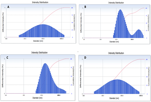
The instrument used in our experiment operates based on the principle of determining the distribution and size of MNP particle diameters. The size distribution of MNP particles in seawater and fish tissues is presented in Figure 1. As observed in Figure 1A, the highest distribution of particles in seawater was dominated by particles ranging from 644.2 to 1435.1 nm, with cumulants results of diameters around 1088.9 nm and differential intensity of 2.2% with a polydispersity index (PI) of approximately 0.501. The highest particle distribution in the gastrointestinal tract was observed at sizes ranging from 168.2 to 219.3 nm, with cumulants results of diameters around 193 nm and differential intensity of 4.7%, along with a polydispersity index (PI) of approximately 0.219 (Figure 1B). Meanwhile, the highest particle distribution in the liver and fish meat occurred at sizes ranging from 147.8 to 189.7 nm and 447.3 to 699.5 nm, respectively. Cumulants results of diameters were approximately 222.5 nm and 627.1 nm, with differential intensity of 4.3% and 2.1%, and polydispersity index (PI) of approximately 0.219 and 0.482 (Figure 1C, D).
The results of measurements of distribution and size of MNP particles in seawater prove that plastic particles present in the water can enter and accumulate in all fish body tissues, including the gastrointestinal tract, liver, and meat (Table 2). The MNP size in water ranges from 23.3 to 14,164.9 nm, capable of penetrating and accumulating
Table 1: Various fish species captured (fishing and trawling) by fishermen on the East Coast of Surabaya, Indonesia. All of the data were displayed by mean ± SD.
Species
Number of individual samples
Weight (grams)
Smallest
Biggest
Means ± SD
Mugil cephalus
12
25
97
48.83 ± 24.29
Johnius trachycephalus
11
48
185
94.59 ± 33.79
Plotosus canius
10
160.4
261
211.21 ± 28.16
Hexanematichthys sagor
14
190
542
276.88 ± 111.35
Synanceia verrucosa
12
67
236
160.91 ± 52.60
Dasyatis sp.
13
19
271.2
106.92 ± 104.97
Mystus nigriceps
10
49
337.1
158.01 ± 94.17
Galeocerdo cuvier
4
268.5
521
394.75 ± 126.25
Total
86
Table 2: Distribution and size of MNP in seawater and fish tissues from fishermen’s catch on the East Coast of Surabaya, Indonesia. All of the data were displayed by mean ± SD.
Parameters
Water
Gastrointestinal tract
Liver
Meat
Smallest size (nm)
23.3
101.0
40.1
34.7
Biggest size (nm)
14,164.9
5,279.4
2,372.1
8,181.6
90% Large amount (nm)
5,605.0 ± 747.04
1,337.2 ± 769.60
962.8 ± 483.06
1,440.8 ± 641.66
50% Medium amount (nm)
1,365.7 ± 640.36
267.4 ± 51.70
187.4 ± 26.33
266.5 ± 198.3
10% Small amount (nm)
330.6 ± 248.76
142.2 ± 60.29
87.4 ± 3.92
95.9 ± 22.3
in tissues as follows: Gastrointestinal tract, liver, and meat ranging from 101 to 5,279.4 nm; 40.1 to 2,372.1 nm; and 34.7 to 8,181.6 nm, respectively. The large particle sizes for 90% of the sample population are: gastrointestinal tract, liver, and meat, measuring 1,337.2 nm, 962.8 nm, and 1,440.8 nm, respectively. Meanwhile, the medium particle sizes for 50% of the population are: 267.4 nm, 187.4 nm, and 266.5 nm, respectively; and the small particle sizes for 10% of the population are: 142.2 nm, 87.4 nm, and 95.9 nm, respectively. As of now, there are no established standards or regulatory limits set by the World Health Organization (WHO) or major health authorities regarding permissible levels of microplastics and nanoplastics in fish, food, or human consumption due to limited data.
Analysis of histopathology of fish tissues
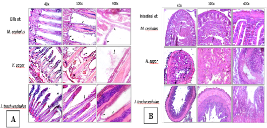
Various forms of histological structures in the gills and intestines of fish caught by fishermen (represented by three species; M. cephalus, H. sagor, and J. trachycephalus) revealed the presence of microplastic particles adhering to the surface of secondary lamellae in the gills of all three species. The presence of MNP hinders the respiratory gas transport process (Figure 2). In the cross-section of the intestines, the epithelial structure on the intestinal villi shows lesions in M. cephalus and J. trachycephalus, while in H. sagor, there are columnar epithelial cells with numerous Goblet cells.
Analysis of malondialdehyde (MDA) measurement
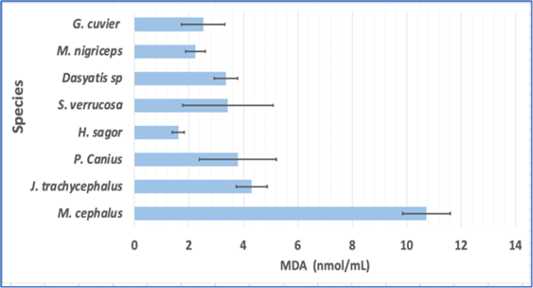
Based on the ELISA test results, the highest MDA levels among the eight fish species caught by fishermen were found in M. cephalus (10.7250 nmol/mL), followed by J. trachycephalus, S. verrucose, Dastis sp., G. cuvier, M. nigriceps, and the lowest level was found in H. sagor (1.6049 nmol/mL). The MDA levels in the other six fish species ranged from 2.2427 to 4.3025 nmol/mL (Figure 3).
Endogenous antioxidant analysis
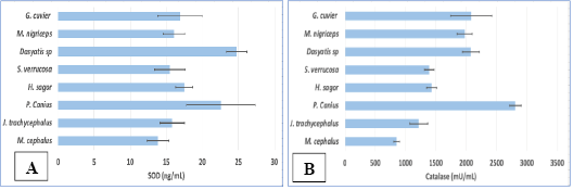
The subsequent ELISA test results indicated that the SOD levels in the captured fish ranged from 13.83 to 24.75 ng/mL. The highest SOD levels were found in Dasyatis sp. (24.75 ng/mL) and P. canius (22.56 ng/mL). The SOD levels in the other six fish species were below the SOD levels of these two species, in the following order: H. sagor, G. cuvier, M. nigriceps, J. trachycephalus, S. verrucose, and M. cephalus, with levels of 17.48, 16.90, 16.05, 15.79, 15.47, and 13.83 ng/mL, respectively (Figure 4A). Meanwhile, the CAT levels, from highest to lowest, were as follows: P. canius, G. cuvier, Dasyatis sp., M. nigriceps, H. sagor, J. trachycephalus, and M. cephalus, with levels of 2807, 2081, 2073, 1971, 1429, 1214, and 851 mU/mL, respectively (Figure 4B).
Immune system analysis
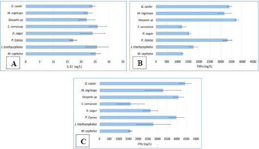
To assess the immune system of fish exposed to MNP, cytokine levels, including IL-12, TNF-α, and IFN-γ, were measured. Seven out of eight species showed nearly similar IL-12 levels ranging from 21.98 to 25.96 ng/L, except for one species (P. canius) with the lowest IL-12 level (17.12 ng/L). This differs from TNF-α and IFN-γ levels, which varied among species. High TNF-α levels were found in Dasyatis sp., G. cuvier, P. canius, and M. nigriceps (3730, 3392, 3315, and 3168 ng/L), while the other four species, J. trachycephalus, H. sagor, S. verrucose, and M. cephalus, had lower levels (1734, 1540, 1209, and 1234 ng/L). Similarly, for IFN-γ, from highest to lowest, it was G. cuvier, Dasyatis sp, P. canius, M. nigriceps, J. trachycephalus, H. sagor, S. verrucosa, and M. cephalus, with levels of 4431, 4083, 3983, 3280, 2796, 2629, 1612, and 1592 ng/L, respectively (Figure 5).
Apoptosis analysis
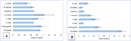
Exposure to MNP in their habitat leads to high pro-apoptotic enzyme activity, including Caspase-9 and Caspase-3. The enzymatic activity, indicated by enzyme levels, showed that Caspase-9 levels were nearly the same for all fish species, ranging from 0.68 to 1.05 ng/mL, except for G. cuvier, which had a lower level of approximately 0.49 ng/mL. Meanwhile, Caspase-3 levels varied significantly. The highest level was found in M. cephalus (4.25 ng/mL), followed by J. trachycephalus, H. sagor, S. verrucose, and G. cuvier, with levels of 4.25, 1.92, 1.65, 1.18, and 0.87 ng/mL, respectively. The lowest Caspase-3 levels were observed in Dasyatis sp, P. canius, and M. nigriceps, at 0.45, 0.42, and 0.33 ng/mL (Figure 6).
Discussion
Improper disposal of plastic waste, especially in water bodies, poses environmental and aquatic health issues, including those affecting fish. PSA analysis results indicate that all examined fish are exposed to MNP. The distribution and particle size analysis demonstrate the accumulation of MNP in the cells and tissues of fish living in MNP-polluted waters (Figure 1). This situation poses a significant threat to the health of humans consuming these fish. Fish are a source of essential amino acids and polyunsaturated fatty acids (PUFA) necessary for maintenance and growth. However, fish are highly vulnerable to contamination by various organic and inorganic compounds, posing a threat to public health . Additionally, plastic is a potent source of pollution in aquatic environments, and although it is challenging to degrade, it eventually breaks down into smaller particles, i.e., MNP. The degradation process depends on environmental factors such as degradation rate, temperature, time, and physical, chemical, or biological agents (catalytic degradation, mechanochemistry, and biodegradation) . The differential intensity in the gastrointestinal and hepatic tissues is significantly higher (47% and 43%) compared to muscle tissue (2.1%). This is likely related to the digestive system, where food entering the gastrointestinal tract is absorbed into the liver through blood vessels for detoxification. This aligns with the opinion of other researchers stating that particles (<150 μm) can be ingested by living organisms, enter the body through the intestinal wall, and reach lymph nodes and other organs .
Although there have been numerous studies on the effects of MNP on fish, knowledge of the histological effects of MNP on fish as a whole, especially in the gills and intestines of marine fish, is still limited. In the present study, accumulated MNP particles in tissues indicate changes in cell and tissue structure. Blood vessels in the primary lamellae, consisting of pillar cells and capillaries, prominently extend towards the secondary lamellae, resembling fingers. Additionally, MNP attached to the surface of the primary lamellae affects the gill structure, causing morphological deformation, shortening, and degeneration of the secondary lamellae (Figure 2). This aligns with other researchers’ opinions regarding the effects of MNP, stating that fish exposed to MNP at specific concentrations and durations can alter gill morphology, causing thickening and proliferation of gill lamellae, erosion and abscission of apical filament areas, hyperplasia, vacuolization, infiltration, irregular cell arrangement, epithelial removal, increased mucus production, flat aneurysms, and fusion of primary and secondary lamellae, leading to hematoma ; ; .
In this study, MNP accumulation in the digestive tract reveals morphological changes in the intestinal epithelium, including degeneration of the basal membrane, goblet cell reduction, submucosal atrophy, and mucosal cell vacuolization. Histopathological changes in the intestine, in addition to MNP concentration and size, are also presumed to be influenced by the duration of fish exposure to MNP. Severe changes include degeneration, shortening, and erosion of villi, and lesions observed in M. cephalus and J. trachycephalus, except for the well-organized goblet cells in the intestinal epithelium of H. sagor (Figure 2). Increased numbers and sizes of goblet cells observed in this study (Figure 2, H. sagor intestine) may be a response to increased mucus secretion, considered a first-line defence strategy against foreign MNP particles. Epithelial layer damage can lead to cytotoxic substances from the intestinal lumen entering the bloodstream, potentially causing functional changes in various organs.
One of the primary mechanisms underlying MNP toxicity is associated with oxidative stress. Oxidative stress is an inevitable aspect of aerobic life, resulting from an imbalance between the production of reactive oxygen species (ROS) and the defence mechanisms of antioxidants in living organisms. This study demonstrates Malondialdehyde (MDA) levels as a marker for oxidative stress induced by MNP in eight fish species. Although MDA levels are not the exclusive indicator of oxidative stress, they are commonly used due to their straightforward procedures. In this research, the highest MDA levels were found in M. cephalus compared to the other seven species (Figure 3). M. cephalus is commonly found in tropical and subtropical estuaries . This species is classified as benthopelagic, adapted for foraging, with its primary diet comprising algae growing on rocks, drifting wood, or the river estuary bottom. The elevated MDA levels indicate a high presence of ROS stimulated by the unintentional ingestion of MNP along with food through the digestive system. Despite its high economic importance due to the delicious taste of its flesh, little is known about the impact of MNP on the health of consumers.
In this study, the levels of Superoxide Dismutase (SOD) and Catalase (CAT) are highlighted as endogenous antioxidants, and they are consistently high across all species. Superoxide radicals, due to MNP, are converted into hydroperoxides by the enzyme SOD. Subsequently, these hydroperoxides are neutralized by the enzyme CAT, effectively countering ROS. Thus, the presence of these antioxidants is crucial to balance external stressors originating from exposure to pollutants, toxins, chemicals, and microplastics. Oxidative stress from these substances can lead to increased ROS, causing oxidative stress in fish . It is important to note that higher levels of oxidative stress can disrupt the health, growth, reproduction, and other aspects of animals, including fish ; . Oxygen molecules act as precursors to the formation of ROS.
Based on previous research, hazardous substances in the environment, including MNP, can induce inflammatory responses in organisms . However, inflammatory responses in fish are often closely related to cytokines such as TNF, IL, and IFN . These three are crucial immune factors induced in the inflammatory response and pathogen clearance. They interact to enhance the organism’s resilience to external environmental factors. TNF-α induces the expression of other genes involved in the immune response, attracting the movement of T lymphocytes, neutrophils, and basophils in the body. Meanwhile, IFN is secreted to regulate apoptosis and cellular immunity. In this study, the expression of these three immune factors indicates varying changes among different fish species. The expression of pro-inflammatory immune response from the captured fish species shows consistently high levels of IL-12, but the levels of TNF-α and IFN-γ vary for each species. This suggests that MNP-contaminated waters increase the levels of pro-inflammatory enzymes. This increased expression indicates that MNP activates the immune system mechanism through the activation of the NF-κB signalling pathway, leading to an increase in the expression of signalling factors within cells .
The contamination of MNP has become a threat to fish health. The toxic impact at the cellular level and molecular mechanisms have been studied in this research. A series of molecular events underlying apoptosis is investigated through the measurement of Caspase-9 and Caspase-3 levels in the fish’s blood serum. Caspases (3 and 9) are crucial molecules in regulating the apoptosis mechanism . Previous studies have also shown that various pollutants, including microplastics, can increase Caspase 3 and Caspase 9 levels and induce apoptosis in fish ; . MNP induces the formation of ROS, which, in turn, activates the oxidative defense mechanism, increasing the regulation of Caspase-9 and Caspase-3 expression, leading to apoptosis. Furthermore, oxidative damage caused by MNP exposure can result in cytological damage, causing structural and functional changes in cells. Thus, the regulation of Caspase-3 and Caspase-9 in all captured fish species plays a crucial role in protecting organisms from stress signals caused by MPN pollutants.
Conclusion
This study confirms that MNP pollution in the waters of East Surabaya, Indonesia, has contaminated fish bodies. MNP found in seawater and fish tissues (gastrointestinal, liver and meat) with varying distribution and sizes in each fish tissue. MNP adhered to the surface of lamellae and caused lesion in intestinal villi. The accumulation of MNP in fish tissues induces health disorders mediated by ROS, as evidenced by the examination of indicators such as MDA, endogenous antioxidants such as SOD, cytokine expression (IL-12, TNF-α, IFN-γ), and apoptotic enzymes (Caspase-9 and Caspase-3), affecting the vitality and life of the fish.
Acknowledgments
The author expresses gratitude to Universitas Airlangga and the Directorate of Research, Technology, and Community Service (DRTPM) in 2023, Indonesia, for providing research funding. NUMBER: 0536/E5/PG.02.00/2023.
Novelty Statement
This study is the first to comprehensively evaluate the toxicological effects of micro- and nanoplastics (MNPs) on wild-caught fish species along the East Coast of Surabaya, Indonesia, integrating particle characterization with biochemical, immunological, and histopathological biomarkers. It provides novel baseline data on the accumulation, size distribution, and tissue-specific effects of MNPs in multiple fish species commonly consumed by local communities. The use of oxidative stress markers, cytokine profiling, and apoptosis indicators in a field-based marine context offers new insights into the ecological and physiological consequences of MNP pollution in tropical coastal environments.
Author’s contribution
Alfiah Hayati: Writing review and editing, writing original draft, investigation, conceptualization.
Manikya Pramudya: Writing review and editing, investigation.
Aunurohim: Writing review and editing.
Abdus Salam Junaedi: Writing review and editing.
Farah Annisa Nurbani, Widi Pangestu Wilujeng and Muhammad Iqbal: Investigation.
Firli Rahmah Primula Dewi: Writing review and editing.
Vuanghao Lim: Writing review and editing.
Conflict of interests
The authors have declared no conflict of interest.
Figures
Figure 1:.
Distribution and size of MNP particles. The distribution and size of MNP particles show specific particle sizes encountered in high quantities, although the differential and cumulative intensity differs for each tissue. A represents seawater, B represents the gastrointestinal tract, C represents the liver, and D represents fish meat.
Figure 2:.
Photomicrograph of Fish Gills (A) showing microplastic particles adhering to the surface of gill lamellae and Intestines (B) displaying degeneration in the intestinal epithelial structure (M. cephalus and J. trachycephalus), except in H. sagor where Goblet cells are neatly arranged on the surface of the intestinal epithelium.
Figure 3:.
Malondialdehyde (MDA) levels in eight fish species caught by fishermen in East Surabaya Coast, Indonesia.
Figure 4:.
Superoxide dismutase (SOD) levels (A) and Catalase (CAT) levels (B) of eight fish species caught by fishermen on the East Coast of Surabaya, Indonesia.
Figure 5:.
Interleukin-12 (IL-12) levels (A), Tumor Necrosis Factor-Alpha (TNF-α) levels (B), and Interferon-Gamma (IFN-γ) Levels (C) of eight fish species caught by fishermen on the East Coast of Surabaya, Indonesia.
Figure 6:.
Caspase-9 (A) and Caspase-3 (B) levels in eight fish species caught by fishermen on the East Coast of Surabaya, Indonesia.
References
A’Yun Q, Musthoza FS, Supartini S, Utari D, Listiani I, Triwahyudi H, Fikriyah N, Suprapti N, Hayati A (2022). Potential of feed supplements on morphometric and gonad weight of fish exposed to microplastics. IOP. C. Ser. Earth. Env., 1036: 012001. https://doi.org/10.1088/1755-1315/1036/1/012001[CrossRef]
Auta HS, Emenike CU, Fauziah SH (2017). Distribution and importance of microplastics in the marine environment A review of the sources, fate, effects, and potential solutions. Environ. Int., 102: 165–176. https://doi.org/10.1016/j.envint.2017.02.013[CrossRef]
Aynard A, Courrèges C, Jiménez-Lamana J, Raad A, Miqueu C, Grassl B, Reynaud S (2023). Trace metal sorption on nanoplastics: An innovative analytical approach combining surface analysis and mass spectrometry techniques. Environ. Pollut., 323: 121229. https://doi.org/10.1016/j.envpol.2023.121229[CrossRef]
Banaee M, Faraji J, Amini M, Multisanti CR, Faggio C (2023). Rainbow trout (Oncorhynchus mykiss) physiological response to microplastics and enrofloxacin: Novel pathways to investigate microplastic synergistic effects on pharmaceuticals. Aquat. Toxicol., 261: 106627. https://doi.org/10.1016/j.aquatox.2023.106627[CrossRef]
Banerjee, A., Shelver, WL (2021). Micro- and nanoplastic induced cellular toxicity in mammals: A review. Sci. Total Environ., 755: 142518. https://doi.org/10.1016/j.scitotenv.2020.142518[CrossRef]
Cai Y, Li C, Zhao Y (2022). A review of the migration and transformation of microplastics in inland water systems. Int. J. Environ. Res. Public Health, 19: 148. https://doi.org/10.3390/ijerph19010148[CrossRef]
Cardona L (2000). Effects of salinity on the habitat selection and growth performance of mediterranean flathead grey mullet Mugil cephalus (Osteichthyes, Mugilidae). Estuar. Coast. Shelf Sci., 50: 727–737. https://doi.org/10.1006/ecss.1999.0594[CrossRef]
Choi D, Bang J, Kim T, Oh Y, Hwang Y, Hong J (2020). In vitro chemical and physical toxicities of polystyrene microfragments in human-derived cells. J. Hazard. Mater., 400: 123308. https://doi.org/10.1016/j.jhazmat.2020.123308[CrossRef]
De-la-Torre GE, Aragaw TA (2021). What we need to know about PPE associated with the COVID-19 pandemic in the marine environment. Mar. Pollut. Bull., 163: 111879. https://doi.org/10.1016/j.marpolbul.2020.111879[CrossRef]
Dong X, Liu X, Hou Q, Wang Z (2023). From natural environment to animal tissues: A review of microplastics (nanoplastics) translocation and hazards studies. Sci. Total Environ., 855: 158686. https://doi.org/10.1016/j.scitotenv.2022.158686[CrossRef]
Dyachenko A, Mitchell J, Arsem N (2017). Extraction and identification of microplastic particles from secondary wastewater treatment plant (WWTP) effluent. Anal. Methods, 9: 1412–1418. https://doi.org/10.1039/C6AY02397E[CrossRef]
Enrichetti F, Bavestrello G, Betti F, Rindi F, Tregrosso A, Bo M (2021). Fate of lost fishing gears: Experimental evidence of biofouling colonization patterns from the northwestern Mediterranean Sea. Environ. Pollut., 268: 115746. https://doi.org/10.1016/j.envpol.2020.115746[CrossRef]
Falcão MAP, Dantas MCS, Rios CT, Borges LP, Serafini MR, Guimarães AG, Walker CIB (2022). Zebrafish as a tool for studying inflammation: A systematic review. Rev. Fish. Sci. Aquacult., 30: 101–122. https://doi.org/10.1080/23308249.2021.1898538[CrossRef]
FAO (2019). Microplastics in fisheries and aquaculture. Fish. Aquac. Tech. Paper. http://www.fao.org/3/a-i7577e.pdf
Feng LJ, Li JW, Xu EG, Sun XD, Zhu FP, Ding Z, Tian H, Dong SS, Xia PF, Yuan XZ (2019). Short-term exposure to positively charged polystyrene nanoparticles causes oxidative stress and membrane destruction in cyanobacteria. Environ. Sci. Nano., 6: 3072–3079. https://doi.org/10.1039/C9EN00807A[CrossRef]
Gajanur AR, Jaafar Z (2022). Abandoned, lost, or discarded fishing gear at urban coastlines. Mar. Pollut. Bull., 175: 113341. https://doi.org/10.1016/j.marpolbul.2022.113341[CrossRef]
Gajanur AR, Jaafar Z (2023). Behaviour of stranded abandoned plastic fishing nets in the equatorial tropics. Nat. Singapore., pp. 1–8.
Gao D, Xu Z, Qiao P, Liu S, Zhang L, He P, Zhang X, Wang Y, Min W (2013). Cadmium induces liver cell apoptosis through caspase-3A activation in purse red common carp (Cyprinus carpio). PLoS One, 8: 1–11. https://doi.org/10.1371/journal.pone.0083423[CrossRef]
Guerrera MC, Aragona M, Porcino C, Fazio F, Laurà R, Levanti M, Montalbano G, Germanà G, Abbate F, Germanà A (2021). Micro and nano plastics distribution in fish as model organisms: Histopathology, blood response and bioaccumulation in different organs. Appl. Sci., 11: https://doi.org/10.3390/app11135768[CrossRef]
Gurjar UR, Xavier KAM, Shukla SP, Deshmukhe G, Jaiswar AK, Nayak BB (2021). Incidence of microplastics in gastrointestinal tract of golden anchovy (Coilia dussumieri) from north east coast of Arabian Sea: The ecological perspective. Mar. Pollut. Bull., 169: 112518. https://doi.org/10.1016/j.marpolbul.2021.112518[CrossRef]
Hale RC, Seeley ME, La Guardia MJ, Mai L, Zeng EY (2020). A global perspective on microplastics. J. Geophys. Res. Ocean., 125: 1–40. https://doi.org/10.1029/2018JC014719[CrossRef]
Hamed M, Soliman HAM, Badrey AEA, Osman AGM (2021). Microplastics induced histopathological lesions in some tissues of tilapia (Oreochromis niloticus) early juveniles. Tissue Cell, 71: 101512. https://doi.org/10.1016/j.tice.2021.101512[CrossRef]
Hayati A, Pramudya M, Soepriandono H, Astri AR, Kusuma MR, Maulidah S, Adriansyah W, Dewi FRP (2022). Assessing the recovery of steroid levels and gonadal histopathology of tilapia exposed to polystyrene particle pollution by supplementary feed. Vet. World, 15: 517–523. https://doi.org/10.14202/vetworld.2022.517-523[CrossRef]
Hayati A, Pramudya M, Soepriandono H, Suhargo L, Dewi FRP, Muchtaromah B, Mwendolwa AA (2023). Supplementary Feed potential on histology and immune response of tilapia (Oreochromis niloticus L.) exposed to microplastics. Sains Malaysiana, 52: 1607–1617. https://doi.org/10.17576/jsm-2023-5206-01[CrossRef]
Hollóczki O, Gehrke S (2020). Can Nanoplastics alter cell membranes? Chemphyschem, 21: 9–12. https://doi.org/10.1002/cphc.201900481[CrossRef]
Hu L, Chernick M, Lewis AM, Lee FP, Hinton DE (2020). Chronic microfiber exposure in adult Japanese medaka (Oryzias latipes). PLoS One, 15: 5–7. https://doi.org/10.1371/journal.pone.0229962[CrossRef]
Hu M, Palić D (2020). Micro and nano-plastics activation of oxidative and inflammatory adverse outcome pathways. Redox Biol., 37: 101620. https://doi.org/10.1016/j.redox.2020.101620[CrossRef]
Jiang JQ (2018). Occurrence of microplastics and its pollution in the environment: A review. Sustain. Prod. Consum., 13: 16–23. https://doi.org/10.1016/j.spc.2017.11.003[CrossRef]
Joksimovic N, Selakovic D, Jovicic N, Jankovic N, Pradeepkumar P, Eftekhari A, Rosic G (2022). Nanoplastics as an invisible threat to humans and the environment. J. Nanomater., 2022: 6707819. https://doi.org/10.1155/2022/6707819[CrossRef]
Lai H, Liu X, Qu M (2022). Nanoplastics and human health: Hazard identification and biointerface. Nanomaterials, 12: 1298. https://doi.org/10.3390/nano12081298[CrossRef]
Lall SP, Kaushik SJ (2021). Nutrition and metabolism of minerals in fish. Animals, 11: 1–41. https://doi.org/10.3390/ani11092711[CrossRef]
Law KL (2017). Plastics in the marine environment. Ann. Rev. Mar. Sci., 9: 205–229. https://doi.org/10.1146/annurev-marine-010816-060409[CrossRef]
Li RR, Wang BL, Nan FR, Lv JP, Liu XD, Liu Q, Feng J, Xie SL (2023). Effects of polystyrene nanoplastics on the physiological and biochemical characteristics of microalga Scenedesmus quadricauda. Environ. Pollut., 319: 120987. https://doi.org/10.1016/j.envpol.2022.120987[CrossRef]
Li Y, Liu Z, Li M, Jiang Q, Wu D, Huang Y, Jiao Y, Zhang M, Zhao Y (2020). Effects of nanoplastics on antioxidant and immune enzyme activities and related gene expression in juvenile Macrobrachium nipponense. J. Hazard. Mater., 398:, 122990. https://doi.org/10.1016/j.jhazmat.2020.122990[CrossRef]
Liu Z, Huang Y, Jiao Y, Chen Q, Wu D, Yu P, Li Y, Cai M, Zhao Y (2020). Polystyrene nanoplastic induces ROS production and affects the MAPK-HIF-1/NFkB-mediated antioxidant system in Daphnia pulex. Aquat. Toxicol., 220: 105420. https://doi.org/10.1016/j.aquatox.2020.105420[CrossRef]
Malinowska K, Sicińska P, Michałowicz J, Bukowska B (2023). The effects of non-functionalized polystyrene nanoparticles of different diameters on the induction of apoptosis and mTOR level in human peripheral blood mononuclear cells. Chemosphere, 335: 139137. https://doi.org/10.1016/j.chemosphere.2023.139137[CrossRef]
Manzoor S, Naqash N, Rashid G, Singh R (2022). Plastic material degradation and formation of microplastic in the environment: A review. Mater. Today Proc., 56: 3254–3260. https://doi.org/10.1016/j.matpr.2021.09.379[CrossRef]
Mason SA, Welch VG, Neratko J (2018). Synthetic polymer contamination in bottled water. Front. Chem., 6: 407. https://doi.org/10.3389/fchem.2018.00407[CrossRef]
Maulu S, Nawanzi K, Abdel-Tawwab M, Khalil HS (2021). Fish Nutritional value as an approach to children’s nutrition. Front. Nutr., 8: 1–10. https://doi.org/10.3389/fnut.2021.780844[CrossRef]
Maw MM, Boontanon N, Fujii S, Boontanon SK (2022). Rapid and efficient removal of organic matter from sewage sludge for extraction of microplastics. Sci. Total Environ., 853: 158642. https://doi.org/10.1016/j.scitotenv.2022.158642[CrossRef]
Monira S, Roychand R, Hai FI, Bhuiyan M, Dhar BR, Pramanik BK (2023). Nano and microplastics occurrence in wastewater treatment plants: A comprehensive understanding of microplastics fragmentation and their removal. Chemosphere, 334: 139011. https://doi.org/10.1016/j.chemosphere.2023.139011[CrossRef]
Nanthini DK, Raju P, Santhanam P, Perumal P (2022). Impacts of microplastics on marine organisms: Present perspectives and the way forward. Egypt. J. Aquat. Res., 48: 205–209. https://doi.org/10.1016/j.ejar.2022.03.001[CrossRef]
Panda F, Pati SG, Das K, Samanta L, Sahoo DK, Paital B (2022). Biochemical and molecular responses of the freshwater snail Pila sp. to environmental pollutants, abiotic, and biotic stressors. Front. Environ. Sci., 10: 1–19. https://doi.org/10.3389/fenvs.2022.1033049[CrossRef]
Peng L, Fu D, Qi H, Lan CQ, Yu H, Ge C (2020). Micro- and nano-plastics in marine environment: Source, distribution and threats. A review. Sci. Total Environ., 698: 134254. https://doi.org/10.1016/j.scitotenv.2019.134254[CrossRef]
Rubio L, Marcos R., Hernández A (2020). Potential adverse health effects of ingested micro- and nanoplastics on humans. Lessons learned from in vivo and in vitro mammalian models. J. Toxicol. Environ. Heal. Part B, 23: 51–68. https://doi.org/10.1080/10937404.2019.1700598[CrossRef]
Shi C, Liu Z, Yu B, Zhang Y, Yang H, Han Y, Wang B, Liu Z, Zhang H (2024). Emergence of nanoplastics in the aquatic environment and possible impacts on aquatic organisms. Sci. Tot. Environ., 906: 167404. https://doi.org/10.1016/j.scitotenv.2023.167404[CrossRef]
Subaramaniyam U, Allimuthu RS, Vappu S, Ramalingam D, Balan R, Paital B, Panda N, Rath PK, Ramalingam N, Sahoo DK (2023). Effects of microplastics, pesticides and nano-materials on fish health, oxidative stress and antioxidant defense mechanism. Front. Physiol., 14: 1–24. https://doi.org/10.3389/fphys.2023.1217666[CrossRef]
Thakur S, Mathur S, Patel S, Paital B (2022). Microplastic Accumulation and degradation in environment via biotechnological approaches. Water (Switzerland), 14: https://doi.org/10.3390/w14244053[CrossRef]
Thushari GGN, Senevirathna JDM (2020). Plastic pollution in the marine environment. Heliyon, 6: e04709. https://doi.org/10.1016/j.envpol.2020.114915[CrossRef]
van Emmerik T, Schwarz A (2020). Plastic debris in rivers. Wiley Interdiscip. Rev. Water, 7: 1–24. https://doi.org/10.1002/wat2.1398[CrossRef]
Wang W, Zhang J, Qiu Z, Cui Z, Li N, Li X, Wang Y, Zhang H, Zhao C (2022). Effects of polyethylene microplastics on cell membranes: A combined study of experiments and molecular dynamics simulations. J. Hazard. Mater., 429: 128323. https://doi.org/10.1016/j.jhazmat.2022.128323[CrossRef]
Wright SL, Kelly FJ (2017). Plastic and human health: A micro issue? Environ. Sci. Technol., 51: 6634–6647. https://doi.org/10.1021/acs.est.7b00423[CrossRef]
Xin X, Chen B, Yang M, Gao S, Wang H, Gu W, Li X, Zhang B (2023). A critical review on the interaction of polymer particles and co-existing contaminants: Adsorption mechanism, exposure factors, effects on plankton species. J. Hazard. Mater., 445: 130463. https://doi.org/10.1016/j.jhazmat.2022.130463[CrossRef]
Xiong X, Liu Q, Chen X, Wang R, Duan M, Wu C (2021). Occurrence of microplastic in the water of different types of aquaculture ponds in an important lakeside freshwater aquaculture area of China. Chemosphere, 282: 131126. https://doi.org/10.1016/j.chemosphere.2021.131126[CrossRef]
Yang H, Lai H, Huang J, Sun L, Mennigen JA, Wang Q, Liu Y, Jin Y, Tu W (2020). Polystyrene microplastics decrease F–53B bioaccumulation but induce inflammatory stress in larval zebrafish. Chemosphere, 255: https://doi.org/10.1016/j.chemosphere.2020.127040[CrossRef]
Yuan Z, Nag R, Cummins E (2022). Human health concerns regarding microplastics in the aquatic environment from marine to food systems. Sci. Total Environ., 823: 153730. https://doi.org/10.1016/j.scitotenv.2022.153730[CrossRef]
Zhang M, Li M, Wang R, Qian Y (2018). Effects of acute ammonia toxicity on oxidative stress, immune response and apoptosis of juvenile yellow catfish Pelteobagrus fulvidraco and the mitigation of exogenous taurine. Fish Shellfish Immunol., 79: 313–320. https://doi.org/10.1016/j.fsi.2018.05.036[CrossRef]
Zhang W, Liu Z, Tang S, Li D, Jiang Q, Zhang T (2020). Transcriptional response provides insights into the effect of chronic polystyrene nanoplastic exposure on Daphnia pulex. Chemosphere, 238: 124563. https://doi.org/10.1016/j.chemosphere.2019.124563[CrossRef]
Zheng Y, Hamed M, De-la-Torre GE, Frias J, Jong MC, Kolandhasamy P, Chavanich S, Su L, Deng H, Zhao W, Shi H (2023). Holes on surfaces of the weathered plastic fragments from coastal beaches. Mar. Pollut. Bull., 193: 115180. https://doi.org/10.1016/j.marpolbul.2023.115180[CrossRef]