Research Article · Volume 13, Issue 2, 28 March 2025 · Open Access
Nanoplastic-Induced Immune Modulation and Histopathological Changes in Oreochromis niloticus at Different Salinity Levels
· · · · · · · · ·
Affiliations & Notes ⌄
1Department of Biology, Faculty of Science and Technology, University of Airlangga, Indonesia
2Department of Biology, Faculty Science and Data Analitics Institut Teknologi Sepuluh Nopember, Indonesia. *Correspondence | Alfiah Hayati, Department of Biology, Faculty of Science and Technology, University of Airlangga, Indonesia
Exposure to NPs in tilapia resulted in alterations in pro-inflamatory cytokines (IL-12, TNF-α and IFN-γ), and oxidative stress markers (MDA) in the spleen.
At the same salinity, NP exposure led to increased MDA, IL-12, and IFN-γ levels, while TNF-α exhibited a slight decrease compared to the control group.
At higher salinity, NP exposure led to an upregulation of IL-12, IFN-γ, and TNF-α, indicating that salinity may modulate the inflammatory response to NP toxicity.
These findings suggest that the combined influence of NPs and salinity plays a critical role in determining immune and physiological responses in tilapia, highlighting the need for further research on the interaction between environmental stressors and NP toxicity in aquatic organisms .
Selected from the article's own text.
Abstract
Nanoplastics (NPs) are ubiquitous environmental pollutants and pose significant ecological and human health risks due to their non-biodegradable nature and bioaccumulation potential. Evaluating their toxicity is crucial to understanding these risks. This study investigates the immunotoxicological and histopathological effects of NPs on Nile tilapia (Oreochromis niloticus) at different salinity levels. Fish were divided into control groups (without NPs) and treatment groups exposed to NPs (2 µL/kg) at salinities of 0 ppt, 2.5 ppt, 5 ppt, 10 ppt, and 15 ppt for 25 days. Immune response biomarkers, including pro-inflammatory cytokines (IL-12, TNF-α, IFN-γ), were measured using enzyme-linked immunosorbent assay (ELISA), while malondialdehyde (MDA) levels were assessed using spectrophotometry. Histopathological analysis of the liver and testes was performed using H&E staining. The results showed that NP exposure induced significant (P < 0.05) cellular degeneration and necrosis, with increasing salinity exacerbating immune activation and oxidative damage. Elevated (P < 0.05) cytokine levels (IL-12, TNF-α, IFN-γ) indicated heightened inflammatory responses, while a marked increase in MDA suggested enhanced lipid peroxidation and oxidative stress. Histological assessments showed structural abnormalities, including hepatocyte vacuolation, nuclear pyknosis, and testicular degeneration, which were more pronounced at higher salinity levels. The findings suggested that salinity modulates NP toxicity by influencing immune and oxidative stress responses, leading to aggravated tissue damage. In conclusion, this study highlighted the combined effects of salinity and NP exposure on aquatic organisms, underscoring the urgent need for further research on mitigation strategies and the broader ecological consequences of NP contamination.
Plastic pollution has become a global crisis, with its accumulation in natural ecosystems posing severe environmental and ecological risks . Despite growing awareness, ineffective waste management and irresponsible disposal practices continue to exacerbate the problem, leading to the release of approximately 8 to 11 million tons of plastic into marine environments annually. Over the past few decades, plastic production has surged, reaching 9.2 billion tons between 1950 and 2017 .
A significant portion of this waste originates in freshwater systems before migrating to estuarine and marine habitats, where it degrades into smaller plastic particles, including microplastics (MPs; 1 µm–5 mm) and nanoplastics (NPs; 1 nm–100 nm) through various mechanisms, such as physical, chemical (corrosion and photo-oxidation), and biological processes ; . Among these pollutants, NPs pose a greater threat due to their smaller size and high surface reactivity, which enable them to infiltrate biological membranes and interfere with cellular processes more effectively than MPs .
Fish are highly susceptible to NP exposure through ingestion, respiration, and skin absorption. Once inside the body, NPs accumulate in key organs such as the gills, liver, and kidneys, leading to potential health effects . Respiration-related uptake occurs when NPs are suspended in water and absorbed through the gills, subsequently entering systemic circulation ; ; . Additionally, fish may indirectly consume MPs and NPs when these particles resemble their natural food sources, increasing the risk of bioaccumulation ; ; ; ; . Recent research has confirmed NP internalization at the cellular level, including within zebrafish embryonic fibroblast cells (ZF4), demonstrating their potential for cellular toxicity and genetic interference .
NP exposure has been shown to induce oxidative stress, leading to the overproduction of reactive oxygen species (ROS). Excess ROS triggers a chain reaction of oxidative damage by stripping electrons from cellular molecules, causing lipid peroxidation, protein oxidation, and DNA damage ; . In immune cells, ROS overproduction activates the NF-κB signaling pathway, a key regulator of inflammatory responses. Upon activation, NF-κB translocates to the nucleus, where it promotes the transcription of pro-inflammatory cytokines such as interleukin-12 (IL-12), tumor necrosis factor-alpha (TNF-α), and interferon-gamma (IFN-γ). These cytokines mediate inflammation by recruiting immune cells to affected tissues and amplifying the inflammatory response. Prolonged NP exposure leads to chronic inflammation, which may result in cellular swelling, tissue necrosis, and organ dysfunction. Studies have shown that excessive ROS production in blood cells can disrupt homeostasis, triggering apoptosis and impairing immune function. Consequently, fish exposed to high NP concentrations experience immunosuppression, making them more susceptible to secondary infections and environmental stressors .
Salinity is a key environmental factor that influences the physicochemical behavior of NPs, affecting their aggregation, dispersion, and bioavailability in aquatic systems. Previous studies have shown that MP accumulation in fish varies depending on salinity, with higher levels detected in saltwater species compared to freshwater species . Similarly, polyamide (PA) MPs have been found in greater concentrations in fish living in high-salinity environments compared to those in low-salinity conditions (0, 3, and 6 ppt) . These findings suggest that salinity may modulate NP toxicity by influencing their interaction with biological membranes and cellular uptake mechanisms. Tilapia (Oreochromis niloticus), a widely distributed freshwater species, frequently encounters varying salinity levels, making it an ideal model organism for studying NP toxicity under different environmental conditions . Due to their euryhaline nature, Nile tilapia can tolerate a wide range of salinities, enabling researchers to assess the combined effects of salinity and NP exposure.
This study focuses on evaluating the impact of NPs on immune response biomarkers (IL-12, TNF-α, IFN-γ), a biomarker of lipid peroxidation and oxidative stress (MDA), as well as histopathological alterations in Nile tilapia across different salinity conditions. By investigating how salinity influences NP toxicity, this research aims to provide a deeper understanding of the ecological risks posed by NP pollution and its potential effects on aquatic organisms.
MATERIALS AND METHODS
Animal and Ethical Clearance
The study followed international ethical guidelines for animal research, ensuring humane treatment and minimal distress. All procedures, including handling, NP exposure, sample collection, and euthanasia, were designed to follow ethical standards. The research was approved by the Health Research Ethical Clearance Commission of Faculty of Dental Medicine, Universitas Airlangga (0885/HRECC.FODM/VIII/2024).
Experimental Design
Twenty-four male tilapia (Oreochromis niloticus), 100 ± 10 g, were obtained from the Umbulan Freshwater Aquaculture Development Technical Implementation Unit in Pasuruan, East Java, Indonesia. The fish were quarantined for seven days prior to the experiment to ensure acclimatization. They were housed in a 40 L glass aquarium under controlled conditions at the Fish Maintenance Laboratory in Universitas Airlangga. Water temperature was maintained at 28-30°C, pH of 7-8, and a 12-hour light/dark cycle was applied. Each aquarium was equipped with a water filtration system (Armada-Aquarium Top Filter, Color Aquatic Indonesia) and an aerator (Amara-AA350, MTA-4362145-Indonesia) to maintain water quality. The fish were fed a commercial pellet diet at 2-3% of their body weight twice daily at 08:00 and 16:00 for 25 days, with uneaten food and waste removed regularly.
To evaluate the effects of NPs under different salinity conditions, the fish were gradually acclimated to 15 ppt over four days, with salinity increased from 2.5 ppt on day one, to 5 ppt on day two, 10 ppt on day three, and 15 ppt on the final day. After acclimation, the fish were randomly divided into six experimental groups, with four replicates per group. Fish were exposed to 100 nm polystyrene NPs (Sigma Aldrich, St. Louis, USA) at a concentration of 2 µL/kg. The NPs were prepared by diluting them in 60 mL distilled water, mixing with 1.95 kg of fish feed, and air-drying the mixture for 24 hours. Spectroscopic analysis confirmed the presence of NPs in the feed after drying .
Anesthesia and Euthanasia Methods
Fish were anesthetized using clove oil (0.1 mL/L), a natural sedative containing eugenol, chosen for its effectiveness and minimal physiological impact . The clove oil was first emulsified in ethanol (1:9 ratio) before dilution in water for even dispersion. Fish were placed in the anesthetic solution and monitored for sedation, indicated by reduced opercular movement and loss of equilibrium. Once fully anesthetized, euthanasia was performed by immersion in an overdose of clove oil (0.3 mL/L) for at least 10 minutes, ensuring complete loss of reflexes before tissue collection. The procedure followed ethical guidelines to minimize stress and discomfort.
Enzyme-Linked Immunosorbent Assay (ELISA) for IL-12, TNF-α, IFN-γ and MDA
After 25 days of NP exposure, spleen samples were collected for cytokine and oxidative stress analysis. Tissue homogenization was performed using sonication (20 kHz for 30 seconds) in an ice-cold lysis buffer with protease inhibitors. Samples were then centrifuged at 13.000 rpm for 10 minutes at 4°C (Eppendorf 5424R), and supernatants were transferred into 1.5 mL microtubes and stored at -80°C. IL-12 and MDA were measured using a sandwich-ELISA kit, while TNF-α and IFN-γ were analyzed using a competitive-ELISA kit (Bioassay Technology Laboratory, Shanghai, China). ELISA procedures followed the manufacturer’s instructions, and absorbance was read using a microplate reader (Bio-Rad Model 680, USA). All samples and standards were run in triplicate.
Histological Analysis of Organ Samples
For histopathological examination, organ samples from tilapia, including detoxification organs (liver), and reproductive organs (testis), were carefully collected and processed. Immediately after dissection, the organs were rinsed in PBS before fixation in 10% neutral-buffered formalin for 24–48 hours. The tissues were dehydrated in a graded ethanol series, cleared in xylene, and embedded in paraffin wax. Thin sections (4 µm) were prepared using a semi-automatic microtome (Leica RM2125, China). Sections were deparaffinized in xylene, rehydrated through an ethanol series, and stained with hematoxylin and eosin (HE) for structural evaluation. Histopathological changes, including cellular degeneration, necrosis, vacuolation, and inflammation, were examined under a light microscope (Olympus CX33, Japan) at various magnifications, with images captured for analysis.
Qualitative and Quantitative Histological Analysis
Histological changes in the liver and testis were assessed by comparing control and NPs-exposed groups for each organ. Qualitative analysis examined tissue integrity, cellular degeneration, necrosis, vacuolation, and inflammation. For quantitative analysis, seminiferous tubule and cyst diameters were measured, and spermatogenic cells (spermatogonia, spermatocytes, and spermatids) were counted. Liver evaluation focused on hepatocyte morphology, nuclear abnormalities, vacuolation, and inflammatory cell infiltration. A digital camera microscope (Optilab, Indonesia) connected to image raster software was used for measurements and analysis.
Statistical Analysis
Data analysis was performed using GraphPad Prism 10.3.1. One-way analysis of variance (ANOVA) was applied to determine significant differences between control and NP-treated groups, with a significance level set at p < 0.05. Prior to ANOVA, data were tested for normality using the Shapiro-Wilk test and homogeneity of variance using Levene’s test to ensure statistical validity. Quantitative histological data, including measurements of seminiferous tubule diameters, cyst sizes, and spermatogenic cell counts, were analyzed using ImageJ software for image quantification. Results were expressed as mean ± standard deviation (SD), and significant differences were further examined using Tukey’s post hoc test, where applicable. Descriptive analysis was used for qualitative histological observations.
RESULTS AND DISCUSSION
Immune Response and Oxidative Stress in Fish Exposed to NPs at Different Salinity Levels
Salinity has been widely recognized as a key environmental factor influencing the accumulation and toxic effects of MPs in aquatic organisms. Previous research has shown that fish in saline environments accumulate significantly higher levels of MPs compared to those in freshwater systems, suggesting that salinity enhances the bioavailability or retention of MPs . For instance, polyamide microplastics were observed in greater quantities in the digestive organs of fish exposed to higher salinity levels (9 ‰ and 12 ‰) compared to lower salinities (0, 3, and 6 ‰) . Moreover, fish exposed to MPs in high-salinity environments exhibited elevated levels of erythrocyte abnormalities, both cellular and nuclear, further emphasizing the role of salinity in modulating the toxic effects of microplastics .
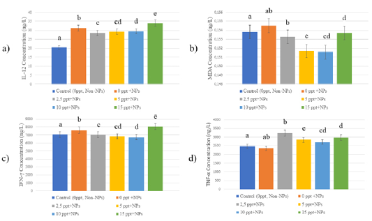
As shown in Figure 1, this study extended these findings to NPs, demonstrating a strong interaction between salinity and NP exposure on immune and oxidative stress responses in tilapia. The results showed that cytokine IL-12 levels in the spleen significantly increased with salinity, reaching a peak at 15 ppt (p < 0.05), indicating enhanced immune activation. This supported the hypothesis that salinity influences NP bioavailability, potentially amplifying their interaction with immune cells. In contrast, MDA levels, an indicator of oxidative stress, decreased in 2.5 – 10 ppt, but exhibited an upward trend at 15 ppt, although the increase was not statistically significant (p > 0.05). This suggested that oxidative stress may have escalated at elevated salinities, possibly due to increased NP accumulation or prolonged exposure effects.
Tumor necrosis factor-alpha (TNF-α), a pro-inflammatory cytokine, exhibited significantly elevated levels in fish exposed to salinities of 2.5, 5, 10, and 15 compared to the control group at 0 ppt (p < 0.05). The highest TNF-α levels were recorded at 2.5 ppt and 15 ppt, indicating that salinity played a critical role in modulating inflammatory responses to NP exposure. Similarly, interferon-gamma (IFN-γ) levels were significantly higher at 15 ppt (p < 0.05), highlighting the strongest immune response at this salinity level (Figure 1). These findings collectively suggested that salinity not only influenced the accumulation and distribution of MPs and NPs but also exacerbated their toxic effects by modulating immune and oxidative stress pathways. The significant increases in IL-12, TNF-α, and IFN-γ at higher salinities underscored the synergistic effect of salinity and NPs in activating inflammatory responses.
NPs and MPs enter fish predominantly via ingestion, passing into the intestines and subsequently entering the circulatory system. Additionally, NPs suspended in the water column can be absorbed during respiration, allowing them to enter the bloodstream. Once in circulation, NPs can accumulate in various organs, causing tissue damage. The spleen, a vital lymphatic organ connected to the circulatory system, is particularly susceptible to NP exposure. It houses lymphocytes, macrophages, and dendritic cells, which are essential for mounting immune responses . This study investigates how NPs influence the immune response in fish.
Pro-inflammatory cytokines such as IL-12, TNF-α, and IFN-γ are widely recognized as reliable markers for assessing immune activation, playing key roles in maintaining immune homeostasis. Malondialdehyde (MDA) serves as a biomarker for oxidative stress, reflecting lipid peroxidation and cellular damage when elevated. Previous research has established that NP exposure can significantly elevate TNF-α and MDA levels in zebrafish intestines . Furthermore, NPs ingested via the food chain have been shown to upregulate inflammatory-related genes, including, including IL6 (interleukin 6), IL8 (interleukin 8), IL1b (interleukin 1b), and TNF-α . Polystyrene NPs have been documented to cross phospholipid membranes via endocytosis, enhancing cellular cytolytic activity ; . However, other findings revealed that while NPs induced histological changes in tilapia testes, they did not significantly affect TNF-α or IFN-γ levels in the blood .
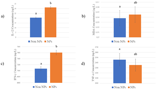
In this study, statistical analysis using a T-test demonstrated that NP exposure significantly increased IL-12 (P < 0.05) and IFN-γ (P < 0.05) levels in the spleen of NP-fed fish compared to the control group, indicating a pronounced immune activation in response to NP exposure (Figure 2). TNF-α levels showed a decreasing trend in NP-exposed fish; however, this change was not statistically significant (P > 0.05). Similarly, MDA levels displayed an upward trend in NP-exposed fish but did not reach statistical significance (p > 0.05), suggesting a potential increase in oxidative stress at the cellular level (Figure 2). These findings highlighted the immunomodulatory effects of NP exposure, particularly the significant upregulation of IL-12 and IFN-γ, which were critical for initiating and maintaining immune defenses.
Histopathological Effects of NPs on the Testis and Liver at Different Salinity Levels
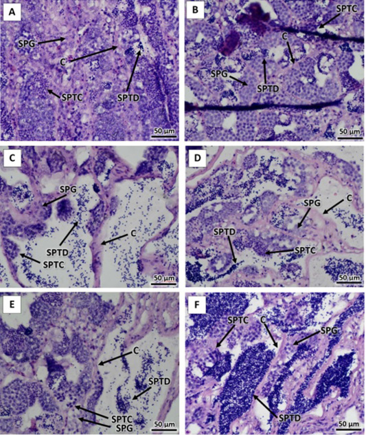
The histopathological analysis of testis tissues, as shown in Figure 3, revealed distinct structural changes across different treatment groups, including the control without NPs, the control with NPs, and NP exposure at salinity levels of 2.5 ppt, 5 ppt, 10 ppt, and 15 ppt. In the control group without NPs, the testis tissue remained structurally intact, with uniform cell distribution and no visible signs of damage or inflammation. In contrast, the control group exposed to NPs exhibited minor structural changes, indicating the initial effects of NP exposure, although these changes were minimal. As salinity increased, the severity of histopathological changes became more evident. At 2.5 ppt, mild structural disruptions and early signs of inflammation appeared, suggesting the initial effects of NP and salinity exposure. By 5 ppt, tissue damage was more pronounced, with increased inflammatory cell infiltration and structural modifications. At 10 ppt, the extent of tissue deterioration intensified, characterized by structural disintegration and moderate to severe inflammation. At 15 ppt, the highest salinity level, testis tissues exhibited severe damage, including widespread inflammation, extensive structural breakdown, and early signs of necrosis. These findings demonstrated a clear relationship between increasing salinity and the severity of NP-induced tissue damage. The progressive rise in inflammation and structural degradation suggested a compounding effect of NP toxicity under higher salinity conditions.
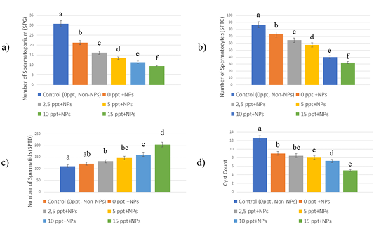
Figure 4 illustrated the adverse effects of NP exposure at varying salinity levels on the spermatogenesis process in tilapia. The results showed a progressive decline in spermatogonia, spermatocytes, and cysts as salinity increased, suggesting that higher salinity exacerbated NP-induced toxicity in the gonads. This decline indicated impaired early-stage germ cell development, which is crucial for normal reproductive function. Spermatogenesis is a tightly regulated process involving the sequential development of spermatogonia (early-stage germ cells), spermatocytes (intermediate-stage cells undergoing meiotic division), and spermatids (late-stage cells that mature into spermatozoa). In control fish, a balanced distribution of these cells was observed, ensuring continuous sperm production. However, exposure to NPs disrupted this balance. At increasing salinity levels, the number of spermatogonia and spermatocytes significantly decreased, indicating a reduction in new cell formation. This suggested that NP exposure interfered with the proliferation and differentiation of early germ cells, possibly through oxidative stress, inflammatory responses, or hormonal disruptions . Interestingly, despite the decline in early-stage germ cells, spermatid numbers increased in groups exposed to higher salinity. This accumulation likely resulted from a reduced capacity of the gonads to generate new spermatogonia and spermatocytes, leading to a bottleneck effect in which spermatids were not replenished at the same rate as they would be under normal conditions. Since spermatids represent the final stage before sperm maturation , their increased presence without a corresponding influx of earlier-stage cells suggested an interruption in the spermatogenic cycle, potentially reducing overall sperm production and fertility. These findings provided strong evidence that NP exposure, in combination with elevated salinity, disrupted normal reproductive processes in tilapia by impairing spermatogenesis at multiple stages.
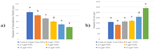
As shown in Figure 5, seminiferous tubule diameter decreased in fish exposed to NPs, with further reduction observed as salinity levels increased. This suggests that NP exposure under higher salinity conditions led to testicular atrophy, likely due to disrupted spermatogenesis and cellular damage. A reduction in seminiferous tubule diameter is often linked to lower sperm production and impaired testicular function ; , highlighting the negative impact of environmental stressors on reproductive health. In contrast, cyst diameter showed a different response to salinity. In fish exposed to NPs at 0 ppt salinity, cyst diameter decreased, suggesting impaired germ cell development and reduced spermatogenic activity ; . However, as salinity increased, cyst diameter progressively expanded. This pattern may indicate a compensatory response, where the gonads attempt to adapt to environmental stress by modifying cyst structure . These findings suggest that NP exposure, particularly under increasing salinity, negatively affects spermatogenesis by reducing seminiferous tubule diameter and altering cyst structure. The combination of NP toxicity and elevated salinity contributes to reproductive dysfunction.
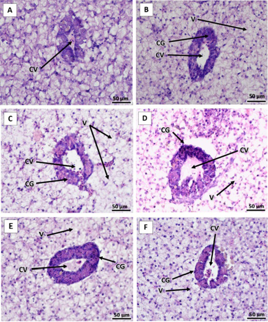
The histopathological analysis of liver tissues, as presented in Figure 6, demonstrated a progressive increase in tissue damage in tilapia exposed to NPs, with severity escalating in response to rising salinity levels. At 2.5 ppt, liver damage was more pronounced than at 0 ppt, suggesting that even a slight increase in salinity heightened hepatocyte congestion and vacuolar degeneration ; ; . This trend indicates that salinity may act as a physiological stressor, exacerbating the hepatotoxic effects of NPs. At 5 ppt, histopathological alterations became more severe, with increased vacuolization and hepatic congestion. This suggests that the combined effects of salinity-induced osmotic stress and NP toxicity intensified hepatic dysfunction. By 10 ppt, liver damage was more extensive, marked by pronounced hepatocyte disintegration and significant vacuolar degeneration. These findings imply that as salinity increased, fish experienced greater physiological strain, further amplifying NP-induced hepatotoxicity. At 15 ppt, the highest salinity level tested, liver damage was most severe, characterized by widespread hepatocyte necrosis, severe congestion, and extensive vacuolar degeneration. These findings suggest that at high salinity, NP exposure leads to compounded hepatic impairment, potentially overwhelming the liver’s capacity for detoxification and repair ; ; . Overall, the results of this study indicate a clear correlation between increasing salinity and the severity of NP-induced hepatic damage. The findings suggest that environmental stressors, such as salinity fluctuations, may enhance the toxicological effects of NPs, leading to greater physiological burden and organ dysfunction in aquatic organisms. These results highlight the importance of considering multiple environmental factors when assessing pollutant toxicity in aquatic ecosystems.
CONCLUSIONS AND RECOMMENDATIONS
Exposure to NPs in tilapia resulted in alterations in pro-inflamatory cytokines (IL-12, TNF-α and IFN-γ), and oxidative stress markers (MDA) in the spleen. At the same salinity, NP exposure led to increased MDA, IL-12, and IFN-γ levels, while TNF-α exhibited a slight decrease compared to the control group. The study also revealed variations in NP-induced cytokine responses across different salinity levels. At higher salinity, NP exposure led to an upregulation of IL-12, IFN-γ, and TNF-α, indicating that salinity may modulate the inflammatory response to NP toxicity. Furthermore, NP exposure had distinct effects on different organs, with both the liver and testis exhibiting variable responses at increasing salinity levels. These findings suggest that the combined influence of NPs and salinity plays a critical role in determining immune and physiological responses in tilapia, highlighting the need for further research on the interaction between environmental stressors and NP toxicity in aquatic organisms
ACKNOWLEDGEMENTS
The author would like to express gratitude to the Directorate of Research, Technology, and Community, Ministry of Education, Culture, Research and Technology, Universitas Airlangga, Indonesia which provides funding for Master Thesis Research Activities in 2024 Grant no. 0667/E5/AL.04/2024 , May 30th 2024.
NOVELTY STATEMENTS
Salinity significantly influences nanoplastics toxicity, with higher salinity levels amplifying immune responses, oxidative stress, and histological damage in fish.
AUTHOR’S CONTRIBUTIONS
Muhammad Iqbal, Muhammad Nuh Fathsyah Siregar and Alfiah Hayati; designed and planned the research, as well as contributed to manuscript writing. Muhammad Iqbal, Muhammad Nuh Fathsyah Siregar, Ghaitsa Zahira Sofa, Manikya Pramudya and Hisyam Rizky Saputra; conducted all experiments and prepared the fish samples. Muhammad Iqbal, wrote the manuscript, performed statistical analysis and created the graphical designs for this paper. All authors read and approved the final manuscript.
Conflict of Interest
The authors declare no conflict of interest.
Figures
Figure 1:.
Immune response and oxidative stress exposed to NPs at different salinity levels. (a) IL-12; (b) MDA; (c) IFN γ; (d) TNF-α levels in fish exposed to various NP concentrations: 0 ppt (control), 0 ppt + NP, 2.5 ppt + NP, 5 ppt + NP, 10 ppt + NP, and 15 ppt + NP. All of the data were displayed by mean ±SD. Different letters indicate statistically significant differences (p<0.05).
Figure 2:.
Cytokine concentrations (IL-12, IFN-γ, TNF-α) and MDA levels in the spleen of fish exposed to (NP) (Orange bars) compared to non-exposed fish (blue bars). a) IL-12, b) MDA, c) IF-γ, d) TNF-α. All of the data were displayed by mean ±SD (α= 0.05). Different letters indicate statistically significant differences (p<0.05).
Figure 3:.
Histological analysis of tilapia testis after 25 days of exposure to NPs. Panel A: control (0 ppt without NPs); Panel B: 0 ppt + NPs; Panel C: 2.5 ppt + NPs; Panel D: 5 ppt + NPs; Panel E: 10 ppt + NPs; Panel F: 15 ppt + NPs. SPG: Spermatogonia; SPTC: Spermatocytes; SPTD: Spermatids; C: Cyst Sections. All samples were stained with hematoxylin and eosin and observed at 400x magnification.
Figure 4:.
The number of spermatogenic and cysts after NPs exposure. (a) Spermatogonium (SPG) (b) Spermatocytes (SPTC) (c) Spermatids (SPTD) (d) Cyst Count. All of the data were displayed by mean ±SD (α= 0.05). Different letters indicate statistically significant differences (p<0.05).
Figure 5:.
Diameter of seminiferous tubules (a) and cysts (b) after NPs exposure. All of the data were displayed by mean ± SD (α = 0.05). Different letters indicate statistically significant differences (p<0.0001).
Figure 6:.
Histology of tilapia liver after exposure to NPs for 25 days. A= control (0 ppt without NPs); B= 0 ppt + NPs; C= 2.5 ppt + NPs; D = 5 ppt + NPs; E = 10 ppt + NPs; F = 15 ppt + NPs. CV: central vein; V: cytoplasmic vacuolation and CG: congestion. Sections were stained with hematoxylin and eosin and observed at 400x magnification.
References
Amponsah AK, Afrifa EA, Essandoh PK, Enyoh CE (2024). Evidence of microplastics accumulation in the gills and gastrointestinal tract of fishes from an estuarine system in Ghana. Heliyon., 10: e25608. https://doi.org/10.1016/j.heliyon.2024.e25608[CrossRef]
Andrady AL (2011). Microplastics in the marine environment. Mar. Pollut. Bull., 62: 1596–1605. https://doi.org/10.1016/j.marpolbul.2011.05.030[CrossRef]
Asmat MS, Zheng XY, Nauman M, Zheng D, Stanley P (2024). Deletion of Mgat2 in spermatogonia blocks spermatogenesis. Front. Cell Dev. Biol., 12: 1–14. https://doi.org/10.3389/fcell.2024.1428715[CrossRef]
Assas M, Qiu X, Chen K, Ogawa H, Xu H, Shimasaki Y, Oshima Y (2020). Bioaccumulation and reproductive effects of fluorescent microplastics in medaka fish. Mar. Pollut. Bull., 158: 4–9. https://doi.org/10.1016/j.marpolbul.2020.111446[CrossRef]
Bai Z, Wang N, Wang M (2021). Effects of microplastics on marine copepods. Ecotoxicol. Environ. Saf., 217: 112243. https://doi.org/10.1016/j.ecoenv.2021.112243[CrossRef]
Cai Y, Li C, Zhao Y (2022). A review of the migration and transformation of microplastics in inland water systems. Int. J. Environ. Res. Public Health., 19: 148. https://doi.org/10.3390/ijerph19010148[CrossRef]
Che X, Geng L, Zhang Q, Wei H, He H, Xu W, Shang X (2024). Selenium-rich Lactobacillus plantarum alleviates salinity stress in Cyprinus carpio: Growth performance, oxidative stress, and immune and inflammatory responses. Aquacult. Rep., 36: 102058. https://doi.org/10.1016/j.aqrep.2024.102058[CrossRef]
Emon FJ, Hasan J, Shahriar SIM, Islam N, Islam MS, Shahjahan M (2024). Increased ingestion and toxicity of polyamide microplastics in Nile tilapia with increase of salinity. Ecotoxicol. Environ. Saf., 282: 116730. https://doi.org/10.1016/j.ecoenv.2024.116730[CrossRef]
Hamed M, Soliman HAM, Badrey AEA, Osman AGM (2021). Microplastics induced histopathological lesions in some tissues of tilapia (Oreochromis niloticus) early juveniles. Tissue Cell., 71: 101512. https://doi.org/10.1016/j.tice.2021.101512[CrossRef]
Hayati A, Wilujeng WP, Pramudya M, Dewi FRP, Sugiharto, Muchtaromah B, Susilo RJK (2024). The Effect of Exposure to Polystyrene Nanoplastics on Cytokine Levels and Reproductive System of Male Tilapia. Trop. J. Nat. Prod. Res., 8: 6300–6303. https://doi.org/10.26538/tjnpr/v8i2.30[CrossRef]
Hoyo-Alvarez E, Arechavala-Lopez P, Jiménez-García M, Solomando A, Alomar C, Sureda A, Moranta D, Deudero S (2022). Effects of pollutants and microplastics ingestion on oxidative stress and monoaminergic activity of seabream brains. Aquatclt. Toxicol., 242: 106048. https://doi.org/10.1016/j.aquatox.2021.106048[CrossRef]
Li L, Gu H, Chang X, Huang W, Sokolova IM, Wei S, Sun L, Li S, Wang X, Hu M, Zeng J, Wang Y (2021). Oxidative stress induced by nanoplastics in the liver of juvenile large yellow croaker Larimichthys crocea. Mar. Pollut. Bull., 170: 112661. https://doi.org/10.1016/j.marpolbul.2021.112661[CrossRef]
Li X, Zheng Y, Lu L, Eom J, Ru S, Li Y, Wang J (2024). Trophic transfer of micro- and nanoplastics and toxicity induced by long-term exposure of nanoplastics along the rotifer (Brachionus plicatilis)-marine medaka (Oryzias melastigma) food chain. Environ. Pollut., 346: 123599. https://doi.org/10.1016/j.envpol.2024.123599[CrossRef]
Lin X, Wang Y, Yang X, Watson P, Yang F, Liu H (2023). Endocrine disrupting effect and reproductive toxicity of the separate exposure and co-exposure of nano-polystyrene and diethylstilbestrol to zebrafish. Sci. Total Environ., 865: 161100. https://doi.org/10.1016/j.scitotenv.2022.161100[CrossRef]
Liu L, Xu K, Zhang B, Ye Y, Zhang Q, Jiang W (2021). Cellular internalization and release of polystyrene microplastics and nanoplastics. Sci. Total Environ., 779: 146523. https://doi.org/10.1016/j.scitotenv.2021.146523[CrossRef]
Palmer RM, Sandbach A, Buckley BA (2024). Tissue-specific effects of temperature and salinity on the cell cycle and apoptosis in the Nile Tilapia (Oreochromis niloticus). Comp. Biochem. Physiol. -Part A Mol. Integr. Physiol., 297: 111706. https://doi.org/10.1016/j.cbpa.2024.111706[CrossRef]
Pratiwi HM, Takagi T, Rusni S, Inoue K (2023). Euryhaline fish larvae ingest more microplastic particles in seawater than in freshwater. Sci. Rep., 13: 1–10. https://doi.org/10.1038/s41598-023-30339-y[CrossRef]
Roch S, Friedrich C, Brinker A (2020). Uptake routes of microplastics in fishes: practical and theoretical approaches to test existing theories. Sci. Rep., 10: 1–12. https://doi.org/10.1038/s41598-020-60630-1[CrossRef]
Rodrigues SM, Espincho F, Elliott M, Almeida CMR, Ramos S (2023). Methodology optimization to quantify microplastic presence in planktonic copepods, chaetognaths and fish larvae. MethodsX., 11: 102466. https://doi.org/10.1016/j.mex.2023.102466[CrossRef]
Soares MO, Matos E, Lucas C, Rizzo L, Allcock L, Rossi S (2020). Microplastics in corals: An emergent threat. Mar. Pollut. Bull., 161: 111810. https://doi.org/10.1016/j.marpolbul.2020.111810[CrossRef]
Solomando A, Cohen-Sánchez A, Box A, Montero I, Pinya S, Sureda A (2022). Microplastic presence in the pelagic fish, Seriola dumerili, from Balearic Islands (Western Mediterranean), and assessment of oxidative stress and detoxification biomarkers in liver. Environ. Res., 212: 4–11. https://doi.org/10.1016/j.envres.2022.113369[CrossRef]
Sun Z, Zhao L, Peng X, Kang B (2024). A comparative study on the toxic effects of lead pollution and nanoplastic-lead mixed pollution on red drum and their detoxification strategies. J. Hazard. Mater., 480: 136018. https://doi.org/10.1016/j.jhazmat.2024.136018[CrossRef]
Thoman TX, Kukulka T, Cohen JH, Boettcher H (2023). Zooplankton-microplastic exposure in Delaware coastal waters: Atlantic blue crab (Callinectes sapidus) larvae case study. Mar. Pollut. Bull., 196: 115541. https://doi.org/10.1016/j.marpolbul.2023.115541[CrossRef]
Walsh CT, Pease BC (2002). The use of clove oil as an anaesthetic for the longfinned eel, Anguilla reinhardtii (Steindachner). Aquac. Res., 33: 627–635. https://doi.org/10.1046/j.1365-2109.2002.00701.x[CrossRef]
Wang X, Liu Q, Li J, Zhou L, Wang T, Zhao N (2023). Dynamic cellular and molecular characteristics of spermatogenesis in the viviparous marine teleost Sebastes schlegelii. Biol. Reprod., 108: 338–352. https://doi.org/10.1093/biolre/ioac203[CrossRef]
Williams AT, Rangel-Buitrago N (2022). The past, present, and future of plastic pollution. Mar. Pollut. Bull., 176: 113429. https://doi.org/10.1016/j.marpolbul.2022.113429[CrossRef]
Yang M, Wang WX (2023). Recognition and movement of polystyrene nanoplastics in fish cells. Environ. Pollut., 316: 120627. https://doi.org/10.1016/j.envpol.2022.120627[CrossRef]
Yu J, Chen L, Gu W, Liu S, Wu B (2022). Heterogeneity effects of nanoplastics and lead on zebrafish intestinal cells identified by single-cell sequencing. Chemosphere., 289: 133133. https://doi.org/10.1016/j.chemosphere.2021.133133[CrossRef]
Zapata AG (2024). The fish spleen. Fish Shellfish Immunol., 144: 109280. https://doi.org/10.1016/j.fsi.2023.109280[CrossRef]
Zheng Y, Hamed M, De-la-Torre GE, Frias J, Jong MC, Kolandhasamy P, Chavanich S, Su L, Deng H, Zhao W, Shi H (2023). Holes on surfaces of the weathered plastic fragments from coastal beaches. Mar. Pollut. Bull., 193: 115180. https://doi.org/10.1016/j.marpolbul.2023.115180[CrossRef]
Zhou Y, Jin Q, Xu H, Wang Y, Li M (2023). Chronic nanoplastic exposure induced oxidative and immune stress in medaka gonad. Sci. Total Environ., 869: 161838. https://doi.org/10.1016/j.scitotenv.2023.161838[CrossRef]
Zhou Y, Zhao L, Xu H, Xu EG, Li M, Wang Y (2022). Long-Term Exposure to Polystyrene Nanoplastics Impairs the Liver Health of Medaka. Water (Switzerland)., 14: 1–11. https://doi.org/10.3390/w14172767[CrossRef]