Research Article · Volume 13, Issue 3, 1 September 2025 · Open Access
Investigation of the In Vitro Therapeutic Potential of Strigosella africana Against Leishmania tropica
· · ·
Affiliations & Notes ⌄
1Center of Desert Studies, University of Anbar, Ramadi 31001, Iraq
2College of Pharmacy, University of Al Maarif, Ramadi 31001, Iraq
3Department of Occupational Safety and Occupational Medicine Techniques, College of Health and Medical Techniques, Northern Technical University, Kirkuk, Iraq. *Correspondence | Mustafa M. Fahad, Center of Desert Studies, University of Anbar, Ramadi 31001, Iraq
This is the first study to utilize S. africana against the L. tropica parasite in vitro.
As shown in Table 1, promastigote counts in the control group increased significantly over time, reaching (1.24, 7.87, 18.01, and 28.54) × 10⁵ cells/cm³ at 24, 48, 72, and 96 h, respectively.
At 96 h, parasite growth decreased from 82.1% to 0% at the highest concentration, showing significant differences from the untreated control, where no inhibition occurred.
This study concludes that the alcoholic extract of S. africana is effective and highly efficient in treating L. tropica parasites and eliminating them through the significant inhibition of promastigote growth of these parasites in the culture media, resulting from numerous active chemicals that kill the parasites.
Selected from the article's own text.
Abstract
Leishmania tropica is a zoonotic protozoan parasite capable of infecting both humans and animals, with dogs identified as primary reservoir hosts. This study evaluated the in vitro effects of the alcoholic extract of Strigosella africana on the growth and development of L. tropica promastigotes. The extract exhibited a clear inhibitory effect on promastigote proliferation, reducing the number of generations and prolonging generation time over a 24–96 hour incubation period. Dose-dependent suppression was observed across concentrations ranging from 0.5 to 5.5 mg/ml, with higher concentrations producing a more pronounced decline in parasite count. Notably, the 5.5 mg/ml concentration completely inhibited promastigote growth by 72 hours. The half-maximal lethal dose (LDâ‚…â‚€) was achieved at 2.5 mg/ml, reducing parasite density to 14.27 × 10ⶠcells/cm³ compared to 28.54 × 10ⶠcells/cm³ in the control group. Moreover, the number of generations decreased significantly, reaching 4.21 after 96 hours of treatment, compared to 8.43 in the untreated control. These results demonstrate that the alcoholic extract of S. africana possesses potent antiparasitic activity against L. tropica promastigotes in vitro. The findings support the potential use of bioactive plant-derived compounds as natural alternatives for developing safer and more effective therapies for parasitic infections. This study highlights the promise of S. africana in contributing to plant-based interventions for managing parasitic diseases in animals.
Leishmania is a genus of single-celled, flagellated blood parasites, and more than 30 species parasitize different vertebrate hosts, particularly mammals, of which 20 species infect humans, causing a range of pathological symptoms . This genus belongs to the Trypanosomatidae family and the Zoomastigophora class, which obligately parasitizes within phagocytic cells in the retinal lining of vertebrate hosts . The most common Leishmania species is L. tropica, which causes skin lesions that start as papules and develop into large ulcers, potentially resulting in permanent scars or severe disFigurement (Peç and da Silva, 2024). This parasite causes cutaneous leishmaniasis, which is known by various local names in endemic regions, such as Delhi boil, Aleppo boil, Baghdad boil, and Oriental boil . The disease is endemic to over 88 countries, with an estimated 350 million people at risk .
The bite of an infected female sandfly transmits the parasite. Transmission may also occur through contact with crushed insects on the skin . The parasite exists in two main stages: the amastigote stage, which is found inside the phagocytic cells of the vertebrate host and has a small, circular, or oval shape measuring 2–4 microns in length , and the promastigote stage, which is found in the midgut of the vector or culture media, which is characterized by its spindle shape and is equipped with a fine flagellum at its anterior end, which is why it is called the promastigote stage, and its length ranges from 10 to 15 µm . Recently, research has shifted towards the use of medicinal herbs and plants to treat various diseases, including parasitic diseases. These offer safe therapeutic alternatives to harmful chemical treatments because of the abundance of beneficial compounds, such as volatile oils, alkaloids, flavonoids, glycosides, phenols, resins, and saponins which target pathogens and facilitate their elimination .
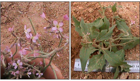
Strigosella africana, locally known as khazima, belongs to the family Brassicaceae (Cruciferae). With highly branched stems at the base, this annual herb grows between 15 and 40 cm in height. Plants prefer moist areas and small irrigation ditches, regardless of the soil type. They are commonly found in tree-lined irrigation ditches, swamps near agricultural fields, meadows, and farming oases. Numerous active compounds are found in plants, including volatile oils, flavones, alkaloids, sterols, terpenes, and saponins . The alcoholic extract of the plant has been shown to treat various conditions, including eczema and cutaneous leishmaniasis . Moreover, it has antimicrobial properties and limits the growth of parasites and fungi . Several studies have investigated the effects of medicinal plants on L. tropica. For example, Shahatha and Saleh used an extract of Rhanterium epapposum to inhibit the proliferation of L. tropica in vitro. In addition, Balikci et al. studied the impact of Pelargonium sidoides on the promastigote forms of the same parasite in vitro. Furthermore, some researchers have conducted studies to determine the effect of Carrichtera annua plant extract on L. donovani activity in vitro .
In light of the widespread prevalence of Leishmania tropica and its severe pathological consequences, this study was conducted to explore the potential of Strigosella africana alcoholic extract as an effective alternative to conventional chemical treatments, which are often costly and associated with serious side effects. The research aimed to evaluate the extract’s in vitro inhibitory effect on L. tropica promastigotes, with the goal of identifying a natural therapeutic option to reduce parasite burden and mitigate the associated health complications.
MATERIALS AND METHODS
Preparing an Alcoholic Plant Extract of S. Africana
A S. africana plant, as shown in Figure 1, was obtained from the Zankoura area, northwest of the Ramadi/Anbar province, and was identified at the University of Anbar’s Herbarium. The plants were then cleaned and dried. Following the Harborne method , 50 g of dried plant material was mixed with 500 mL of 70% ethanol (10:1 ratio) to prepare an alcoholic extract. The mixture was stirred using a medium-speed magnetic stirrer and was maintained at 40–45°C for 72 h. Subsequently, a Buchner funnel was used to filter it, and the resulting filtrate was collected.
The solvent was eliminated by evaporation at a moderate speed and 45°C in a rotating evaporator. Chlorophyll pigments were removed using filter sheets, after which the solution was re-evaporated to eliminate residual water; the extract underwent pasteurization at 62°C for ten minutes to obtain a concentrated form, after receiving the standard concentration of the extract, the required experimental concentrations (0.5, 1.5, 2.5, 3.5, 4.5, and 5.5 mg/mL) were made.
Identification of the Plant’s Active Ingredients Chemically
Glycoside identification: One milligram of the dried plant extract was combined with ten millilitres of distilled water, filtered, and then a few drops of Fehling’s reagent were added. Glycosides were present when red hues appeared.
Tannins identification: Five milliliters of the plant extract were mixed using several drops of lead acid (1%); tannins are available when a white, gelatinous deposit forms.
Volatile oils identification: The presence of volatile oils is shown by the formation of a grey hue when drops of the extract are applied to paper filters until saturated and exposed to UV light.
Flavonoids identification: The desiccated extract (10 g) was solubilized in 50 mL of 95% ethyl alcohol, followed by filtration of the solution. Subsequently, a solution was prepared by combining ten milliliters of 50% ethyl alcohol with ten milliliters of KOH at 50%, and equivalent volumes of the two solutions were mixed. Flavonoids can be identified by their yellow hue.
Alkaloids identification: Plant extracts (10 g) were heated with 50 mL of purified water that had been acidified with 4% HCl, and the mixture was then filtered. After cooling, half a milliliter of this mixture was examined.
Phenol identification: One milliliter of the dry plant extract was combined with one milliliter of a 1% FeCl3 solution. Phenols were indicated when blue or olive hues appeared.
Terpene identification: The existence of terpenes is shown by the formation of a brown precipitate after one gram of the dried extract was diluted in two milliliters of chloroform, and concentrated H2SO4and anhydrous acetic acid were added.
Saponin identification: When 5 mL of the plant extract was combined with 3 mL of a 1% mercuric chloride (HgCl₂) solution, a white precipitate formed, demonstrating the presence of saponins. The identification methods used were based on the procedures described by Han et al. (2024).
Development and Calculation of L. tropica Promastigotes
The original stock culture of L. tropica (strain MHOM/IQ/1992/MREC3) containing promastigote stages, was acquired from the University of Nahrain’s Medical Research Center, Medicine College. It was enzymatically characterized using the isoenzyme method . The parasites were cultured in Tobie’s medium . To cultivate Leishmania parasites, 0.1 cm³ of Locke’s solution containing live promastigotes (four days old) was added to glass bottles containing 1.9 cm³ of liquid medium. The initial cell density was 2 × 10⁵ cells/cm³. The cultures were incubated at 26°C for four days. A Neubauer chamber was used to count promastigotes in each culture. The parasites were fixed while counting by adding 0.1 mL of 10% formalin (0.9 mL to a culture). Normal saline was used as a diluent to promote dense growth. Promastigotes were counted using a light microscope at 40× magnification, yielding a count of promastigote stages per 1 cm³ of culture .
Impact of Extract Concentrations on the Leishmania Promastigote
Alcoholic extract concentrations of S. africana (0.5, 1.5, 2.5, 3.5, 4.5, and 5.5 mg/mL) were prepared and added to sterile glass bottles containing Tryptone-Soya broth (Tobie’s medium). Each solution was inoculated with 2 × 10⁵ cells/cm³ of the Leishmania culture.
A control group without extract was also included, where 1.9 cm³ of sterile Tobie’s medium (Tryptone-Soya broth) was placed in sterile glass bottles and inoculated with 0.1 cm³ of Locke’s solution containing 2 × 10⁵ L. tropica promastigotes per cm³, without any addition of the plant extract. This setup mirrored the conditions of the treated groups but excluded the S. africana extract. The bottles were incubated at 26°C for four days, and promastigote counts were taken at 24, 48, 72, and 96 hours.
And all bottles were incubated at 26°C for four days. Parasite counts were assessed at different growth intervals (24, 48, 72, and 96 h), and the generation number and time were estimated following the method of Benjamin and Garman . Each experiment was performed in triplicate (n = 3) to ensure statistical reliability.
Statistical Analysis
Data were analyzed using the Statistical Analysis System (SAS, 2012). A one-way analysis of variance (ANOVA) was performed to determine significant differences among treatment groups. Means were compared using Duncan’s multiple range test at a significance level of p < 0.05. Results were expressed as mean ± standard deviation (SD), based on triplicate measurements (n = 3). Furthermore, the median inhibitory concentration (LD₅₀) of S. africana alcoholic extract against L. tropica promastigotes was determined using nonlinear regression analysis performed in IBM SPSS Statistics, version 25.0 , based on the dose–response curve generated from parasite counts at various extract concentrations.
RESULTS AND DISCUSSION
This is the first study to utilize S. africana against the L. tropica parasite in vitro. This lays the groundwork for investigating the potential for therapy using plants and herbs against parasitic infections due to their content of bioactive compounds that can inhibit or eliminate pathogenic parasites.
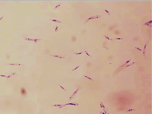
This study revealed a clear inhibitory effect of the S. africana alcoholic extract on L. tropica promastigote growth at varying concentrations, in contrast to the control (untreated) group, as illustrated in Figure 2, where promastigotes appeared abundant, slender, and exhibited active motility under the microscope, indicating normal growth and viability.
Table 1: Impact of various concentrations of alcohol extract of S. Africana on the number of L. tropica (×106) at different growth times (number of promastigotes utilised in culture 2×10⁵ cells/cm3).
Concentration
(mg/ml)
24 h (AV ± SD)
GR %
48 h (AV ± SD)
GR %
72 h (AV ± SD)
GR %
96 h (AV ± SD)
GR
%
Control (0)
1.24 ± 0.12ᵃ
100
7.8 ± 0.98ᵃ
100
18.01 ± 0.15ᵃ
100
28.54 ± 0.04ᵃ
100
0.5
1.04 ± 0.22ᵇ
82.2
6.5 ± 0.23ᵇ
81.0
17.03 ±0.33ᵇ
80.5
23.66 ± 0.58ᵇ
82.1
1.5
0.89 ± 0.02ᶜ
68.5
5.6 ± 0.91ᶜ
63.0
13.79 ± 0.18ᶜ
58.9
20.80 ± 1.03ᶜ
55.2
2.5
0.62 ± 0.35ᶜ
54.2
3.5 ± 0.16ᵈ
52.9
8.18 ± 0.14ᵈ
49.0
14.27 ± 0.09ᵈ
50.1
3.5
0.51 ± 0.09ᵈ
41.3
2.5 ± 0.78ᵉ
38.2
5.34 ± 0.39ᵉ
25.3
7.08 ± 0.78ᵉ
24.5
4.5
0.38 ± 0.11ᵉ
31.0
1.9 ± 0.10ᶠ
18.6
2.91 ± 0.02ᶠ
14.1
0.00 ± 0.00ᶠ
0.0
5.5
0.20 ± 0.43ᶠ
21.5
1.1 ± 0.22ᵍ
12.6
0.00 ± 0.00ᵍ
0.0
0.00 ± 0.00ᶠ
0.0
* Values are means ± standard deviations (n=3). Different superscript letters within columns indicate significant differences (p < 0.05). AV: Average; SD: Standard Deviation; GR%: Growth Rate Percentage.
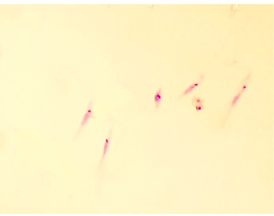
As shown in Table 1, promastigote counts in the control group increased significantly over time, reaching (1.24, 7.87, 18.01, and 28.54) × 10⁵ cells/cm³ at 24, 48, 72, and 96 h, respectively. In contrast, treatment with various extract concentrations (0.5, 1.5, 2.5, 2.0, 3.5, 4.5, and 5.5 mg/ml) inhibited the growth of promastigotes. A marked reduction in parasite numbers was observed as the extract concentration increased across all time intervals, as the maximum concentration of 5.5 mg/ml entirely stopped parasite growth 72 h after treatment, as demonstrated in Figure 3, including morphological changes and a significant reduction in parasite numbers.
There were noticeable variations (P< 0.05) in the growth rates of the promastigotes after treatment compared with the untreated group. After 96 h, 50% inhibition was observed at a 2.5 mg/mL concentration (LD < > < B-B->). The growth percentage also declined at higher doses. At 24 hours, the reduction ranged from 82.2% to 21.5% for 0.5 and 5.5 mg/mL, respectively. At 48 h, the decrease was from 81% to 12.6%, and by 72 h, the growth was reduced from 80.5% to 0%. At 96 h, parasite growth decreased from 82.1% to 0% at the highest concentration, showing significant differences from the untreated control, where no inhibition occurred. A standard antileishmanial drug (e.g., Amphotericin B) was not included as a positive control; future comparative studies are recommended to validate the relative efficacy of the extract.
This aligns with the results of Akya et al. (2020) and Badirzadeh et al. (2020). The pronounced inhibitory effect of the alcoholic extract of S. africana on L. tropica promastigote growth is attributed to its rich composition of bioactive substances, including sterols, saponins, volatile oils, alkaloids, flavonoids, and terpenes, which directly interfere with vital cellular functions, including enzyme synthesis and mitochondrial activity, ultimately resulting in cytotoxicity and parasite destruction . It may also lead to inactivation of the enzyme dihydrofolate reductase (DHFR), which is responsible for building thymine nucleotides through the de novo pathway, forming the primary sources of DNA, RNA, protein, and phospholipids. In addition, it converts dihydrofolate to tetrahydrofolate, as inhibition of this enzyme leads to inhibition of tetrahydrofolate, thus causing cell poisoning and parasite death . Furthermore, the extract also appears to suppress acetylcholinesterase enzyme activity, which governs essential physiological processes in the parasite and regulates ion permeability of the cell membrane, ultimately resulting in its death . The proposed mechanism, involving inhibiting enzymes such as dihydrofolate reductase (DHFR) and acetylcholinesterase, is hypothetical. These mechanistic interpretations are based on prior literature related to similar phytochemicals, and no direct biochemical assays were performed in the present study to confirm these effects.
Effect of S. Africana Extract on L. tropica Promastigote Generation Number
These findings confirmed the effect of S. africana’s alcoholic extract of S. africana on the promastigote generation number, showing a reduction proportional to the increase in concentration. Notable variances (P<0.05) were noted in the number of promastigote generations between the treated and control groups during different incubation periods.
Complete inhibition of generation was observed after 72 h of treatment with 5.5 mg/mL, similarly, 4.5 mg/mL inhibited generation entirely after 96 h. Within 24 hours, the number of generations decreased from 2.62 to 0.38 when using the concentration from 0.5 to 5.5 mg/ml, compared to 2.98 in the control. After 48 h, the number dropped from 4.49 to 0.98 compared to 4.92 in the control group. At 96 hours, 2.5 mg/mL resulted in 4.21 generations, representing the (LD50) calculated using nonlinear regression analysis with IBM SPSS Statistics version 25.0 , compared to 8.43 in the control group; these generation differences are detailed in Table 2.
Table 2: Impact of various concentrations of alcohol extract of S. Africana on the generation number of L. tropica promastigote (x106) at different times (number of promastigotes utilised in culture 2×10⁵ cells/cm3).
Concentration (mg/ml)
24 h
(AV ± SD)
48 h
(AV ± SD)
72 h
(AV ± SD)
96 h
(AV ± SD)
Control (0)
2.98±0.31ᵃ
4.92±0.53ᵃ
7.97±0.63ᵃ
8.43±0.20ᵃ
0.5
2.62±0.21ᵇ
4.49±0.01ᵇ
6.64±0.03ᵇ
7.85±0.65ᵇ
1.5
2.30±0.03ᵇ
4.31±0.12ᵇ
5.71±0.73ᶜ
6.32±0.09ᶜ
2.5
1.45±0.04ᶜ
3.78±0.03ᶜ
3.85±0.06ᵈ
4.21±0.80ᵈ
3.5
1.25±0.10ᶜ
3.11±0.22ᵈ
3.20±0.23ᵉ
2.58±0.24ᵉ
4.5
1.08±0.12ᵈ
2.14±0.93ᵉ
2.23±0.12ᶠ
0.00±0.00ᶠ
5.5
0.38±0.05ᵉ
0.98±0.44ᶠ
0.00±0.00ᵍ
0.00±0.00ᶠ
* Values are means ± standard deviations (n=3). Different superscript letters within columns indicate significant differences (p < 0.05).
These results align with those of , which showed similar effects of various plant extracts on the generation rate of promastigotes in vitro. The inhibitory effect of S. africana alcoholic extract on the generation number is attributed to its significant suppression of parasite growth, likely due to disrupted reproductive mechanisms, inhibition of genetic material production, and impaired formation of new generations. The bioactive constituents of the extract create suboptimal conditions for parasite development and reproduction .
Effect of S. africana Extract on L. tropica Promastigote Generation Time
Table 3 shows the impact of the S. Africana on the parasite’s generation time. An increase in the extract concentration was associated with a prolonged generation time. The results demonstrated an apparent influence of extract concentration on promastigote generation time, resulting in extended generation periods due to growth inhibition compared to the control group.
Table 3: Impact of concentrations of alcohol extract of S. Africana on the generation time (hours) of the L. tropica promastigote (x106) at different times (number of promastigotes utilised in culture 2×10⁵ cells/cm3)
Concentration (mg/ml)
24 h
(AV ± SD)
48 h
(AV ± SD)
72 h
(AV ± SD)
96 h
(AV ± SD)
Control(0)
9.50±0.11ᵃ
13.60±0.18ᵃ
15.16±0.05ᵃ
17.35±0.18ᵃ
0.5
10.21±0.60ᵇ
14.12±0.31ᵇ
15.84±0.03ᵇ
17.46±0.11ᵇ
1.5
14.80±0.03ᶜ
16.81±0.72ᶜ
19.31±0.50ᶜ
21.03±0.15ᶜ
2.5
16.77±0.71ᵈ
20.08±0.03ᵈ
23.15±0.06ᵈ
25.64±0.80ᵈ
3.5
19.85±0.09ᵉ
25.21±0.12ᵉ
30.10±0.13ᵉ
32.10±0.34ᵉ
4.5
20.01±0.72ᶠ
29.55±0.33ᶠ
33.27±0.62ᶠ
0.00±0.00ᶠ
5.5
28.10±0.55ᵍ
34.20±0.05ᵍ
0.00±0.00ᵍ
0.00±0.00ᶠ
* Values are means ± standard deviations (n=3). Different superscript letters within columns indicate significant differences (p < 0.05).
Concentrations ranging from 0.5 to 5.5 mg/ml increased the generation time from 10.21 hours to 28.10 hours, compared to 9.50 hours in the control. At 48 h, the generation time ranged from 14.12 to 34.20 hours, while the control remained at 13.60 hours. At 72 hours, generation time increased from 15.84 to 33.27 hours at concentrations from 0.5 to 4.5 mg/mL, compared to 15.16 hours in the control. At 96 hours, generation time ranged from 17.46 to 32.10 hours for concentrations between 0.5 and 3.5 mg/mL. These findings align with those of , who demonstrated that fruit and leaf extracts from Melia azedarach and Nerium oleander extended the generation time of L. tropica by disrupting its physiology and development.
Phytochemical analysis of S. africana identified bioactive substances, such as sterols, flavones, alkaloids, saponins, terpenes, and volatile oils. These compounds completely inhibited promastigote development at the maximum tested concentrations. Yang et al. (2024) noted that various medicinal plants contain active ingredients that have antibiotic-like effects on parasites, bacteria, and fungi. However, only qualitative analysis was performed in this study; future work should include quantitative phytochemical characterization using advanced methods such as GC-MS or HPLC.
These results indicate that S. africana extracts are essential for the treatment of leishmaniasis. Our results are consistent with some of the findings of previous studies mentioned above and thus confirm the potential future use of medicinal plants as an alternative to anti-leishmaniasis treatments and other parasitic diseases . The current study did not include cytotoxicity testing of the extract on mammalian host cells; it is advisable to assess this using cell lines such as Vero or HepG2 in subsequent investigations.
CONCLUSIONS AND RECOMMENDATIONS
This study concludes that the alcoholic extract of S. africana is effective and highly efficient in treating L. tropica parasites and eliminating them through the significant inhibition of promastigote growth of these parasites in the culture media, resulting from numerous active chemicals that kill the parasites. Hence, it is vital to employ medicinal herbs and plants as safe therapeutic methods to eliminate pathogens and the serious health problems they cause.
ACKNOWLEDGEMENTS
The authors would like to express their sincere gratitude to the Center of Desert Studies, University of Anbar, for providing the laboratory facilities and technical support throughout this study. The authors gratefully acknowledge the support and collaboration of the University of Nahrain’s Medical Research Center, Medicine College, which contributed significantly to the success of this research.
NOVELTY STATEMENTS
This study is the first to report the in vitro antileishmanial activity of Strigosella africana alcoholic extract against Leishmania tropica promastigotes. The research not only demonstrates a dose-dependent inhibitory effect on parasite growth but also quantifies the impact on generation number and duration, providing mechanistic insights. These findings highlight the potential of S. africana as a novel source of bioactive compounds for developing safer, plant-based therapies for parasitic infections in both humans and animals.
AUTHOR’S CONTRIBUTIONS
Suad S. Shahatha: Conceived and designed the study, performed the experimental work, and contributed to data analysis.
Abdulwahab Mahmood Abdulazeez: Assisted in methodology development, supervised laboratory procedures, and reviewed the manuscript.
Ayad J. Dhulkefl: Contributed to data interpretation, prepared figures and tables, and participated in manuscript editing.
Mustafa M. Fahad: Led the overall research project, analyzed and interpreted the results, drafted the manuscript, and performed the final critical revisions.
All authors read and approved the final manuscript.
Generative AI or AI-assisted Technology Statement
The author(s) declare that no Genrative AI was used in the creation of this manuscript.
Conflict of Interest
The authors have no conflicts of interest to declare.
Figures
Figure 1:.
The Khazima plant (Strigosella africana).
Figure 2:.
L. tropica promastigote, untreated with extract concentrations (control group), observed under 40x magnification.
Figure 3:.
L. tropica promastigote treated with 5.5 mg/mL of S. africana extract, observed under 40x magnification.
References
ACSAD (2008). Atlas of the Syrian Badia plants, Damascus, Syria, The Arab Center for the Studies of Arid Zones and Dry Lands (ACSAD) ACSAD (2008). Atlas of the Syrian Badia Plants. The Arab Center for the Studies of Arid Zones and Dry Lands, Damascus, Syria.
Aghaei M, Aghaei S, Ghelichzadeh A, Ansari N, Fakhar M, Hosseini SM, Hejazi SH (2024). The therapeutic effect of Shirvan herbal ointment on human cutaneous leishmaniasis compared with Glucantime. Adv. Biomed. Res., 13: 74. https://doi.org/10.4103/abr.abr_243_23[CrossRef]
Ahmed AO, Ali AA, Mardan HJ (2023). Hematological change in type II Diabetic mellitus (TIIDM) patient infected with intestinal parasites in Kirkuk/Iraq. NTU J. Pure Sci., 2: 1-4. https://doi.org/10.56286/ntujps.v2i1.340.[CrossRef]
Akya A, Lorestani RC, Zhaleh H, Zargaran FN, Ghadiri K, Rostamian M (2020). Effect of Vigna radiata, Tamarix ramosissima and Carthamus lanatus extracts on Leishmania major and Leishmania tropica: An in vitro study. Chin. Herb. Med., 12: 171–177. https://doi.org/10.1016/j.chmed.2019.12.006[CrossRef]
Al-Doori MAM (2020). Isolation and diagnosis of the fatty acids from Curcuma longa and the seeds of Prunus avium L. (cherry plants) and studying their effect on the growth of Leishmania tropica promastigotes in vitro. Syst. Rev. Pharm., 11(6): 702–709.
Albuquerque-Wendt A, McCoy C, Neish R, Dobramysl U, Alagöz Ç, Beneke T, Gluenz E (2025). TransLeish: Identification of membrane transporters essential for survival of intracellular Leishmania parasites in a systematic gene deletion screen. Nat. Commun., 16: 299. https://doi.org/10.1038/s41467-024-55538-7[CrossRef]
Aljeboori TI, Evans DA (1980). Leishmania spp. in Iraq. Electrophoretic isoenzyme patterns. II. Cutaneous leishmaniasis. Trans. R. Soc. Trop. Med. Hyg., 74: 178–184. https://doi.org/10.1016/0035-9203(80)90239-4[CrossRef]
Badirzadeh A, Heidari-Kharaji M, Fallah-Omrani V, Dabiri H, Araghi A, Salimi Chirani A (2020). Antileishmanial activity of Urtica dioica extract against zoonotic cutaneous leishmaniasis. PLoS Negl. Trop. Dis., 14: e0007843. https://doi.org/10.1371/journal.pntd.0007843[CrossRef]
Balikci E, Güngör N, Kolayli F, Hökelek M (2021). In vitro effect of Pelargonium sidoides on promastigote forms of Leishmania infantum and Leishmania tropica. Kafkas Univ. Vet. Fak. Derg., 27(1): 21–26.
Ben Miri Y (2025). Essential oils: Chemical composition and diverse biological activities: A comprehensive review. Nat. Prod. Commun., 20: 1934578X241311790. https://doi.org/10.1177/1934578X241311790[CrossRef]
Benjamin CL, Garman GR (1993). Student’s study guide to accompany Pelczar/Chan/Krieg microbiology: Concepts and Applications. McGraw-Hill, New York, USA.
Dalimi A, Sadeghi SH, Moghadamizad Z, Pirestani M (2024). Effects of silver nanoparticle based on ginger extract on Leishmania infantum and Leishmania tropica parasites: in vitro. Arch. Razi Inst., 79: 335. https://doi.org/10.32592/ARI.2024.79.2.335[CrossRef]
Gonzalez-Garcia LN, Rodríguez-Guzmán AM, Vargas-León CM, Aponte S, Bonilla-Valbuena LA, Matiz-González JM, Echeverry MC (2025). Genomic characterization of Leishmania (V.) braziliensis associated with antimony therapeutic failure and variable in vitro tolerance to amphotericin B. Sci. Rep., 15: 12973. https://doi.org/10.1038/s41598-025-96849-z[CrossRef]
Han C, Qu F, Wang X, Zhai X, Li J, Yu K, Zhao Y (2024). Terahertz spectroscopy and imaging techniques for herbal medicinal plants detection: a comprehensive review. Crit. Rev. Anal. Chem., 54: 2485–2499. https://doi.org/10.1080/10408347.2023.2183077[CrossRef]
Harborne JB (1984). Phytochemical Methods: A Guide to Modern Techniques of Plant Analysis. Chapman and Hall, London, UK. https://doi.org/10.1007/978-94-009-5570-7_1[CrossRef]
IBM Corp (2017). IBM SPSS Statistics for Windows, Version 25.0. Armonk, NY: IBM Corp.
Mousa MO, Shahatha SS, AlFahdawy HSM, Abood NM, Mukhlif FH (2024). Identifying and check names of wild plants from Brassicaceae family in Western Desert District of Iraq. IOP Conf. Ser. Earth Environ. Sci., 1325(1): 012047. https://doi.org/10.1088/1755-1315/1325/1/012047[CrossRef]
Noor W, Zafar M, Ahmad M, Sadia B, Gillani SW, Manzoor M, Ameen M (2025). Taxonomic significance of palyno-morphic markers for the delimitation of some Brassicaceous taxa in Balochistan Province (Pakistan). Genet. Resour. Crop Evol., 72(4): 587–614. https://doi.org/10.1007/s10722-024-01997-2[CrossRef]
Othman OM, Hamad RM (2022). Pollen micromorphological study of ten genera of Brassicaceae in West Iraq Desert. IOP Conf. Ser. Earth Environ. Sci., 1060(1): 012103. https://doi.org/10.1088/1755-1315/1060/1/012103[CrossRef]
Özpinar H, Culha G, Ozpinar N, Kaya T, Kara B, Yucel H (2024). Effect of Hypericum thymbrifolium Boiss. et Noe, Hypericum scabrum L. and Eryngium creticum Lam. plant extracts on Leishmania major, Leishmania tropica and Leishmania infantum/donovani strains and their cytotoxic potential. Trop. Biomed., 41(1): 36–44. https://doi.org/10.47665/tb.41.1.005[CrossRef]
Palacios Cano MA, Zamora Herrera NY (2024). Prevalencia de Leishmaniasis cutánea en pacientes atendidos en el Hospital primario Jorge Navarro en el Municipio de Wiwili-Jinotega, durante el período mayo 2022 a mayo 2023. Doctoral dissertation, Universidad Nacional Autónoma de Nicaragua, Nicaragua.
Peç R, da Silva AVM (2024). Leishmania in an Ecotourism Area in Rio de Janeiro: Still a Concern for Public Health. Open J. Anim. Sci., 14(4): 288–301. https://doi.org/10.4236/ojas.2024.144020[CrossRef]
Pescher P, Douché T, Giai Gianetto Q, Druart K, Kovářová J, Li B, Späth GF (2025). Mapping regulatory networks underlying Leishmania stage differentiation reveals an essential role for protein degradation in parasite development. BioRxiv., 2025-03. https://doi.org/10.1101/2025.03.24.644963[CrossRef]
Previti A, Biondi V, Sicuso DA, Pugliese M, Passantino A (2024). Trends and Gaps in the Scientific Literature about the Effects of Nutritional Supplements on Canine Leishmaniosis. Pathogens. 13(10): 901. https://doi.org/10.3390/pathogens13100901[CrossRef]
Salih TA, Shahatha SS, Alwan A (2019). The effect study of adding extracts of some medicinal plants in the bloody, serological and histological changes of Eimeria necatrix and compared with amprolium in broiler chickens Ross 308. Plant Arch., 19: 1529–1537.
SAS Institute Inc (2012). Statistical Analysis System: User’s Guide, Version 9.1. Cary, NC: SAS Institute Inc.
Shahatha SS (2019). An epidemiological and diagnostic study of Anaplasma ovis parasite in native goats in Anbar province, Iraq. Int. J. Drug Deliv. Technol., 9(1): 34–38. https://doi.org/10.25258/ijddt.9.1.6[CrossRef]
Shahatha SS, AlFahdawy HSM, Dhulkefl AJ, Mousa MO, Abood NM, Othman OM, AlFahdawy MIK (2024). Effect of alcoholic extract of Carrichtera annua plant on the growth of Leishmania donovani parasite: In vitro study. IOP Conf. Ser. Earth Environ. Sci., 1325: 012027. https://doi.org/10.1088/1755-1315/1325/1/012027[CrossRef]
Shahatha SS, Ayyed IM, Mousa MO (2022). An epidemiological and therapeutic study of Sarcoptes scabiei parasite in cows of Anbar province, Iraq. Iraqi J. Vet. Sci., 36(1): 103–109. https://doi.org/10.33899/ijvs.2021.129420.1646[CrossRef]
Shahatha SS, Saleh TA (2018). An epidemiological, diagnostic, and therapeutic study of the Leishmania tropica parasite in Iraq’s Anbar Province. Baghdad Sci. J., 15(4): 392–400. https://doi.org/10.21123/bsj.15.4.392-400[CrossRef]
Sultan NM, Ali AA, Mardan HJ (2023). Hematological changes in IBS patients with intestinal parasites. NTU J. Pure Sci., 2(1): 39–42. https://doi.org/10.56286/ntujps.v2i1.341[CrossRef]
Tobie EJ, von Brand T, Mehlman B (1950). Cultural and physiological observations on Trypanosoma rhodesiense and Trypanosoma gambiense. J. Parasitol., 36(1): 48–54. https://doi.org/10.2307/3273491[CrossRef]
Yang W, Li J, Yao Z, Li M (2024). A review on the alternatives to antibiotics and the treatment of antibiotic pollution: Current development and future prospects. Sci. Total Environ., 171757. https://doi.org/10.1016/j.scitotenv.2024.171757[CrossRef]