Research Article · Volume 13, Issue 3, 6 August 2025 · Open Access
Efficacy of Nano Lotus Leaves Extract on the 2nd Degree Cutaneous Burns
· ·
Affiliations & Notes ⌄
1Department of Surgery and Obstetrics, College of Veterinary Medicine, University of Al-Qadisiyah, Al-Qadisiyah, Iraq. *Correspondence | Muslim F. Diwan, Department of Surgery and Obstetrics, College of Veterinary Medicine, University of Al-Qadisiyah, Al-Qadisiyah, Iraq
This suggests lotus extract might be a good natural alternative to antibiotic ointments for burns.
This study showed that Nano lotus leaves extract improved healing in second-degree burns in rats.
Based on these results, Nano lotus extract may serve as a safe and natural option for treating burns.
Selected from the article's own text.
Abstract
Burn injuries can cause extensive skin damage and elevate the risk of infection due to the compromised skin barrier. While conventional treatments may prevent infection, they can sometimes delay wound healing. This study aimed to evaluate the healing efficacy of Nano lotus (Nelumbo nucifera) leaf extract on second-degree burn wounds in rats. Fifteen male albino rats were randomly assigned to three groups (n=5 each): Group I (untreated control), Group II (treated with 5% Nano lotus leaf extract ointment), and Group III (treated with 2% Fucidin ointment as a reference standard). Standardized second-degree burns were induced on the dorsal surface of each rat using a heated metal device. The Nano lotus extract was prepared via ethanol extraction and nanoparticle synthesis, followed by characterization using UV-Vis spectroscopy, FE-SEM, FTIR, and X-ray diffraction. Wound area and contraction percentage were measured on days 0, 7, and 14. Histological samples were collected for microscopic analysis. The Nano lotus group demonstrated a statistically significant reduction in wound size and greater wound contraction compared to the control group (p < 0.05). No significant difference was observed between the Nano lotus and Fucidin groups (p > 0.05). Histopathological evaluation revealed enhanced collagen fiber organization, increased granulation tissue, and neovascularization in both treated groups, while the control group showed sparse collagen deposition and wider scar formation. These findings suggest that Nano lotus leaf extract possesses potent antioxidant and anti-inflammatory properties that promote effective burn wound healing. It may serve as a safe and natural alternative for managing second-degree burns.
Burn injuries remain a major health problem because they destroy the skin’s protective barrier, making infections more likely ; . Infections after burns can lead to serious complications and even death. The main goals in burn care are to stabilize the patient, prevent infections, and promote proper wound healing . Burn healing takes place in three main stages. First is the inflammation phase, where white blood cells and immune cells move to the injury to clear dead tissue and bacteria. Next is the proliferation phase, during which new skin cells, blood vessels, and connective tissue form. Finally, the remodeling phase begins after two weeks, where new tissue becomes organized and stronger . Burns can result from heat, electricity, or chemicals. Current treatments often include topical drugs, wound cleaning, dressings, skin grafts, nutrition, and pain management (Elizbita et al., 2023).
While topical antibacterial agents help prevent infections, some may slow healing. For example, silver sulfadiazine can delay wound healing by inhibiting granulation tissue formation. In contrast, many plant-based treatments show fewer side effects and may even improve healing . Over 40 medicinal plants from various families are used to treat burns, with leaves being the most common part applied directly to wounds .
Lotus leaves offer many health benefits, including anti-inflammatory and antioxidant properties. They can also regulate cytokines by increasing IL-10 and IL-12 and reducing IL-16 and IL-1β levels . The plant, which belongs to the Nelumbonaceae family, contains numerous valuable phytochemicals distributed across its leaves, roots, and flowers . Nanoparticles derived from plants have attracted increasing attention for their potential roles in drug delivery, antimicrobial therapy, and tissue repair . The use of nanoparticles may allow for lower dosing, thereby reducing the likelihood of side effects and toxicity . These nanoparticles are remarkably small, typically measuring less than 100 nanometers in diameter . With these properties, they are becoming essential tools in modern medicine, providing advantages such as targeted delivery, improved therapeutic efficiency, reduced adverse effects, and enhanced bioavailability of drugs .
Based on these considerations, the present study was conducted to evaluate the healing effects of Nano lotus leaves extract in the treatment of second-degree burns in a rat model.
MATERIALS AND METHODS
Ethical Approval
The experimental procedures were reviewed and approved by the Scientific Board of the College of Veterinary Medicine at the University of Al-Qadisiyah. The ethical approval was granted under reference number 1454, dated March 27, 2025.
Induction of Burns
A total of fifteen healthy male albino rats, each weighing approximately 250 ± 1.2 grams, were included in the experiment. Anesthesia was achieved by intramuscular administration of ketamine at a dose of 80 mg/kg, combined with xylazine at 10 mg/kg body weight. The dorsal skin of each rat was shaved and thoroughly disinfected to prepare the area for burn induction. Burns were created using a circular metal device, 20 mm in diameter, heated in boiling water to reach 100℃. The device was then applied to the skin for 15 seconds to produce second-degree burns .
Experimental Design
The rats were randomly assigned into three groups, with five rats in each group. Group one (G1) served as the control and received no treatment. Group two (G2) was treated with 5% Nano lotus leaves extract ointment applied once daily for seven days. Group three (G3) received 2% Fucidin ointment (LEO Pharma, Ireland) in the same manner, serving as the reference treatment.
The 5% Nano lotus ointment was prepared by mixing 5 grams of Nano lotus leaves powder into 95 grams of Vaseline. The mixture was stirred continuously until fully blended.
Preparation of Nano Lotus Leaves Extract
Nelumbo nucifera leaves were collected from a river in Al-Diwaniyah province. The leaves were washed three times with distilled water, dried in the shade for two weeks, and then ground into powder using an electric grinder. After extraction, the solution was concentrated using a rotary evaporator. The final concentration was adjusted to 5 mg/ml. The extract was stored at 4℃. For nanoparticle synthesis, 80 ml of 1 mM metal precursor solution was prepared. It was heated to 70℃ and stirred for 30 minutes in a 250 ml beaker. After that, 10 ml of the plant extract solution was added. The mixture was stirred for one more hour. Next, the nanoparticles were separated by centrifugation at 10,000 rpm. The collected nanoparticles were dried . The dried Nano lotus samples were sent for further analysis. Characterization included UV-visible spectroscopy, Scanning Electron Microscopy (FE-SEM), Fourier Transform Infrared Spectroscopy (FTIR), and X-ray Diffraction (XRD). All tests were conducted at AL-Khora Laboratory in Baghdad.
Morphological Evaluation of Burns
The burn wound sizes were measured on day 0, day 7, and day 14. Wound contraction was calculated using this formula: (Wound area on day 0 − Wound area on day n) ÷ Wound area on day 0 × 100 .
Histopathological Evaluation
Small skin samples (about 5 mm³) were taken from the healed areas on days 7 and 14. The tissues were fixed in 10% formalin. They were then embedded in paraffin. Thin sections were cut and stained with hematoxylin and eosin (H and E). The slides were examined under a light microscope to study the healing process.
Statistical Analyses
Data were analyzed using one-way analysis of variance (ANOVA). When differences were found, the least significant difference (LSD) test was used for comparisons between groups. The data were shown as mean ± standard deviation (SD). A p-value less than 0.05 was considered significant.
RESULTS
UV-Visible Spectroscopy
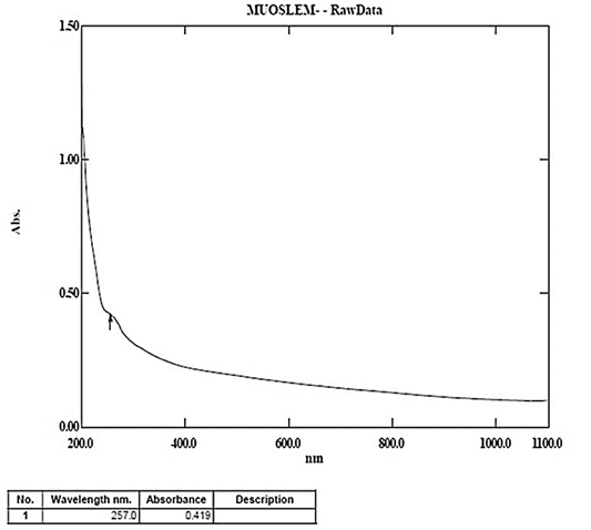
The UV-visible spectroscopy confirmed the successful preparation of Nano lotus leaves extract. A strong absorption peak appeared at 257 nm. This peak indicated nanoparticle formation (Figure 1). The sharp peak reflects the surface plasmon resonance linked with nanoparticle presence.
FE-SEM Analysis
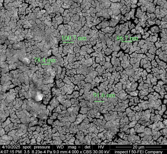
The morphology and size of Nano lotus extract were studied using Scanning Electron Microscopy (FE-SEM). The images showed that the nanoparticles had irregular shapes. They did not have uniform geometry. To measure particle size, 100 nanoparticles were selected randomly. The particle sizes ranged from 78.4 nm to 100.7 nm. The average size was 89.35 ± 1.26 nm (Figure 2).
These results confirm that the particles were within the nanoscale range. This small size helps improve cell penetration. It also increases their effectiveness in biological systems like wound healing.
FTIR Analysis
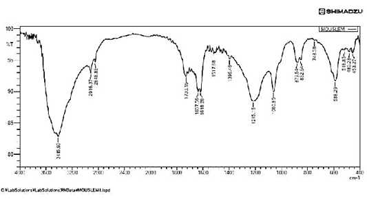
Further structural analysis of the Nano lotus extract was performed using Fourier Transform Infrared Spectroscopy (FTIR). The resulting spectrum revealed several characteristic absorption peaks, each corresponding to specific functional groups present within the extract. A prominent peak was observed at 3415.38 cm⁻¹, which is typically associated with phenolic groups. Additional peaks appeared at 2916.37 cm⁻¹ and 2848.86 cm⁻¹, reflecting C-H stretching vibrations. The presence of C=C double bonds and amide groups was indicated by peaks at 1730.15 cm⁻¹ and 1618.28 cm⁻¹, while the signal at 1517.98 cm⁻¹ represented N-H bending vibrations characteristic of amide II bonds. Further peaks were noted at 1396.46 cm⁻¹, corresponding to C-H and O-H bending, and at 1215.15 cm⁻¹ and 1060.85 cm⁻¹, which are indicative of C-O stretching vibrations, likely originating from acids or phenolic compounds. In the fingerprint region, peaks at 879.54 cm⁻¹ and 852.54 cm⁻¹ confirmed the presence of aromatic C-H bending vibrations (Figure 3). Collectively, these FTIR findings suggest that the Nano lotus extract contains a variety of bioactive compounds, including polyphenols, alkaloids, and phenolic acids, all of which may play a role in promoting the wound healing process.
X-RAY Diffraction (XRD) Analysis
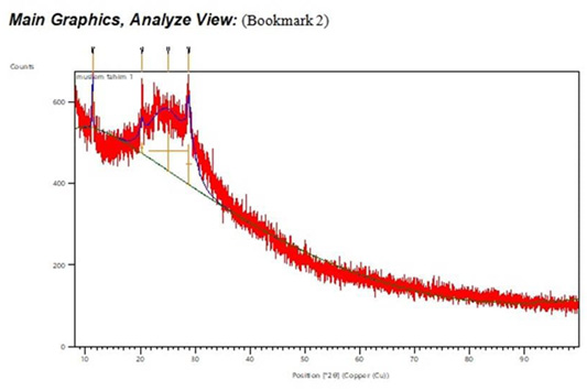
The crystalline nature of the prepared Nano lotus extract was examined using X-ray Diffraction (XRD). The analysis showed that the extract contained both crystalline and amorphous phases. The major diffraction peaks were observed at 2θ values of 11.29°, 20.26°, 25.02°, and 28.81°. The corresponding full width at half maximum (FWHM) values for these peaks were 0.101, 0.579, 7.41, and 1.054, respectively (Figure 4). These results confirm that the Nano lotus extract displayed a crystalline structure that may enhance its stability and biological activity.
Surface Area and Wound Size Reduction
Wound surface areas were measured on days 0, 7, and 14 after burn induction. At day 0, all groups showed an identical wound area of 314 mm². On day 7, wound size in the untreated control group (G1) was 289.02 ± 28.87 mm², while in the Nano lotus group (G2) it was significantly reduced to 168.13 ± 11.58 mm². The Fucidin group (G3) showed further reduction to 156.36 ± 18.89 mm². Statistical analysis revealed that both treated groups had significantly smaller wound areas compared to G1 (p < 0.05), while no significant difference was observed between G2 and G3 (p = 0.087; Table 1).
Table 1: Surface area (mm2) of wound.
Groups
Period
0 time
7 days
14 days
G1
314±0Aa
289.02±28.87Aa
271.04±35.78Ab
G2
314±0Aa
168.13±11.58Bb
133.60±11.61Bb
G3
314±0Aa
156.36±18.89Bb
88.45±8.81Cc
LSD (P<0.05)
40.75
The different letters between any two mean denote to the significant difference at p<0.05. Capital letters denote to the vertical statistical reading. Small letters denote to the horizontal statistical reading.
On day 14, the untreated group (G1) still had a larger wound area of 271.04 ± 35.78 mm². The Nano lotus group (G2) showed a substantial reduction to 133.60 ± 11.61 mm², and the Fucidin group (G3) had the smallest wound area at 88.45 ± 8.81 mm². The differences between G1 and both treated groups were highly significant (p < 0.001). Additionally, G3 showed a statistically significant difference compared to G2 at this stage (p = 0.032), indicating slightly more effective healing with Fucidin by day 14 (Table 1).
Wound Contraction
As shown in Table 2 the wound contraction percentages reflected a similar pattern of healing improvement. On day 7, G1 exhibited minimal contraction at 7.95 ± 1.68%, whereas G2 and G3 showed much higher contractions of 46.45 ± 2.36% and 50.20 ± 2.12%, respectively. The difference between G1 and both treated groups was highly significant (p < 0.001). However, there was no significant difference between G2 and G3 at this stage (p = 0.064).
Table 2: Wound contraction data of rats.
Groups
Period
0 time
7 days
14 days
G1
0 ± 0Aa
7.955 ± 1.68Aa
30.78 ± 1.21Ab
G2
0 ± 0Aa
46.45 ± 2.36Bb
57.45 ± 1.13Bc
G3
0 ± 0Aa
50.20 ± 2.12Bb
71.83 ± 2.18Cc
LSD (P<0.05)
8.14
The different letters between any two mean denote to the significant difference at p<0.05. Capital letters denote to the vertical statistical reading. Small letters denote to the horizontal statistical reading.
By day 14, wound contraction increased across all groups. The control group (G1) reached 30.78 ± 1.21%. Meanwhile, G2 achieved 57.45 ± 1.13%, and G3 reached the highest contraction rate at 71.83 ± 2.18%. The differences between all groups were statistically significant at p < 0.001 (Table 2), confirming the superior healing response in both treated groups compared to the untreated control.
Macroscopic Observations
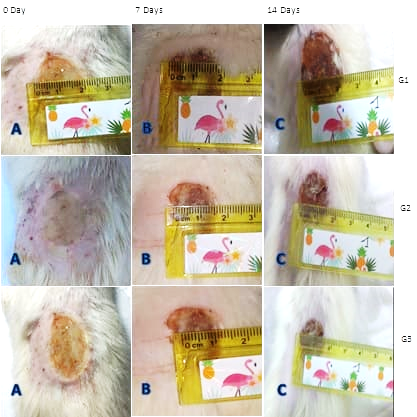
Macroscopic evaluations supported the quantitative measurements. On day 0, all burns appeared similar across groups. By day 7, the untreated group (G1) still displayed open, inflamed wounds with little contraction. In contrast, G2 and G3 showed visible shrinkage of the wound edges and less inflammation. By day 14, G3 demonstrated the most advanced healing with near-complete wound closure, followed by G2, which also showed marked reduction in wound size. The untreated group continued to exhibit larger, poorly contracted wounds with incomplete healing (Figure 5).
Histopathological Findings
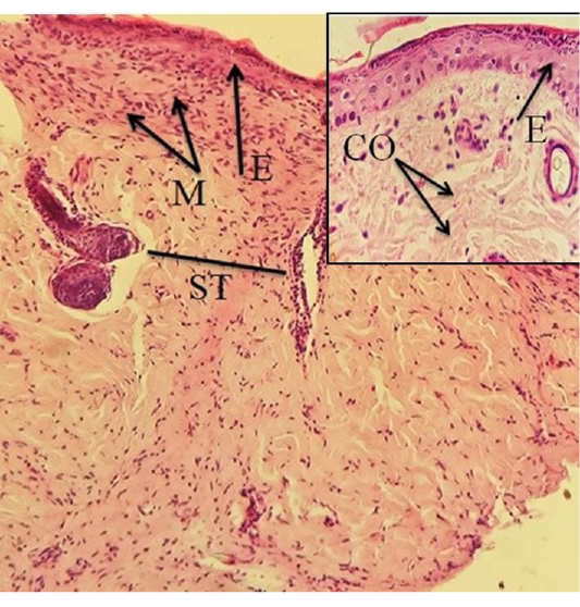
Day 7 histology: Histopathological analysis at day 7 showed clear differences among groups. In the untreated group (G1), the epidermis remained thin, with scant collagen fibers and wide scar tissue present. Minimal granulation tissue was observed, indicating slow healing (Figure 6).
In the Nano lotus-treated group (G2), mild epidermal hyperplasia was seen with irregular collagen fiber distribution in the dermis. Early granulation tissue formation was evident, and scar tissue was narrower compared to G1 (Figure 7).
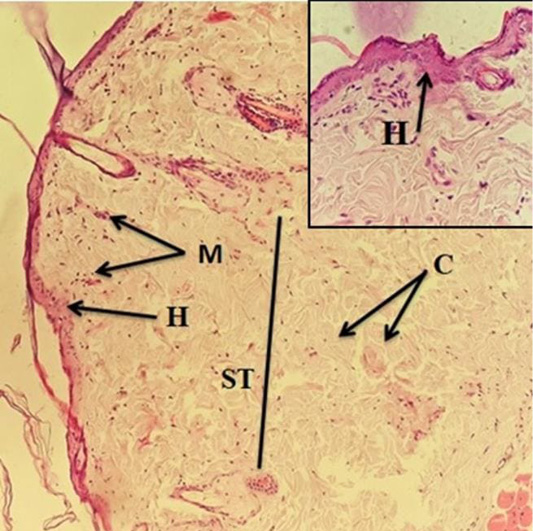
The Fucidin-treated group (G3) displayed more advanced epidermal hyperplasia, thicker epidermis, and upward proliferation of the stratum basale. Granulation tissue formation was more prominent, with irregular collagen fibers visible (Figure 8).
Day 14 histology: By day 14, further improvements were observed. The untreated group (G1) still exhibited wide scar tissue, disorganized and scant collagen fibers, and little evidence of new blood vessel formation. The epidermis remained thin with limited remodeling (Figure 9).
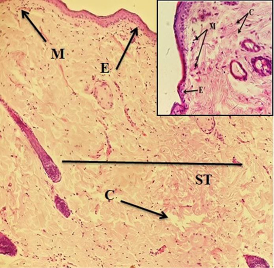
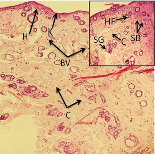
In contrast, the Nano lotus group (G2) showed a well-organized collagen fiber network, clear formation of new blood vessels, and the development of dermal appendages such as new hair follicles and sweat glands. The scar tissue was narrower, and epidermal layers showed downward hyperplasia (Figure 10).
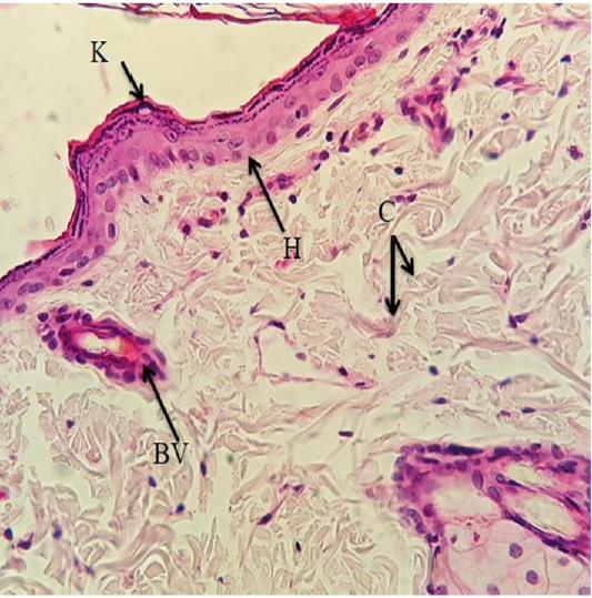
In the Fucidin-treated group (G3), healing reached its most advanced stage. The dermis displayed densely packed collagen fibers, while newly formed blood vessels were abundant throughout the tissue. The epidermis was completely regenerated, showing a noticeably thick keratinized layer. Granulation tissue remained prominent, indicating ongoing tissue remodeling and near-complete restoration of normal skin architecture (Figure 11).
DISCUSSION
Burn wound healing is a dynamic process that depends on several interconnected biological events. Inflammation, cell growth, tissue rebuilding, and restoration of the skin’s structure all play vital roles. In this study, the application of Nano lotus leaves extract produced clear improvements in healing for second-degree burns. This outcome supports existing research that recognizes the wide range of pharmacological activities found in lotus (Nelumbo nucifera). Multiple bioactive compounds present in lotus leaves, such as polyphenols, alkaloids, flavonoids, and polysaccharides, may work together to promote healing ; . Lotus contains many bioactive substances. These include polyphenols, flavonoids, alkaloids, and polysaccharides. Together, they provide antioxidant, anti-inflammatory, antibacterial, and tissue-regenerating effects. These actions may help the skin heal and also reduce infection risks ; .
One major challenge in burn healing is managing oxidative stress. After burns, the body produces high levels of reactive oxygen species (ROS). These can worsen tissue damage if not controlled . The polyphenols and flavonoids in lotus help neutralize these free radicals. This limits oxidative injury ; . In this study, faster wound contraction was seen in the Nano lotus group. This suggests the antioxidants played a strong role. Similar results were found in other studies using polyphenol-rich plant extracts . The polysaccharides in lotus also help regulate the immune response. They lower excess inflammation. At the same time, they support fibroblast and keratinocyte activity, both important for new tissue formation ; .
Microscopic results gave more evidence of healing. The Nano lotus-treated animals showed better collagen organization. Blood vessel growth increased. Skin structures such as sweat glands and hair follicles reappeared. These signs point to the effects of lotus polysaccharides and alkaloids. They help form extracellular matrix, promote new vessel growth, and support cell renewal ; . The Nano formulation likely improved how well these compounds reached the damaged tissue. Nanoparticles may improve absorption and keep the active ingredients in place longer ; . Recent studies also showed that nanoparticles help plant extracts penetrate the skin better. They allow slow and steady delivery ; .
Lotus also shows strong antibacterial properties. Burns often become infected, which can delay healing. The alkaloids and phenolic compounds in lotus have been shown to fight many types of bacteria ; . In this experiment, both the Nano lotus and Fucidin groups had similar wound size reduction and tissue repair. This suggests lotus extract might be a good natural alternative to antibiotic ointments for burns. Using plant-based treatments may also help lower the risk of antibiotic resistance, which is a growing problem in medicine.
Other parts of the lotus plant may also help healing. Research has found useful compounds in lotus seedpods and rhizomes. These include polysaccharides, quinic acid, and plant starches. They may work with polyphenols and alkaloids to support tissue repair, control inflammation, and regulate healing processes ; ; ; . In this study, the healing seen likely came from the combined effects of these many compounds .
In summary, this study showed that Nano lotus leaves extract improved healing in second-degree burns. It helped wounds contract faster, improved collagen structure, and supported full skin regeneration. The active compounds provide antioxidant, anti-inflammatory, and antibacterial effects. Nanoparticles may also help deliver these compounds more effectively. These findings suggest that Nano lotus extract may be a safe and promising option for burn treatment. Still, more research is needed to confirm its effects in humans and explore its full potential ; ; .
CONCLUSIONS AND RECOMMENDATIONS
This study showed that Nano lotus leaves extract improved healing in second-degree burns in rats. The extract helped wounds contract faster. It also reduced wound size. Organized collagen fibers and new tissue structures formed. Microscopic study confirmed better skin repair. Granulation tissue developed well. New blood vessels appeared. Skin appendages such as hair follicles and sweat glands reappeared. The healing effects likely came from antioxidant, anti-inflammatory, and antibacterial actions. These are provided by polyphenols, flavonoids, alkaloids, and polysaccharides found in lotus. The Nano-formulation may have helped these compounds penetrate the wound better and stay active longer. Based on these results, Nano lotus extract may serve as a safe and natural option for treating burns. Its effect may be similar to that of Fucidin ointment. Further studies are needed in human patients with different types and depths of burns. More research should check long-term safety. Future work should also find the best doses. Combination therapies with other wound care agents may be useful. More studies can help explain how the compounds work. They may clarify how inflammation, oxidative stress, and tissue repair are controlled. Other parts of the lotus, like seedpods and rhizomes, might also offer healing benefits. These parts could be studied for new uses in skin repair.
ACKNOWLEDGEMENTS
The author is grateful to the university of Al-Qadisiyha/College of Veterinary Medicine for all the facilities to achieve this study.
NOVELTY STATEMENTS
First time usage of lotus extract nanoparticles in 2nd degree skin-burns.
AUTHOR’S CONTRIBUTIONS
Muslim F. Diwan conceptualized and design the experiment. Qayes T. Ali and Ali Ismail Jassim carried out the experimental work. All authors analyzed the data and drafted the manuscript. All authors reviewed and approved the final version of the manuscript.
Conflict of Interest
The authors declare that there is no conflict of interest related to this study.
Figures
Figure 1:.
UV-visible absorption of Nano lotus leaves extract.
Figure 2:.
Image for FE-SEM of Nano lotus leave extract.
Figure 3:.
FT-IR for Nano lotus leaves extract.
Figure 4:.
X- ray image of Nano lotus leaves extract.
Figure 5:.
Macroscopic evaluation of burn wound healing at different time points in the three experimental groups. (G1): Control group (non-treated); (A) at day 0, (B) at day 7, and (C) at day 14 post-burn induction, showing slow wound contraction and incomplete healing. (G2): Nano lotus leaves extract-treated group; (A) at day 0, (B) at day 7, and (C) at day 14 post-burn induction, demonstrating progressive wound size reduction, improved healing, and narrowed scar tissue formation. (G3): Fucidin-treated group; (A) at day 0, (B) at day 7, and (C) at day 14 post-burn induction, showing advanced wound closure, minimal remaining wound area, and well-organized wound healing compared to the control group.
Histological section of Fucidin-treated group (G3) at day 7 post-burn induction. The section shows irregular collagen fiber network (C) in the dermis, with thickened epidermis and upward proliferation of the stratum basale (E). Moderate granulation tissue is also present, indicating active dermal repair and ongoing wound healing progression. Stained with Hematoxylin and Eosin (H and E), magnification 20X.
Figure 9:.
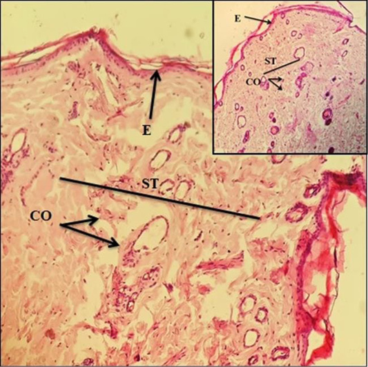
Histological section of control group (G1) at day 14 post-burn induction. The section demonstrates wide scar tissue (ST), sparse and disorganized collagen fibers (CO), and limited presence of granulation tissue (M). Mild hyperplasia of the epidermis is observed with minimal dermal regeneration. Denaturation of collagen and absence of new blood vessels are evident, reflecting delayed and incomplete wound healing. Stained with Hematoxylin and Eosin (H and E), magnification 20X.
Figure 10:.
Microscopic section from the Nano lotus leaves extract-treated group (G2), examined on day 14 after burn induction. The epidermis appears fully developed, showing a thin keratinized layer (K) and downward growth of the stratum basale (SB). In the dermis, collagen fibers (C) are arranged in thick and organized bundles. Several skin structures, including newly formed hair follicles (HF) and sweat glands (SG), have reappeared, indicating tissue regeneration. The scar tissue (ST) is noticeably narrow, while a rich network of new blood vessels (BV) is clearly visible throughout the dermis. These changes reflect advanced stages of skin healing and remodeling. Samples were stained using Hematoxylin and Eosin (H and E) and observed under both 10X and 40X magnification.
Figure 11:.
In the Fucidin-treated group (G3), the tissue displayed almost complete healing by day 14. The dermal collagen fibers were densely packed, forming a strong supportive network. Numerous newly formed blood vessels were present throughout the tissue. The epidermal layer was fully restored, with a thick keratinized surface clearly visible. Granulation tissue was abundant, reflecting active tissue repair. Altogether, these features demonstrate that the healing process in this group had progressed to an advanced and nearly complete state.
References
Abuzer AY, Cetinel S, Zuvin M, Kosar A, Kutlu O (2020). Therapeutic nanoparticles and their targeted delivery applications. Molecules, 25(9):2193. https://doi.org/10.3390/molecules25092193[CrossRef]
Arun K, Jassim AY, Mohammed AA, Sharma V, AL-Tawaha ARMS, Sivanesan I (2024). Pharmaceuticals, 17(12):1711. https://doi.org/10.3390/ph17121711[CrossRef]
Benali T, Bakrim S, Ghchime R, Benkhaira N, El Omari N, Balahbib A, Taha D, Zengin G, Hasan MM, Bibi S, Bouyahya A (2024). Pharmacological insights into the multifaceted biological properties of quinic acid. Biotechnol. Genet. Eng. Rev., 40(4):3408-3437. https://doi.org/10.1080/02648725.2022.2122303[CrossRef]
Bencheikh N, Radi FZ, Fakchich J, Elbouzidi A, Ouahhoud S, Ouasti M, Bouhrim M, Ouasti I, Hano C, Elachouri M (2023). Ethnobotanical, phytochemical, toxicological, and pharmacological properties of Ziziphus lotus (L.) Lam.: A comprehensive review. Pharmaceuticals (Basel), 16(4):575. https://doi.org/10.3390/ph16040575[CrossRef]
Bishayee A, Patel PA, Sharma P, Thoutireddy S, Das N (2022). Lotus (Nelumbo nucifera Gaertn.) and its bioactive phytocompounds: A tribute to cancer prevention and intervention. Cancers (Basel), 14(3):529. https://doi.org/10.3390/cancers14030529[CrossRef]
Chong L, He Y, Yang Y, Gou Y, Li S, Zing RS, Zhao X (2021). Antioxidant and inflammatory effects of Nelumbo nucifera Gaertn. Leaves. Oxid. Med. Cell. Longev., 2012:1-14. https://doi.org/10.1155/2012/8375961[CrossRef]
Dai G, Wang J, Zheng J, Xia C, Wang Y, Duan B (2023). Bioactive polysaccharides from lotus as potent food supplements: a review of their preparation, structures, biological features and application prospects. Front. Nutr., 10:1171004. https://doi.org/10.3389/fnut.2023.1171004[CrossRef]
Dhrisya C, Sadhasivam S, Sankar GR, Swaminathan K, Musthafa ME, Feng-Huei LIN and M. Walid Q 2019. Therapeutic efficacy of nanoparticles and routes of adminsterration. Biomaterials Research, 23(2019): 20-49. Doi.:10.1186/s40824-019-0166-x
Dhull SB, Chandak A, Chawla P, Goksen G, Rose PK, Rani J (2023). Modifications of native lotus (Nelumbo nucifera G.) rhizome starch and its overall characterization: A review. Int J Biol Macromol, 253(Pt 8):127543. https://doi.org/10.1016/j.ijbiomac.2023.127543[CrossRef]
Duangjai T, Pinthong DW, Hano C (2018). Flavonoids from Nelumbo nucifera Gaertn., a medicinal plant: Uses in traditional medicine, photochemistry and pharmacological activities. Medicines (Basel), 5(4):127. https://doi.org/10.3390/medicines5040127[CrossRef]
Elizbieta RB, Lopuszynska I, Flieger W, Tobiasz M, Maciejewski R, Flieger J (2023). An overview of recent development in the management of burn injuries. Int. J. Mol. Sci., 24(22):16357. https://doi.org/10.3390/ijms242216357[CrossRef]
Fukui H, Mizuguchi H, Kitamura Y, Takeda N (2022). Patho-pharmacological research of anti-allergic natural products targeting antihistamine-sensitive and -insensitive allergic mechanisms. Curr. Top. Behav. Neurosci., 59:77-90. https://doi.org/10.1007/7854_2021_255[CrossRef]
Francesco DF, Riccio M, Jimi S (2022). Contribution of topical agents such as hyaluronic acid and silver sulfadiazine to wound healing and management of bacterial biofilm. Medicina, 58(6):835. https://doi.org/10.3390/medicina58060835[CrossRef]
Hanae NM, Doudach L, Mekkaoui M, Khalil Z, Harraqui K, Fozia F, Naceiri Mrabti N, EL-Shazly M, Alotaibi A, Ullah R, Faouz MEA, Bouyahya A (2022). Profile of medicinal plants traditionally used for the treatment of skin burns. Evid. Based Complement Alternat. Med., 2022:1-10. https://doi.org/10.1155/2022/3436665[CrossRef]
Kakar MU, Karim H, Shabir G, Iqbal I, Akram M, Ahmad S, Shafi M, Gul P, Riaz S, Rehman RU, Salari H (2023). A review on extraction, composition, structure, and biological activities of polysaccharides from different parts of Nelumbo nucifera. Food Sci. Nutr., 11(7):3655-3674. https://doi.org/10.1002/fsn3.3376[CrossRef]
Limwachiranon J, Huang H, Shi Z, Li L, Luo Z (2018). Lotus flavonoids and phenolic acids: Health promotion and safe consumption dosage. Compr. Rev. Food Sci. Food Saf., 17(2):458-471. https://doi.org/10.1111/1541-4337.12333[CrossRef]
Liu H, Wang S, Qiu K, Zheng C, Tan H (2024). Preparation, structural characterization, and biological activities of lotus polysaccharides: A review. Int. J. Biol. Macromol., 279(Pt 2):135191. https://doi.org/10.1016/j.ijbiomac.2024.135191[CrossRef]
Mathew PR, Cancio LC, Elster EA, Burmeister DM, Rose LF, Natesan S, Christy RJ, Chung KK (2015). Burn wound healing and treatment: A review and advancements. Crit. Care, 19:243. https://doi.org/10.1186/s13054-015-0961-2[CrossRef]
Meng D, Wang RP, Phei ES, Zhiyi C. 2024. Advancing burn wound treatment: exploring hydrogel as a trans dermal drug delivery system. Drug Delivery, 31(1): 1-15. DOI: 10.1080/10717544.2023.2300945[CrossRef]
Monika GD, Paulina P, Francesco C, Alicja KJ, Antonella MG, Narcyz K, Michal W, Magdalena GP 2018. Potential health benefit of olive oil and plant polyphenols. International Journal of Molecule, 19(3): 686. DOI: 10.3390/ijms19030686[CrossRef]
Omar AB, Jasim AM, Jawad MJ, Nahi HH (2022). The role of PLGA/TPGS nanoparticle on xylazine-ketamine anesthetic activity in male albino rabbits. Iraqi J. Vet. Sci., 36(1):201-206. https://doi.org/10.33899/IJVS.2021.129688.1679[CrossRef]
Patil MV, Kandhare AD, Bhise SD (2012). Pharmacological evaluation of ethanolic extract of Daucus carota Linn root formulated cream on wound healing using excision and incision wound model. Asian Pac. J. Trop. Biomed., 2(2): S646-S655. https://doi.org/10.1016/S2221-1691(12)60290-1[CrossRef]
Qaisar A, 2019. Understanding the UV-Vis spectroscopy for Nanoparticles. Journal of Nanomaterials and Molecular Nanotechnology, 8(3): 1-3. Doi: 10.4172/2324-8777.1000268[CrossRef]
Renuga D, Jeyasundari J, Sakthi Athithan AS (2022). Green synthesis and characterization of metal oxide nanoparticles using Nelumbo nucifera flower and its antimicrobial activity. Int. J. Innov. Res. Sci. Eng. Technol., 11(1):519-526. https://doi.org/10.15680/IJIRSET.2022.110175[CrossRef]
Tavares DDS, Lima-Ribeiro MHM, Pontes-Filho NT, Carneiro-Leao AMDA, Correia MTDS (2012). Development of animal model for studying deep second-degree thermal burns. J. Biomed. Biotechnol., 2012:1-7. https://doi.org/10.1155/2012/46084[CrossRef]
Tuti L, Sumaryati S, Gusti R, Rauza SR and Rini R. 2023. The burn wound healing process A review. International Journal of Progressive Sciences and Technologies, 40(1): 77-88. DOI: 10.52155.
Wang M, Hu WJ, Wang QH, Yang BY, Kuang HX (2023). Extraction, purification, structural characteristics, biological activities, and application of the polysaccharides from Nelumbo nucifera Gaertn. (lotus): A review. Int. J. Biol. Macromol., 226:562-579. https://doi.org/10.1016/j.ijbiomac.2022.12.072[CrossRef]
Wang YF, Shen ZC, Li J, Liang T, Lin XF, Li YP, Zeng W, Zou Q, Shen JL, Wang XY (2022). Phytochemicals, biological activity, and industrial application of lotus seedpod (Receptaculum nelumbinis): A review. Front. Nutr., 9:1022794. https://doi.org/10.3389/fnut.2022.1022794[CrossRef]
Wang Z, Li Y, Ma D, Zeng M, Wang Z, Qin F, Chen J, Christian M, He Z (2023). Alkaloids from lotus (Nelumbo nucifera): recent advances in biosynthesis, pharmacokinetics, bioactivity, safety, and industrial applications. Crit. Rev. Food Sci. Nutr., 63(21):4867-4900. https://doi.org/10.1080/10408398.2021.2009436[CrossRef]
Wu Q, Zhang F, Niu M, Yan J, Shi L, Liang Y, Tan J, Xu Y, Xu J, Wang J, Feng N (2023). Extraction methods, properties, functions, and interactions with other nutrients of lotus procyanidins: A review. J. Agric. Food Chem., 71(40):14413-14431. https://doi.org/10.1021/acs.jafc.3c05305[CrossRef]
Zhao X, Zhao R, Yang X, Sun L, Bao Y, Shuai Liu Y, Blennow A, Liu X (2023). Recent advances on bioactive compounds, biosynthesis mechanism, and physiological functions of Nelumbo nucifera. Food Chem., 412:135581. https://doi.org/10.1016/j.foodchem.2023.135581[CrossRef]
Zheng H, Han L, Shi W, Fang X, Hong Y, Cao Y (2022). Research advances in lotus leaf as Chinese dietary herbal medicine. Am. J. Chin. Med., 50(6):1423-1445. https://doi.org/10.1142/S0192415X22500616[CrossRef]
Zhenyu W, Yong C, Maomao Z, Zhaojun W, Fang Q, Yongzhi W, Jie C, Zhiyong H 2021. Lotus (Nelumbo nucifera Gaertn) leaf: A narrative review of its phytoconstituents, health benefits and food industry applications. Trends in Food Science & Technology, 112: 631-650. Doi: 10.1016/j.tifs.2021.04.033[CrossRef]
Zwieretto W, Piorun K, Skorka-Majewicz M, Maruszewska A, Antoniewski J, Gutowska I (2023). Burn: classification, pathophysiology and treatment: A review. Int. J. Mol. Sci., 24(4):37-49. https://doi.org/10.3390/ijms240437449[CrossRef]