
Visceral leishmaniasis is a neglected zoonotic protozoal disease caused by Leishmania infantum and Leishmania donovani that is transmitted by sandflies (Phlebotamine flies). In November 2020, a case of leishmaniasis was diagnosed in a captive tiger through microscopy and L. infantum was confirmed by PCR and sequencing analysis. DNA sequencing of the amplicon revealed close homology with Leishmania sequences available in GenBank. Alignments and phylogenetic analyses of the Leishmania infantum from a tiger in Pakistan indicated 94-100 % identity with Leishmania from animals and 98.8-100% with Leishmania from humans origins suggesting the need for screening of animals before transporting, and of humans before taking care of captive animals, in order to prevent transboundary spread of Leishmania.
Short Communication · Volume 55, Issue 6, 31 October 2023 · Open Access
First Report of Leishmania infantum in a Captive Panther from Pakistan
· · · · ·
Affiliations & Notes ⌄
- 1Department of Parasitology, Faculty of Veterinary Science, University of Veterinary and Animal Sciences, Lahore, Pakistan 2Institute of Microbiology, Faculty of Veterinary Science, University of Veterinary and Animal Sciences, Lahore, Pakistan 3Lahore Zoological Garden, Lahore, Pakistan. * Corresponding author: drharoonakbar@uvas.edu.pk, imran.rashid@uvas.edu.pk
Article Info ⌄
- Journal:
- Pakistan Journal of Zoology
- Published:
- 31 October 2023

From the article
- The sequence alignments indicated the highest percent identity with L. infantum of animals (94-100%; Fig. 2a) and humans (98.8-100%; Fig. 2b).
- Moreover, reverse zoonoses, is suspected in this case, suggesting that human caretakers could be a source of infection for captive wild animals.
- The tigers should be screened for Leishmania infantum especially before transboundary transportation as tigers can be reservoirs of visceral leishmaniasis.
- Moreover, the health status of the caretakers and other human beings working in close proximity of captive animals should be carefully monitored and only Leishmania free caretakers should be allowed access to captive animals.
Selected from the article's own text.
Abstract
Materials and methods
In November 2020, a blood sample of a male brown tiger (Panthera tigris tigris) from Lahore Zoological Garden (Lahore, Pakistan) was received at the Diagnostic Laboratory of the Department of Parasitology, University of Veterinary and Animal Sciences, Lahore, Pakistan for the evaluation of hemoparasites. The tiger was imported from United Arab Emirates (UAE) in April, 2019 and was healthy. In October 2020, the tiger received a wound and a blood sample was collected at that time. The approximate age of tiger was 3-4 years. Later on the tiger died possibly due to the previous injury or Leishmaniasis in November 2020. Blood smears were prepared, staining with Giemsa stain, and examined under oil emersion lens (100X).
The blood sample was subjected to PCR. Briefly DNA was extracted by Phenol-Chloroform-Isoamyl alcohol method . PCR was performed with L. infantum primer pair targeting a 570 bp internal transcribed spacer region of rRNA . The amplification was performed in a thermocycler (Thermo Fisher Scientific, Gloucester, UK) in a final volume of 20 µl containing 10 µl of dream taq master mix (Thermo Fisher Scientific, Waltham, MA, USA), 2 µl of each primer, 2 µl DNA template, and 4 µl diethyl pyrocarbonate (DEPC) water. The DNA was amplified using standard amplification settings with initial denaturation at 95°C for 5 min followed by 35 cycles (denaturation at 95 °C for 30 seconds, annealing at 61.5 °C for 1 min, and extension at 72 °C for 1 min) followed by final extension at 72°C for 10 min. The PCR product was subjected to gel electrophoresis using gel apparatus (BioBase, Shandong, China). The gel was analyzed under Ultraviolet illuminator (Mupid-One, Nippon Genetics, Japan). The gel band was excised with a sterile scalpel blade and sent to Advance Bioscience International, Lahore for sequencing. The obtained sequence was aligned using online BLASTN tool (blastn; ncbi.nlm.nih.gov) using default parameters. The partial sequences (n=7) of 18S subunit ribosomal RNA of Leishmania (from animals and humans) were retrieved from public data base (GenBank; ncbi.nlm.nih.gov) and used to align with the sequence obtained in the current study using laser gene (version 11, DNASTAR Inc., Madison, WI). Phylogenetic trees were constructed by maximum likelihood method and Tamura-Nei model using the molecular evolutionary genetics analysis software: MEGA X (version 10.2.2) with 1000 bootstrap replications ; . 18S RNA sequence of L. braziliensis and L. amazonensis was used as an out-group with sequences of L. infantum from animal’s and human’s origins for phylogenetic analyses, respectively.
Results
The microscopic examination revealed the presence of inclusion bodies consistent with amastigote stage of Leishmania (Fig. 1a). As a result of PCR gel analysis, a band was visualized at expected size (Fig. 1b) that was sequenced. The sequence obtained from the tiger was identified as L. infantum. The sequence has been deposited in public data base, GenBank with an accession number (MW730712).
The Blastn analysis revealed top ten hits with 18S small subunit ribosomal RNA of Leishmania species with the query cover of 99-100% and identity of 98.7-99.5%. The sequence alignments indicated the highest percent identity with L. infantum of animals (94-100%; Fig. 2a) and humans (98.8-100%; Fig. 2b).
Discussion
L. infantum infects a range of hosts including domestic and wild canines and felines . In Pakistan, leishmaniasis has been reported previously in a wide variety of hosts including dog (Canis lupis), cattle (Bos taurus), goat (Capra aegagrus hircus), sheep (Ovis aries), buffalo (Bubalus bubalis), donkey (Equus asinus), wild rats (Rattus), and Indian gerbils (Tatera indica) . This is the first report of Leishmaniasis in a captive tiger from Pakistan. Unfortunately, the tiger died of an injury and an underlying Leishmaniasis, in November, 2020 thus closing down the case follow-up whereas the efforts to collect an evidence for the presence of vector continued. We evaluated the tigers’ enclosure and its periphery for the presence of sandflies. As no sandflies could be retrieved during several attempts of fly-capture using electric traps from the vicinity of tiger’s cage, thus it is highly likely that tiger caught the infection before being shifted to the facility. Other possibilities include the occurrence of this infection at the place where the tiger was born and lived (in UAE) before being imported, or during the transport. It’s well known that this disease appears in the host, much longer after the introduction of the parasite by the bite of an infected sandfly, thus occurrence of infection during transport seems less likely as against its occurrence in UAE before being imported to Pakistan. The UAE is located in Middle East a region known to be endemic for Leishmaniasis, predominantly for the cutaneous form whereas it’s not absolutely free from VL. The VL has also been reported in feline and canine hosts in the Middle Eastern countries that are considered endemic for this disease owing to vast-population displacements from Leishmania-infected regions like from Iraq . Challenges in the sandfly population control are also contributing to a rise in the case numbers of leishmaniasis in the Middle East . Interestingly, all Leishmania sequences from humans that clustered with L. infantum sequence of the current study, were from India (Fig. 2b), indicating a potential case of zooanthroponosis. Large part of workforce in Middle East is sourced from India, another endemic region for this disease. It can be hypothesized that a Leishmania-infected care taker from India might be the source of infection for this tiger born in UAE in a captive facility.
Leishmania infantum is not as common as L. major and L. tropica in Pakistan . However, disease caused by Leishmania infantum in Pakistan is not reported suggesting the finding could be coincidental or least important as a potential reservoir, if host, parasite, and environment factors were appropriate. Unfortunately, we could not evaluate the tiger’s caretakers in UAE or Pakistan which should constitute a crucial step in establishing the source of infection as asymptomatic human infection is common .
Leishmaniasis, a neglected tropical disease, is a public health as well as an animal health challenge. The zoonotic potential Leishmaniasis is particularly problematic in a captive environment located in an endemic region. High sequence identity and clustering of our sequence with the sequences of L. infantum from animals and humans from GenBank database highlights that infected captive animals may constitute a risk to other susceptible animals and humans kept in close proximity. Moreover, reverse zoonoses, is suspected in this case, suggesting that human caretakers could be a source of infection for captive wild animals. Vector control based on chemical or electrical insect killers especially during the fly-season, is an important tool to mitigate such a risk. As part of an active surveillance as well as a preventive strategy, thorough-screening of animals must be ensured before their movement across the regions.
Conclusion
The tigers should be screened for Leishmania infantum especially before transboundary transportation as tigers can be reservoirs of visceral leishmaniasis. Moreover, the health status of the caretakers and other human beings working in close proximity of captive animals should be carefully monitored and only Leishmania free caretakers should be allowed access to captive animals.
Acknowledgements
The authors would like to thank Prof Dr. Mohamed Gharbi (Laboratoire de Parasitologie, Université de la Manouba, École Nationale de Médecine Vétérinaire de Sidi Thabet, 2020 Sidi Thabet, Tunisia) for providing the L. infantum control positive DNA.
Funding
This research received no specific funding from any Public, Private or Non-profit Organization.
Ethical statement
The samples were taken by authorized veterinarians for diagnostic purposes according to the protocol of the zoo.
Statement of conflict of interest
The authors have declared no conflict of interest.
Figures
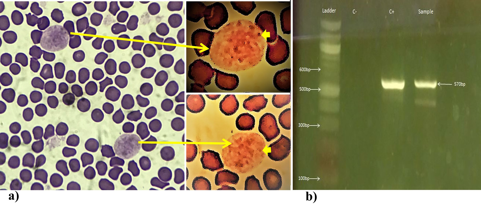
(a) Microscopic image captured with an i-Phone-7 from stained blood smear of tiger blood sample examined at 1000x magnification in an Olympus CX-21 microscope. The inset contains images of two monocytes that were edited with photos editor of windows, with a Zeke Filter on it, for a clearer view of the multiple intracellular Leishmanial amastigotes (Thick yellow arrow pointed to one of those) that actually helped in diagnosis. (b) An image of agarose gel indicating a band at the size expected for L. infantum detection in a tiger blood. C+ and C- represent positive and negative controls, respectively.
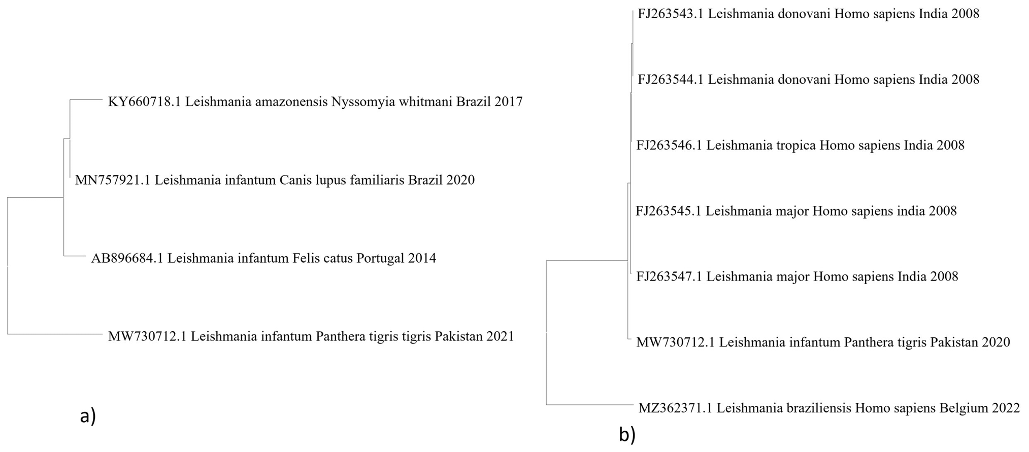
Phylogenetic analyses of L. infantum from a tiger in Pakistan (a) with Leishmania sequences from animals and (b) humans.
References
- Alves, F., Bilbe, G., Blesson, S., Goyal, V., Monnerat, S., Mowbray, C., Muthoni Ouattara, G., Pécoul, B., Rijal, S., Rode, J., Solomos, A., Strub-Wourgaft, N., Wasunna, M., Wells, S., Zijlstra, E.E., Arana, B., Alvar, J., 2018. Clin. Microbiol. Rev., 31: e00048-00018. https://doi.org/10.1128/CMR.00048-18[CrossRef]
- Asfaram, S., Fakhar, M., and Teshnizi, S.H., 2019. J. Venomous Anim. Toxins Incl. Trop. Dis., 25: e20190012. https://doi.org/10.1590/1678-9199-jvatitd-2019-0012[CrossRef]
- Cavalera, M.A., Iatta, R., Laricchiuta, P., Passantino, G., Abramo, F., Mendoza-Roldan, J.A., Otranto, D., and Zatelli, A., 2020. BMC Vet. Res., 16: 214. https://doi.org/10.1186/s12917-020-02419-y[CrossRef]
- Chacon-Cortes, D., and Griffiths, L.R., 2014. J. Biorepos. Sci. appl. Med., 2: 1-9.
- Durrani, A.Z., Durrani, H.Z., Kamal, N., and Mehmood, N., 2011. Pakistan J. Zool., 43: 263-271.
- Herwaldt, B.L., 1999. Lancet (London, England), 354: 1191-1199. https://doi.org/10.1016/S0140-6736(98)10178-2[CrossRef]
- Kumar, S., Stecher, G., Li, M., Knyaz, C., and Tamura, K., 2018. Mol. Biol. Evol., 35: 1547. https://doi.org/10.1093/molbev/msy096[CrossRef]
- Lima, C., Colella, V., Latrofa, M.S., Cardoso, L., Otranto, D., and Alho, A.M., 2019. Parasit. Vectors, 12: 1-4. https://doi.org/10.1186/s13071-019-3394-y[CrossRef]
- Lindoso, J.A.L., Costa, J.M.L., Queiroz, I.T., and Goto, H., 2012. Res. Rep. Trop. Med., 3: 69-77.
- Michel, G., Pomares, C., Ferrua, B., and Marty, P., 2011. Acta Trop., 119: 69-75. https://doi.org/10.1016/j.actatropica.2011.05.012[CrossRef]
- Murray, H.W., Berman, J.D., Davies, C.R., and Saravia, N.G., 2005. Lancet (London, England), 366: 1561-1577. https://doi.org/10.1016/S0140-6736(05)67629-5[CrossRef]
- Ribeiro, R.R., Michalick, M.S.M., da Silva, M.E., Dos Santos, C.C.P., Frézard, F.J.G., and da Silva, S.M., 2018. BioMed. Res. Int., 2018: 3296893. https://doi.org/10.1155/2018/3296893[CrossRef]
- Salam, N., Al-Shaqha, W.M., and Azzi, A., 2014. Leishmaniasis in the Middle East: Incidence and epidemiology. PLoS Negl. Trop. Dis., 8: e3208. https://doi.org/10.1371/journal.pntd.0003208[CrossRef]
- Serafim, T.D., Iniguez, E., and Oliveira, F., 2020. Trends Parasitol., 36: 80-81. https://doi.org/10.1016/j.pt.2019.10.006[CrossRef]
- Stoops, C.A., Heintshcel, B., El-Hossary, S., Kaldas, R.M., Obenauer, P.J., Farooq, M., and Villinski, J.T., 2013. J. Vector Ecol. 38: 411-414. https://doi.org/10.1111/j.1948-7134.2013.12059.x[CrossRef]
- Tamura, K., and Nei, M., 1993. Mol. Biol. Evol., 10: 512-526.
- Tiwananthagorn, S., Bhutto, A.M., Baloch, J.H., Soomro, F.R., Kawamura, Y., Nakao, R., Aoshima, K., Nonaka, N., Oku, Y., and Katakura, K., 2012. Parasitol. Res., 111: 125-133. https://doi.org/10.1007/s00436-011-2808-3[CrossRef]
- Tolentino, N., Pinheiro, G.R.G., Ottino, J., de Oliveira, A.R., Coelho, C.M., Tinoco, H.P., Fujiwara, R.T., Santos, R.L., and Ribeiro, V.M., 2019. Vet. Parasitol. Reg. Stud. Rep., 17: 100308. https://doi.org/10.1016/j.vprsr.2019.100308[CrossRef]
Services
Article usage statistics
Loading…