
Ten out of 30 rats were randomly selected as normal group. Remaining 20 rats were used to establish rat model of mesangial proliferative glomerulonephritis (MsPGN), and were equally divided into model group and total glucoside of paeony (TGP) group. TGP group was given 150 mg/kg TGP by gavage, normal group and model group were given the same amount of saline by gavage. After 12 weeks of intervention, 24-h urine protein quantity, serum levels of IL-6, IFN-γ, and TNF-α ,kidney ROS and MDA levels, relative content of TGF-β1 and CTGF in TGP group were lower than that in model group (P <0.05); serum levels of IL-2 and CTLA-4,SOD levels in TGP group were higher than that in model group (P <0.05).These results indicated that TGP could reduce urinary protein quantity, inhibit inflammatory response, and reduce expression of TGF-β1 and CTGF in kidney tissue of MsPGN rats.
Short Communication · Volume 53, Issue 4, 7 June 2021 · Open Access
Intervention Effect of Total Glucoside of Paeony on Mesangial Proliferative Glomerulonephritis in Rats
· · · ·
Affiliations & Notes ⌄
- 1Departments of Paediatrics, Shandong Provincial Hospital Affiliated to Shandong First Medical University, Shandong Province 250021, China. 2Departments of Combine Tracitional Chinese and Western Medicine, The Fifth Hospital of Jinan, 250000, China. 3Departments of Anesthesia, Shandong Provincial Hospital Affiliated to Shandong First Medical University, Shandong Province 250021, China. 4Urology Departmengt, Hengshui Peoples Hospital, Hebei Province 053000, China. , * Corresponding author: nv1h58@
Article Info ⌄
- Journal:
- Pakistan Journal of Zoology
- Published:
- 7 June 2021

Abstract
Materials and methods
Thirty male SD rats weighing 180-210 g were selected as experimental animals. Ten out of 30 rats were randomly selected as the normal group. The remaining 20 rats were sterilized by intraperitoneal anesthesia before they were removed with the left kidney from the back side and rested for 1 week. After pre-immunization, the experimental rats were injected intraperitoneally with bovine serum albumin every day. The immunization dose started from 1 mg per animal, and increased by 1 mg daily until 5 mg. The dose was continued to increase by 1 mg weekly until 10 mg. Urine protein test was performed on the 5th weekend of modeling. Positive urine protein showed that the MsPGN rat model was successfully established. The remaining 20 rats in this experiment showed significant proteinuria. They were randomly divided into the model group and the TGP group each of 10 rats according to body weight.
The TGP group was given 150 mg/kg TGP by gavage once a day at the 6th week of modeling. The normal group and the model group were given the same amount of saline by gavage. All rats were treated for 12 weeks. The 24-h urine was collected in metabolic cages before and after 12th week of administration intervention. After 12 weeks, the animals were sacrificed. Blood was collected aseptically, serum was separated. Kidney tissue was taken.
Routine biochemical method was used to detect 24-h urine protein quantity in rats. The serum levels of IL-2, IL-6, IFN-γ, CTLA-4, and TNF-α were detected by ELISA. The kidney was cut into two roughly equal halves along the coronal plane, and either half of the kidney tissue was taken to detect the content of ROS, MDA, and SOD in the kidney tissue by ELISA. The other half of the kidney tissue was fixed in 4% paraformaldehyde, dehydrated, transparentized, paraffin-embedded, and sectioned (thickness: 3 μm). The expression of TGF-β1 and CTCF in the kidney tissue was detected by indirect immunofluorescence. A computer image analysis system (Image-Pro Plus) was used for semi-quantitative analysis. The 10 fields of view (×200) were randomly and non-repeatedly selected from each section. Semi-quantitative scoring was determined as the percentage of positive area of the selected glomerulus in view occupied in the whole glomerulus area, and the average was taken.
SPSS 25 statistical software (SPSS, Chicago, IL, USA) was used for statistical processing. The measurement data were expressed by Mean±SD, and the comparison among multiple groups was performed by one-way analysis of variance, and between two groups was assessed by the LSD-t test. The P<0.05 was considered statistically significant.
Results and discussion
Urine protein is a main clinical indicator of renal damage . Before the intervention, the 24 h urine protein quantity in the TGP group and the model group was higher than that in the normal group (P <0.05, Fig. 1). After 12 weeks of intervention, the 24 h urine protein quantity in the TGP group was significantly lower than that in the model group (P <0.05, Fig. 1). This result indicated that TGP could reduce urinary protein quantity in MsPGN rats, which was basically consistent with the report of Zhang et al. (2009).
After 12 weeks of intervention, the serum levels of IL-6, IFN-γ, IL-2, CTLA-4, and TNF-α were significantly different in the three groups (P <0.05, Table I). Among them, the serum levels of IL-6, IFN-γ, and TNF-α in the model group were higher than those in the normal group (P <0.05, Table I), and the levels of IL-2 and CTLA-4 were lower (P <0.05, Table I). The levels of IL-6, IFN-γ, and TNF-α in the TGP group were lower than those in the model group (P <0.05, Table I), and the levels of IL-2 and CTLA-4 were higher (P <0.05, Table I). This result indicated that TGP could increase the content of CTLA-4 and IL-2, inhibit the release of inflammatory factors and control the progression of MsPGN.
After 12 weeks of intervention, the differences in kidney ROS, MDA, and SOD levels were statistically significant among the three groups (P <0.05, Fig. 2). Among them, the kidney ROS and MDA levels in the model group were higher than those in the normal group (P <0.05, Fig. 2), and the SOD levels were lower (P <0.05, Fig. 2). The kidney ROS and MDA levels in the TGP group were lower than those in the model group (P <0.05, Fig. 2),
Table I. Effect of total glucoside of paeony on serum biochemical indicators in each rat group after 12 weeks of intervention.
| Index | Normal group (n=10) | Model group (n=10) | TGP group (n=10) | P value |
| IL-6(pg/mL) | 31.47±2.54 | 66.85±4.33* | 38.02±3.94# | <0.05 |
| IFN-γ(pg/mL) | 43.28±6.79 | 113.46±9.21* | 57.35±4.77# | <0.05 |
| IL-2 (pg/mL) | 352.84±12.95 | 88.03±7.52* | 201.24±8.61# | <0.05 |
| CTLA-4 (pg/mL) | 273.77±15.05 | 152.36±8.94* | 213.87±12.72# | <0.05 |
| TNF-α(ng/mL) | 0.325±0.048 | 0.517±0.031* | 0.396±0.055# | <0.05 |
*P<0.05, compared with the normal group. #P<0.05, compared with the model group.
and the SOD levels were higher (P <0.05, Fig. 2). This study revealed that TGP could reduce ROS and MDA content and increase SOD content in MsPGN rats, indicating that TGP could improve the antioxidant capacity of MsPGN rats in vivo, adjust the disorder of the body’s oxidant-antioxidant system, and inhibit lipid peroxidation.
The CTGF is an important downstream effector molecule that promotes fibrosis mediated by TGF-β1, playing part of the biological role of TGF-β1 . After 12 weeks of intervention, the relative content of TGF-β1 and CTGF in the kidney tissue of the model group was higher than that in the normal group (P <0.05, Fig. 3); and compared with the model group, the relative content of TGF-β1 and CTGF was reduced in the TGP group (P <0.05, Fig. 3). It revealed that TGP could significantly reduce the expression of TGF-β1 and CTGF in renal tissue of MsPGN rats, suggesting that TGP could delay the occurrence and development of glomerular fibrosis.
In conclusion, TGP can reduce urinary protein quantity, inhibit inflammatory response, and reduce the expression of TGF-β1 and CTGF in kidneys of MsPGN rats.
Acknowledgement
This study was supported by Shandong province key research and development plan(2018GSF118163), Science Technology Development Plan of Shandong Province Medical and Health (2017WS009).
Statement of conflict of interest
The authors have declared no conflict of interest.
Figures
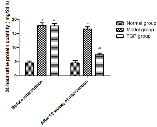
Effect of total glucoside of paeony on 24 h urine protein quantity in different rat groups (n=10). *P<0.05, compared with the normal group. #P<0.05, compared with the model group.
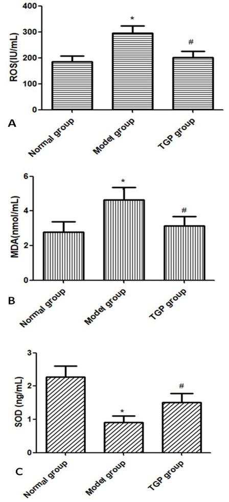
Effect of total glucoside of paeony on kidney ROS (A), MDA (B), and SOD (C) content in the three rat groups after 12 weeks of intervention (n=10). *P<0.05, compared with the normal group. #P<0.05, compared with the model group.
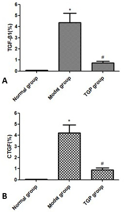
Effect of total glucoside of paeony on kidney TGF-β1 (A) and CTGF (B) in the three groups after 12 weeks of intervention (n=10). *P<0.05,compared with the normal group. #P<0.05,compared with the model group.
References
- Cagdas, D.N., Gucer, S., Kale, G., Duzova, A. and Ozen, S., 2005. Pediatr. Nephrol., 20: 1352-1354. https://doi.org/10.1007/s00467-005-1991-9[CrossRef]
- Darlington, P.J., Baroja, M.L., Chau, T.A., Siu, E., Ling, V., Carreno, B.M. and Madrenas, J., 2002. J. exp. Med., 195: 1337-1347. https://doi.org/10.1084/jem.20011868[CrossRef]
- Düzgün, N., Duman, T., Haydardedeoğlu, F.E. and Tutkak, H., 2009. Tissue Antigens, 4: 539-542. https://doi.org/10.1111/j.1399-0039.2009.01347.x[CrossRef]
- Iida, Y., Yasudo, H., Fukano, R., Azuma, Y., Ichimura, T., Ohga, S. and Hasegawa, S., 2020. Pediatr. Blood Cancer, e28131.
- Ito, Y., Goldschmeding, R., Kasuga, H., Claessen, N., Nakayama, M., Yuzawa, Y., Sawai, A., Matsuo, S., Weening, J.J. and Aten, J., 2010. Am. J. Physiol. Renal Physiol., 299: F545-F558. https://doi.org/10.1152/ajprenal.00120.2009[CrossRef]
- Iwano, M., Akai, Y., Fujii, Y., Dohi, Y., Matsumura, N. and Dohi, K., 1994. Clin. exp. Immunol., 7: 309-314.
- Jin, M., Yin, Z., Wei, K., Xie, Y., Bai, X., Fu, B., Feng, Z., Li, Q. and Chen, X., 2019. J. mol. Med. (Berl), 97: 553-561. https://doi.org/10.1007/s00109-019-01749-1[CrossRef]
- Ležaić, V., Ristić, S., Dopsaj, V. and Marinković, J., 2010. Srp. Arh. Celok. Lek., 138: 726-731. https://doi.org/10.2298/SARH1012726L[CrossRef]
- Lin, J., Xiao, L., Ouyang, G., Shen, Y., Huo, R., Zhou, Z., Sun, Y., Zhu, X., Zhang, J., Shen, B. and Li, N., 2012. Cell. Immunol., 280:156-163. https://doi.org/10.1016/j.cellimm.2012.12.005[CrossRef]
- Liu, D.F., Wei, W. and Song, L.H., 2006. Clin. exp. Pharmacol. Physiol., 33: 332-339. https://doi.org/10.1111/j.1440-1681.2006.04371.x[CrossRef]
- Liu, N., Makino, T., Nogaki, F., Kusano, H., Suyama, K., Muso, E., Honda, G., Kita, T. and Ono, T., 2004. Am. J. Physiol. Renal. Physiol., 287: F612-F620. https://doi.org/10.1152/ajprenal.00322.2003[CrossRef]
- Naveed, M., Han, L., Hasnat, M., Baig, M.M.F.A., Wang, W., Mikrani, R., Zhiwei, L., Sembatya, K.R., Xie, D. and Zhou, X., 2018. Biomed. Pharmacother., 108: 1460-1468. https://doi.org/10.1016/j.biopha.2018.09.168[CrossRef]
- Schuerwegh, A.J., Dombrecht, E.J., Stevens, W.J., Van, Offel, J.F., Bridts, C.H. and De, Clerck, L.S., 2003. Osteoarth. Cart., 11: 681-687. https://doi.org/10.1016/S1063-4584(03)00156-0[CrossRef]
- Tufano, M.A., Cipollaro, de, l’Ero, G., Ianniello. R., Galdiero, M. and Galdiero, F., 1991. Eur. Cytokine Netw., 2: 361-366.
- Zhang, P., Zhang, J.J., Su, J., Qi, X.M., Wu, Y.G. and Shen. J.J., 2009. Am. J. Chin. Med., 37: 295-307. https://doi.org/10.1142/S0192415X09006850[CrossRef]
Services
Article usage statistics
Loading…