
Angelica sinensis polysaccharide (ASP) is a biomacromolecule that isolated from the roots of Angelica sinensis. This study aims to investigate its protective effect on kidney injury and its influence on BMP-7/Smads/TGF-β1 signal pathway in irradiated rats. Total 60 Sprague Dawley (SD) rats were randomly divided into 5 groups: the normal (normal saline), model (normal saline), and low, medium, high dose of ASP groups (9.0, 18.0 and 36.0 mg/mL, 2.0 mL/kg·d, intragastric gavage once a day for 14 days). On the 15th day, all other groups received 60Co γ-ray irradiation with a total dose of 4.0 Gy except the normal group. The levels of NO synthase (NOS) and NO in serum, the contents of malondialdehyde (MDA) and superoxide dismutase (SOD) in kidney of each group were detected with ELISA after 24 h of irradiation, and the protein expression levels of TGF-β1, phosphorylated (p-)Smad2, p-Smad2, p-Smad1, p-Smad5 and BMP7 in kidney were detected by western blotting. In the results, compared with the model group, NOS, NO and MDA contents were decreased in the middle and high dose groups while SOD contents were increased in low, middle and high dose groups. The levels of TGF-β, p-Smad2 and p-Smad3 were increased in low, middle and high dose groups while the levels of BMP7, p-Smad1 and p-Smad5 were decreased in middle and high dose groups. In conclusion, ASP can reduce the expression levels of TGF-β, p-Smad2 and p-Smad3 in kidney of rats induced by radiation, increase the expression levels of BMP7, p-Smad1 and p-Smad5, and resist the body injury caused by radiation by regulating BMP-7/Smads/TGF-β1 signal pathway.
Short Communication · Volume 54, Issue 5, 16 July 2022 · Open Access
Regulatory Effect of Angelica sinensis Polysaccharide on BMP-7/Smads/TGF-β1 Signal Pathway in Kidney of Rats with Radiation Injury
· · · ·
Affiliations & Notes ⌄
- 1School of Pharmacy, Changchun University of Chinese Medicine, Changchun 130117, China 2The Third Affiliated Hospital of Changchun University of Chinese Medicine, Changchun 130117, China * Corresponding author: duanduan-2007@163.com, linzhe1228@163.com
Article Info ⌄
- Journal:
- Pakistan Journal of Zoology
- Published:
- 16 July 2022

From the article
- The results showed that when the drug concentration was up to 18.0 mg/mL, the contents of MDA in kidney decreased (Fig. 2A); while the drug concentration up to 9.0 mg/mL, the contents of SOD in kidney increased (Fig. 2B).
- To verify whether the protective effect of ASP on radiation injury is associated with regulating BMP-7/Smads/TGF signaling pathway, the content of TGF-β1, p-Smad2, p-Smad3, p-Smad1, p-Smad5 and BMP7 were estimated.
- ASP treatment can improve the structure of kidney cells, which had a protective effect on the kidney of radiation injury rats.
- These results indicate that A. sinensis polysaccharide plays a protective role in radiation-induced kidney injury in rats by acting on the TGF-β1 signaling pathway.
Selected from the article's own text.
Abstract
Materials and methods
A. sinensis polysaccharide (ASP) was purchased from Shanghai Yuanye Bio-Technology Co., Ltd (Shanghai, China), and was analyzed using Ultraviolet/Visible (UV/Vis) spectra (UV-2501PV, Shimadzu, Japan). The percentage of total sugar was determined to be 91.14% by phenol sulfuric acid method. Total sixty Sprague Dawley (SD) rats (weighing 300±50 g) were raised under specific-pathogen-free (SPF) conditions at 25±1°C and 55±5 % humidity.
The initial body weight of all the rats was measured, then the different grouping treatments were performed. The SD rats were randomly divided into five groups (12 rats in each group): The normal group (normal saline), the model group (normal saline), the low (9.0 mg/mL), medium (18.0 mg/mL) and high (36.0 mg/mL) dose groups for the drug groups. Rats in each dose group were given intragastric gavage (2.0 mL/kg·d) once a day for 14 days. On the 15th day, except for the normal group, all the rats received γ-ray irradiation once, the exposure time was set at 517s, the distance of irradiation target was set at 60 cm, with a total dose of 4.0 Gy.
After the irradiation for 24 h, the body weight was measured again. After the rats were anesthetized with pentobarbital sodium, the middle finger gently pressed the left forelimb on the sternum heart, and the ring finger on the abdomen. Then, twiddle the thumb and gently press the eye skin on the blood side to make the eyeball congestion protruding. The eyeballs were taken out with forceps and turned the thumb and index fingers to make the blood flow from the eye socket into the centrifuge tube at different speeds. Meanwhile, the left middle finger gently pressed the mouse heart to speed up the heart pumping speed. When the blood was about to run out, the rats were killed by inhaling carbon dioxide. The centrifuge tubes were placed in a 37°C incubator for one hour, then placed in a refrigerator at 4°C for 3 h. After the blood clots contracted, the samples were centrifuged at 4000 rpm for 10 min; the supernatant was placed in a clean centrifuge tube and stored at -20°C for later use. The rats in each group were killed by taking off their necks, and the kidney tissues of the rats were stored at -80 °C.
The contents of NOS and NO in serum were detected by the NOS ELISA Kit and NO ELISA Kit, (Shanghai Yuan Mu Biological Technology Co. Shanghai, China, 96T, YM1436B (SenBeija Biological Technology Co. Nanjing China, 96T, SBJR0469)) according to the instructions. In general, 50µL samples and the labeled antibodies were added to each well, incubated at 37°C for 1 h, washed 3 times; then added 80 µL affinity enzyme-HRP, incubated at 37 °C for 30 min, washed 3 times. After that, 50 µL of substrate A and B were added and incubated for 10 min at 37 °C in dark. The OD value was measured at 450 nm after 50 µL of termination solution was added.
The levels of malondialdehyde (MDA) and superoxide dismutase (SOD) in the kidney tissue were determined according to the instructions of the rat MDA ELISA kit (Wuhan Saipei Biotechnology Co. Wuhan, China, SP30131) and SOD ELISA kit (Wuhan Saipei Biotechnology Co. Wuhan, China, SP12914).
Kidney tissue homogenate was cracked and homogenized with RIPA protein lysate to obtain protein samples. The protein concentration was detected using the BCA protein concentration determination kit (QPBCA, Sigma-Aldrich (Shanghai) Trading, Co. Shanghai, China). After sodium dodecyl sulphate–polyacrylamide gel electrophoresis (SDS-PAGE), the protein concentration was electrophoresed with polyvinylidene fluoride (PVDF) membrane and cleaned by TBST (Thermo Fisher Scientific, Waltham, MA, USA, 28360). The membrane was incubated overnight at 2-8 ºC with TGF-β1 (bs-0086R), phosphorylated (p-)Smad1 (bs-19917R), p-Sma2 (bs- Beiging Biosynthesis Biotechnology Co., Beijing, China 7464R), p-Smad3 (bs-5235R), p-Smad5 (bs-19918R), BMP7 (bs-2242-R), β-actin (bs-0061R), and the primary antibodies, then added goat anti-rabbit immunoglobulin G-horseradish peroxidase antibodies (Beyotime Institue of Bio Technology Shanghai, China, AD208). After detection, exposure, development and fixation of ultra-sensitive enhanced chemiluminescence (ECL) reagents (Sigma-Aldrich (Shanghai) Trading Co. China, HVWP04756), the optical density was analyzed using gel-pro-analyzer software (Medi Cybernetics Inc. Rockville, MD, USA).
Data were expressed as Mean ± Standard deviation. The Bonferroni test was used for multiple comparisons, one-way ANOVA was used for inter-group comparison. Graphpad Prism 5.0 was used to process the data.
Results and discussion
The initial body weight of rats in each group was similar and has no significance. After 14 days of continuous intragastric administration, the body weight in the normal group was slightly higher than that of other groups, however, with no significant difference (Fig. 1A). The effects of ASP on NOS and NO were investigated via the ELISA assay. The results showed that when the concentration of drug up to 18.0 mg/mL, the contents of NOS and NO in serum decreased (Fig. 1B, C).
The effects of ASP on MDA and SOD were investigated via the ELISA assay. The results showed that when the drug concentration was up to 18.0 mg/mL, the contents of MDA in kidney decreased (Fig. 2A); while the drug concentration up to 9.0 mg/mL, the contents of SOD in kidney increased (Fig. 2B).
To verify whether the protective effect of ASP on radiation injury is associated with regulating BMP-7/Smads/TGF signaling pathway, the content of TGF-β1, p-Smad2, p-Smad3, p-Smad1, p-Smad5 and BMP7 were estimated. When the concentration of drug was up to 9.0 mg/mL, the contents of TGF-β1 in kidney decreased. The drug concentration up to 18.0 mg/mL, the contents of BMP-7 and p-Smad1 in kidney increased (Fig. 3A, B), the contents of p-Smad2 and p-Smad3 in kidney decreased, while the contents of p-Smad4 in kidney increased (Fig. 3A, C).
In this study, to explore the effect of ASP on the bodyweight of rats with radiation injury, the bodyweight of rats was measured before and after the radiation, ASP can improve the changes of characteristic of peripheral hemogram caused by radiation injury and improve the appetite of rats, so ASP can maintain the bodyweight of rats with radiation injury. The serum levels of NOS and NO are vasoactive substances released by cells . After radiation, the changes in rat blood cells lead to the increase of the content of NOS and NO . The results showed that ASP could reduce the increase of serum NOS and NO levels induced by radiation.
MDA is one of the final products of membrane lipid peroxidation . After radiation, the content of MDA in the kidney tissue increases, and the content of SOD in the kidney tissue decreases . ASP treatment can improve the structure of kidney cells, which had a protective effect on the kidney of radiation injury rats. TGF-β1 is one of the most effective cytokines known to promote renal fibrosis . BMP-7 (Pravoverov et al., 2018) is an important cytokine that inhibits renal fibrosis and has antagonistic effect on TGF-β1-induced fibrosis . TGF-β1 and BMP-7 (Liu et al., 2018) can regulate each other through the downstream of Smad signaling pathway to maintain the balance of their biological activities . In kidney injury, this balance is disrupted, leading to the upregulation of TGF-β1, activation of Smad3 , down-regulation of BMP-7 and its downstream Smad1/5/8 expression, ultimately leading to renal fibrosis . These results indicate that A. sinensis polysaccharide plays a protective role in radiation-induced kidney injury in rats by acting on the TGF-β1 signaling pathway.
Conclusion
In summary, Angelica sinensis polysaccharide has good protection and recovery effects on radiation-induced kidney injury, and has the potential to prevent and treat radiation-induced nephropathy.
Acknowledgment
This study was supported by 2019 Annual Scientific Research and Development Fund Project of Changchun University of Traditional Chinese Medicine (No. 11091) and Science and Technology Development Program of Jilin Province (Nos. 20210101035JC and 20210402034GH).
Ethical compliance
Research experiments conducted in this article with animals or humans were approved by the Ethical Committee and responsible authorities of Changchun University of Chinese Medicine (Certify No.: 2020039) following all guidelines, regulations, legal, and ethical standards as required for animals.
Statement of conflict of interest
The authors have declared no conflict of interest.
Figures
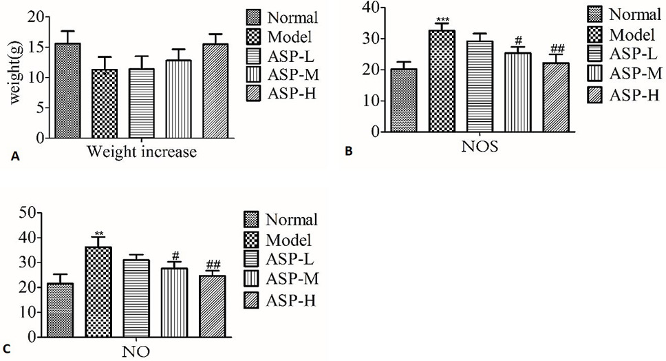
Body weight changes of ASP on SD rats and effects of ASP on NOS and NO levels in serum. Values are expressed as means ± SEM. Notes: Compared with normal group: **p < 0.01, ***p < 0.001; compared with model group: #p < 0.05, ##p < 0.01.
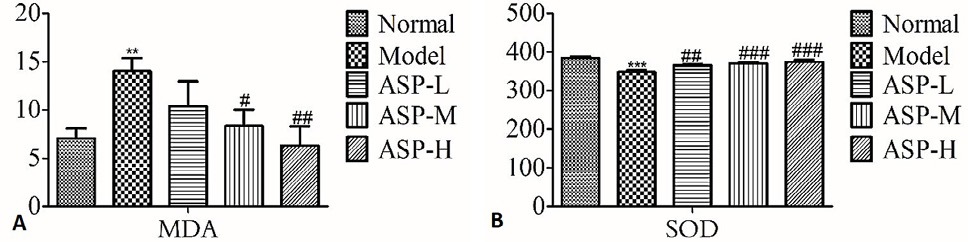
Effects of ASP on MDA and SOD levels in kidney. ###p < 0.001. For statistical details, see Fig.1.
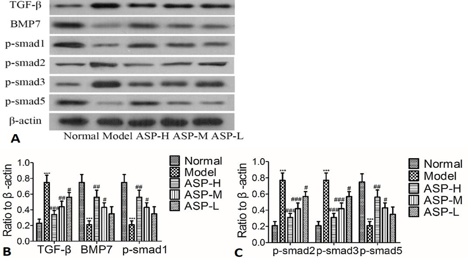
The effect of ASP on TGF-β signaling pathway in rat. For statistical details, see Figs. 1 and 2.
References
- Belpomme, D., Hardell, L., Belyaev, I., Burgio, E. and Carpenter, D.O., 2018. Environ. Pollut., 242: 643-658. https://doi.org/10.1016/j.envpol.2018.07.019[CrossRef]
- Cao, P., Sun, J.L., Sullivan, M.A., Huang, X., Wang, H.X., Zhang, Y., Wang, N. and Wang, K.P., 2018. Int. J. Biol. Macromol., 111: 1133-1139. https://doi.org/10.1016/j.ijbiomac.2018.01.139[CrossRef]
- Chen, Z.T., Cheng, L., Zhang, J. and Cui, J., 2020. BMC Complement. Med. Ther., 20: 192. https://doi.org/10.1186/s12906-020-02902-x[CrossRef]
- Dondelinger, R.M., 2012. Biomed. Instrum. Technol., 46: 219-223. https://doi.org/10.2345/0899-8205-46.3.219[CrossRef]
- Gao, W., Liang, J.X., Ma, C., Dong, J.Y. and Yan, Q., 2017. Biomed. Res. Int., 2017: 4176170(1)-4176170(11). https://doi.org/10.1155/2017/4176170[CrossRef]
- Ghiselli, R., Lucarini, G., Orlando, F., Ortenz, M., Cardinali, L., Provinciali, M., Primio, R.D. and Guerrieri, M., 2017. Neuromodulation, 20: 761-766.
- Gupta, S., Silveira, D.A. and Mombach, J.C.M., 2018. PLoS One, 13: e0200768(1)- e0200768(10). https://doi.org/10.1371/journal.pone.0200768[CrossRef]
- King, M., Joseph, S., Albert, A., Thomas, T.V., Nittala, M.R., Woods, W.C., Vijayakumar, S. and Packianathan, S., 2020. Oncology, 98: 61-80. https://doi.org/10.1159/000502979[CrossRef]
- Li, J.H., Zheng, H.Y. and Feng, C.J., 2018. Front. Biosci. (Landmark Ed), 23: 1803-1821. https://doi.org/10.2741/4674[CrossRef]
- Liu, N., Feng, J., Lu, X.Y., Yao, Z.L., Liu, Q., Lv, Y., Han, Y.R., Deng, J.F. and Zhou, Y.Q., 2019. Mediators Inflamm., 2019: 6175091(1)- 6175091(14). https://doi.org/10.1155/2019/6175091[CrossRef]
- Liu, X.F., Zhou, M., Yang, Y.L., Wu, J. and Peng, Q.S., 2018. Oncotarget, 9: 9596-9607. https://doi.org/10.18632/oncotarget.24073[CrossRef]
- Lodyga, M. and Hinz, B., 2020. Semin. Cell Dev. Biol., 101: 123-139. https://doi.org/10.1016/j.semcdb.2019.12.010[CrossRef]
- Ma, T.T. and Meng, X.M., 2019. Adv. exp. Med. Biol., 1165: 347-364. https://doi.org/10.1007/978-981-13-8871-2_16[CrossRef]
- Marazziti, D., Piccinni, A., Mucci, F., Baroni, S., Loganovsky, K. and Loganovskaja, T., 2016. Probl. Radiac. Med. Radiobiol., 21: 64-90.
- Miousse, I.R., Kutanzi, R. and Koturbash, I., 2017. Int. J. Radiat. Biol., 93: 457-469. https://doi.org/10.1080/09553002.2017.1287454[CrossRef]
- Narasimhulu, C.A. and Singla, D.K., 2020. Cells, 9: 280(1-30). https://doi.org/10.3390/cells9020280[CrossRef]
- Nicolatou-Galitis, O., Sarri, T., Bowen, J., Palma, M.D., Kouloulias, V.E., Niscola, P., Riesenbeck, D., Stokman, M., Tissing, W., Yeoh, E., Elad, S. and Lalla, R.V., 2013. Support Care Cancer, 21: 357-364. https://doi.org/10.1007/s00520-012-1613-6[CrossRef]
- Pravoverov, K., Whiting, K., Thapa, S., Bushong, T., Trang, K., Lein, P.J. and Chandrasekaran, V., 2019. Cell Mol. Neurobiol., 39: 917-934. https://doi.org/10.1007/s10571-019-00688-2[CrossRef]
- Schwartz, A.G. and Cote, M.L., 2016. Adv. Exp. Med. Biol., 893: 21-41. https://doi.org/10.1007/978-3-319-24223-1_2[CrossRef]
- Sun, Q., Guo, Y., Liu, X.H., Czauderna, F., Carr, M.I., Zenke, F.T., Blaukat, A. and Vassilev, L.T., 2019. Mol. Cancer Res., 17: 2457-2468. https://doi.org/10.1158/1541-7786.MCR-19-0362[CrossRef]
- Tsikas, D., 2017. Anal. Biochem., 524: 13-30. https://doi.org/10.1016/j.ab.2016.10.021[CrossRef]
- Wakeford, R., 2019. Hypertension, 73: 1170-1171.
- Wang, B., Liu, T., Wu, J.C., Luo, S.Z., Chen, R., Lu, L.G. and Xu, M.Y., 2018. Biomed. Pharmacother., 98:214-221. https://doi.org/10.1016/j.biopha.2017.12.035[CrossRef]
- Wu, H.Y., Wang, D., Shi, H., Liu, N.N., Wang, C.H., Tian, J.Y., Wang, X. and Zhang, Z.H., 2021. Mol. Immunol., 137: 1-10. https://doi.org/10.1016/j.molimm.2021.06.005[CrossRef]
- Yeh, C.H., Chang, C.K., Cheng, K.C., Li, Y.X., Zhang, Y.W. and Cheng, J.T., 2011. Evid. Based Complement. Altern. Med., 2011: 796723(1)-796723(9). https://doi.org/10.1155/2011/796723[CrossRef]
- Zhuang, C., Wang, Y.J., Zhang, Y.K. and Xu, N.W., 2018. Int. J. Biol. Macromol., 115: 281-286. https://doi.org/10.1016/j.ijbiomac.2018.04.083[CrossRef]
- Zou, G.L., Zuo, S., Lu, S., Hu, R.H., Lu, Y.Y., Yang, J., Deng, K.S., Wu, Y.T., Mu, M., Zhu, J.J., Zeng, J.Z., Zhang, B.F., Wu, X., Zhao, X.K. and Li, H.Y., 2019. World J. Gastroenterol., 25: 4222-4234. https://doi.org/10.3748/wjg.v25.i30.4222[CrossRef]
Services
Article usage statistics
Loading…