Research Article · Volume 13, Issue 3, 18 July 2025 · Open Access
Infestation Rate and Molecular Detection of Hard Ticks in Pet Cats from Hilla City, Iraq
· · ·
Affiliations & Notes ⌄
- 1Department of Parasitology, College of Veterinary Medicine, Al-Qasim Green University, Babylon 51013, Iraq
- 2Aesthetic and Laser techniques Department, College of Health and Medical technique, Al-Mustaqbal University, Babylon 51001, Iraq
- 3Department of Internal and Preventive Veterinary Medicine, College of Veterinary Medicine, AL-Qasim Green University, Babylon 51013, Iraq. *Correspondence | Ahmed Hamzah Mosa, Department of Parasitology, College of Veterinary Medicine, Al-Qasim Green University, Babylon 51013, Iraq
- 4Email: ahmedvet@vet.uoqasim.edu.iq
Article Info ⌄
- Journal:
- Journal of Animal Health and Production
- Published:
- 18 July 2025

From the article
- The incidence of hard tick (Rhipicephalus) infestation among pet cats in Hillah City was 34 out of 70 cats, representing a prevalence rate of 48.5%.
- The present study demonstrated that the infection rate of Rhipicephalus ticks was significantly higher in younger cats compared to older cats.
- Further epidemiological studies on the distribution of Rhipicephalus species in pet cats across various provinces of Iraq are needed.
Selected from the article's own text.
Abstract
INTRODUCTION
Understanding parasitic infections in pet cats is essential for both medical and veterinary fields, as these animals can serve as reservoirs for parasites that are transmissible to humans and other animals . Identifying tick species infesting pet cats is critical for developing effective prevention and control strategies that protect public and animal health . Both stray and pet cats are commonly infested with a variety of parasites .
Ticks, as obligate blood-feeding ectoparasites, can cause a range of clinical issues, including skin disorders, irritation, allergic reactions, weight loss, and anemia . Moreover, they are capable of transmitting numerous bacterial, viral, and protozoal pathogens to both animals and humans .
Despite the recognized importance of tick infestations in cats, there is a notable lack of comprehensive global data on their prevalence, associated disease burdens, and mortality rates. This data gap is particularly evident in Iraq, where systematic studies on tick infestations in pet cats are scarce. Understanding the epidemiology of these parasites is vital for formulating effective control and preventive measures.
Regional studies underscore the importance of this issue: a study in Turkey reported that approximately 40% of cats were infested with Rhipicephalus spp., with significant health impacts , while research in Egypt found a 35% infestation rate in domestic and stray cats, with PCR techniques revealing the presence of pathogens such as Babesia and Ehrlichia .
Given this context, the present study represents the first report on tick infestation in pet cats from Hilla City, Iraq. It aims to estimate the infestation rate and identify tick species using both morphological and molecular (PCR) methods, as visual inspection alone is often insufficient for accurate species classification.
MATERIALS AND METHODS
A total of 70 pet cats of various ages and sexes were collected from different locations in Hilla City, Iraq, through visits to veterinary clinics. The collection and handling of animals were conducted in accordance with the guidelines of the International Animal Ethics Committee or the Institutional Ethics Committee, as well as local laws and regulations . Of the total cats examined, 47 were female and 23 were male.
Each cat was visually examined to determine its sex and age, with age estimated based on dentition. Cats were then grouped into two age categories: less than one year and more than one year. Ticks were also classified according to their location on the host’s body.
Tick collection was performed using non-toothed forceps, and specimens were examined under a dissecting microscope for morphological identification . All collected ticks were preserved in 70% ethanol and later subjected to DNA extraction and molecular identification using 16S rDNA-based PCR techniques .
Identification of Collected Ticks
Identification was accomplished using a stereomicroscope at 40x magnification and compared to features listed in the taxonomic key. Two specimens for each sample were placed at the Iraq Natural History Research Center and Museum’s Department of Vertebrates.
DNA Extraction of Ticks
Genomic DNA was extracted from 20 adult tick samples using the gSYNCTM Genomic DNA Purification Kit (Geneaid, Canada), with minor protocol adjustments. Each tick was incubated in a mixture of 300 μl comprising 200 μl of GST Buffer, 40 μl of Proteinase K solution, and 5 μl of RNase A solution at 60 °C for 2–3 hours, depending on the size of the specimen. After incubation, the ticks were mechanically ground using a sterile glass mortar and pestle. The homogenized material was centrifuged at 10,000 × g to remove insoluble debris.
The resulting supernatant was transferred to a new tube, followed by the addition of 200 μl of GSB Buffer. The mixture was vortexed vigorously for 10 seconds, then mixed with 200 μl of 100% ethanol. The solution was passed through a spin column by centrifugation at 13,000 rpm for 30 seconds.
The column was washed twice: first with 400 μl of W1 Buffer, and then with 600 μl of Wash Buffer, each followed by centrifugation at 14,000 rpm for 30 seconds. For elution, 100 μl of pre-warmed Elution Buffer was added to the center of the column membrane and incubated for 5 minutes at room temperature. DNA was finally eluted into sterile 1.5 ml Eppendorf tubes by centrifugation at 14,000 rpm.
Polymerase Chain Reactions
PCR was employed to confirm the identification of Rhipicephalus spp. using species-specific primers: forward primer 5′-TTGGGCAAGAAGACCCTATGAA-3′ and reverse primer 5′-CCGGTCTGAACTCAGATCAAGT-3′, which amplify a 495 base pair fragment . The PCR protocol began with an initial denaturation step at 95 °C for 5 minutes. This was followed by 10 amplification cycles, each consisting of denaturation at 92 °C for 1 minute, annealing at 72 °C for 1 minute and 30 seconds, and a final extension at 72 °C for 7 minutes.
Statistical Analysis
The Chi-square (X2) test was employed to compare the outcomes. According to Petrie and Watson (2006), differences were deemed statistically significant when P < 0.05.
RESULTS AND DISCUSSION
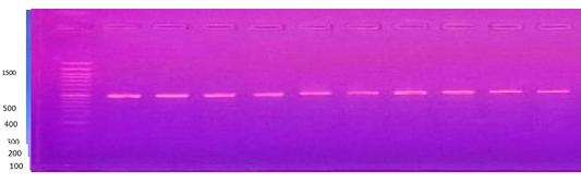
The incidence of hard tick (Rhipicephalus) infestation among pet cats in Hillah City was 34 out of 70 cats, representing a prevalence rate of 48.5%. Species identification was confirmed using polymerase chain reaction (PCR) analysis (Figure 1).
Rhipicephalus infestation Rate by Host Sex and Age
Examination of 70 pet cats revealed an overall Rhipicephalus infestation rate of 34 (48.5%) across both sexes. This prevalence varies considerably compared to other countries, such as 4.2% in Dubai , 80.1% in cats from Egypt , 0.8% in the USA , 1.79% in Ireland , 52.4% in Hungary , and 1.6% in Brazil (Mendes et al., 2011). Differences in infestation rates among nations may be influenced by the effectiveness of control programs, seasonal and environmental factors, as well as the molecular techniques employed for species identification, since morphological identification alone is often insufficient .
In this study, female cats exhibited a higher infestation rate (51.06%) compared to males (43.4%), although this difference was not statistically significant. These findings align with Kamaruddin et al. (2020), who reported no significant sex-based difference in infestation rates, suggesting that both males and females are equally affected by this parasite.
Table 1: Infection rate of Rhipicephalus turanicus according to host sex and age.
| Sex / Age | Male | Female | ||||
| No. of cats examined | No. of cats infected | Infection rate, % | No. of cats examined | No. of cats infected | Infection rate, % | |
| ≤ 1 year | 9 | 6 | 66.6a | 18 | 14 | 73.6a |
| >1 year | 14 | 4 | 28.5b | 28 | 10 | 42.4b |
| Total | 23 | 10 | 43.4a | 47 | 24 | 51.06a |
Different superscript letters in column differ significantly at level of p<0.05.
Age appeared to be a significant factor in infestation rates, with younger cats showing a higher prevalence of tick infestation than older cats, as confirmed by statistical analysis (Table 1). The current study also observed significant differences in infestation rates between nymphal and adult ticks, with adults, particularly females, being more susceptible to infection. This may be attributed to their prolonged feeding periods and higher blood consumption, which increase the risk of acquiring and transmitting pathogens . Städel (2024) supports this by noting that the extended attachment of female ticks to their hosts increases their susceptibility to bloodborne diseases. In contrast, males tend to have a lower infection rate, possibly due to shorter feeding durations, although infection rates may rise during tick mating seasons (Ding et al., 2024).
Adult ticks generally exhibit higher infection rates than nymphs, likely because they feed longer on infected hosts, increasing their exposure to pathogens . The elevated infection rates in female ticks are particularly concerning for public health due to their greater potential for transmitting infections to humans and other animals. Consequently, tick control efforts should prioritize targeting adult female ticks.
Overall, multiple factors, including environmental conditions, seasonal variations, and host characteristics, influence tick infestation levels. These findings highlight the need for further research to better understand these dynamics and develop more effective control strategies.
Rhipicephalus Infestation by Body Site in Cats
This study examined the infestation patterns of Rhipicephalus ticks on domestic cats, focusing on the preferred attachment sites on the host’s body. The data revealed a non-random distribution of ticks, with certain anatomical locations favored significantly over others. The ears and neck were the most commonly infested areas, consistent with previous reports of tick attachment in domestic animals. These sites feature thinner skin, richer blood supply, and are less accessible for grooming, making them ideal for tick attachment and feeding. The ears, in particular, provide a warm microclimate that protects ticks and enhances their feeding success.
Moderate infestation rates were also observed in the perineal and inguinal regions, areas that are less frequently groomed by cats and benefit from protective flesh folds combined with moisture, creating favorable conditions for tick survival. In contrast, limbs and dorsal parts of the body exhibited considerably lower infestation rates. These regions are more exposed and frequently groomed, reducing tick attachment and facilitating their removal through grooming behavior.
Kohler et al. (2018) noted that tick site selection is influenced by host traits such as hair density, skin thickness, and grooming habits. Understanding these attachment preferences is crucial for effective tick control strategies in cats. Focused inspection and treatment of known preferred sites can lead to timely tick removal, thereby reducing the risk of disease transmission. The concentration of ticks in specific areas underscores the importance of thorough examinations by veterinarians and pet owners, particularly around the ears, neck, and groin.
These findings also suggest that tick repellents could be applied more efficiently by targeting these strategic body regions rather than using uniform whole-body treatments. In this study, significant tick burdens were found on the ears (79.4%), back (64.7%), and abdomen (61.7%). Table 2 details the variation in tick concentrations across different body parts.
Table 2: Infection rate of Rhipicephalus turanicus according to attachment sites on the cat’s body.
| Attachment site | No. of infected cats | Infection rate, % |
| Head | 15 | 44.1b |
| Ears | 27 | 79.4a |
| Neck | 5 | 18.7c |
| Abdomen | 21 | 61.7a |
| Back | 22 | 64.7a |
| Legs and feet | 8 | 23.5c |
| Tail and perianal | 7 | 20.5c |
Different superscript letters in column differ significantly at level of p<0.05.
Rhipicephalus ticks were especially prevalent on the ears, head, legs, and feet, consistent with Salant et al. (2013), who reported the highest Rhipicephalus infestations in these areas, and Geurden (2018), who identified the toes as common tick attachment sites.
CONCLUSIONS AND RECOMMENDATIONS
The present study demonstrated that the infection rate of Rhipicephalus ticks was significantly higher in younger cats compared to older cats. Ticks were predominantly concentrated on specific body sites, such as the ears, back, and abdomen. Further epidemiological studies on the distribution of Rhipicephalus species in pet cats across various provinces of Iraq are needed. Additionally, a comprehensive examination of imported animals, particularly those in close contact with local pets, is essential to identify carrier animals that may harbor and transmit various diseases, especially parasitic infections.
ACKNOWLEDGEMENTS
We extend our sincere thanks to the veterinary clinics in Babylon for their assistance in taking medical histories from the breeders, collecting samples, and recording data. We also thank everyone who helped us.
NOVELTY STATEMENTS
This study was conducted in Babylon province and provided data on ectoparasites infecting small animals. It showed that Rhipicephalus spp. is the predominant tick species infecting domestic cats, as it can transmit a variety of pathogens and negatively impact animal health.
AUTHOR’S CONTRIBUTIONS
Safaa Mohamed Karim conducted, supervised, and analyzed the study, while Ali Rabeea contributed to manuscript preparation. Ahmed Mosa participated in sample collection, laboratory testing, and epidemiological data collection. Fatima Hashim Abbas contributed to molecular analysis and carefully reviewed the manuscript. All authors approved the final version of the manuscript.
Conflict of Interest
The authors have declared no conflict of interest.
Figures

Gel electrophoresis of PCR products amplified using 16S rDNA primers, showing the DNA band of Rhipicephalus turanicus at 496 bp isolated from pet cats.
References
- Aboelhadid SM, Arafa WM, Mahrous LN, Fahmy MM, Kamel AA (2018). Molecular detection of Rhipicephalus (Boophilus) annulatus resistance against deltamethrin in middle Egypt. Vet. Parasitol. Reg. Stud. Rep., 13: 198–204. https://doi.org/10.1016/j.vprsr.2018.06.005[CrossRef]
- Baker AS, Persinger KA, Olafson PU, Mulenga AO, Johnson TL (2024). Feeding and reproductive parameters of Ixodes scapularis and Amblyomma americanum on deer. J. Med. Entomol., 61(1): 245–249. https://doi.org/10.1093/jme/tjad193[CrossRef]
- Beugnet F, Bourdeau P, Chalvet-Monfray K, Cozma V, Farkas R, Guillot J (2014). Parasites of domestic cats in Europe: co-infestations and risk factors. Parasites Vect., 7: 1–13. https://doi.org/10.1186/1756-3305-7-87[CrossRef]
- Capari B, Hamel D, Visser M, Winter RP, Fister K, Rehbein S (2013). Parasitic infections of domestic cats in western Hungary. Vet. Parasitol., 192(1–3): 33–42. https://doi.org/10.1016/j.vetpar.2012.11.003[CrossRef]
- Deng YP, Fu YT, Elsheikha HM, Cao ML, Zhu XQ, Wang JL (2024). Global impact and distribution of tick paralysis. Clin. Microbiol. Rev., 37(4): e00074-24. https://doi.org/10.1128/cmr.00074-24[CrossRef]
- Ebani VV, Guardone L, Marra F, Altomonte I, Nardoni S, Mancianti F (2020). Arthropod-borne pathogens in stray cats from northern Italy. Animals, 10(12): 2334. https://doi.org/10.3390/ani10122334[CrossRef]
- El-Sherbini G, El-Hawy A, Farag A (2021). Tick infestation and tick-borne pathogens in cats in Egypt. Parasites Vect., 14(1): 220. https://doi.org/10.1186/s13071-021-04739-z[CrossRef]
- Faouzi D, M’Zoughi N, Laarif J, Darghouth MA (2018). Rhipicephalus spp. identification in Tunisia. Exp Appl. Acarol., 74(1): 55–66. https://doi.org/10.1007/s10493-018-0220-6[CrossRef]
- Fourie JJ, Joubert A, Labuschagné M, Beugnet F (2014). qPCR method to assess acaricide anti-feeding effect. Comp. Immunol. Microbiol. Infect. Dis., 37(3): 181–187. https://doi.org/10.1016/j.cimid.2014.01.003[CrossRef]
- Geurden T (2018). Tick infestation patterns in companion animals. Parasitol. Rep., 7(4): 45–50.
- Ghafar A, Khan A, Cabezas-Cruz A, Gauci CG, Niaz S, Ayaz S (2020). Molecular diversity of ticks and pathogens in Pakistan. Microorganisms, 8(9): 1428. https://doi.org/10.3390/microorganisms8091428[CrossRef]
- Jatav RS, Pratap A, Vaishnav N, Sharma N (2024). Disease overview in dogs and cats. In: Introduction to Diseases, Diagnosis, and Management of Dogs and Cats. Academic Press. https://doi.org/10.1016/B978-0-443-12932-1.00002-6[CrossRef]
- Kalayou S, Gort G, Bron GM, Koenraadt CJM, Ten Bosch QA, Masiga D (2024). Tickoff® trial in zebu cattle in Kenya. Parasite Epidemiol. Control, 27: e00384. https://doi.org/10.1016/j.parepi.2024.e00384[CrossRef]
- Kamaruddin MA, Abdullah S, Mohd Zain SN, Osman AY (2020). Gender-based parasitism in cats. J. Vet. Parasitol., 34(2): 120–125.
- Kohler A, Smith B, Jones, C (2018). Host traits affecting tick attachment preferences. Journal of Parasitology Research., 124(3): 215–223. https://doi.org/10.1016/j.jparres.2018.04.012[CrossRef]
- Mendes-de-Almeida F, Crissiuma AL, Gershony LC, Willi LMV, Paiva JP, Guerrero J, Labarthe N (2011). Ectoparasites in cats in Rio de Janeiro. Parasitol. Res., 108: 1431–1435. https://doi.org/10.1007/s00436-010-2180-3[CrossRef]
- Morelli S, Diakou A, Di Cesare A, Colombo M, Traversa D (2021). Canine and feline parasitology for human health. Clin. Microbiol. Rev., 34(4): e00266-20. https://doi.org/10.1128/CMR.00266-20[CrossRef]
- Mustafa BHS (2019). Ectoparasites in small ruminants in Sulaimani. Iraqi J. Vet. Sci., 33(2): 303–309. https://doi.org/10.33899/ijvs.2019.163507[CrossRef]
- Petrie A, Watson P (2006). Statistics for veterinary and animal science (2nd ed.). Blackwell Publishing.
- Rochlin I, Toledo A (2020). Emerging tick-borne pathogens: Mini-review. J. Med. Microbiol., 69(6): 781–791. https://doi.org/10.1099/jmm.0.001212[CrossRef]
- Salant H, Mumcuoglu KY, Baneth G (2013). Ticks on cats in Israel. Vet. Parasitol., 191(1–2): 123–128. https://doi.org/10.1016/j.vetpar.2012.08.022[CrossRef]
- Saleh MN, Sundstrom KD, Duncan KT, Ientile MM, Jordy J, Ghosh P, Little SE (2019). Tick survey on pets in USA. Parasites Vect., 12: 1–11. https://doi.org/10.1186/s13071-019-3697-4[CrossRef]
- Schuster RK, Thomas K, Sivakumar S, O’Donovan D (2009). Parasites of cats in Dubai. Parasitol. Res., 105: 125–134. https://doi.org/10.1007/s00436-009-1380-5[CrossRef]
- Städele C (2024). Ixodes scapularis detects CO₂ without Haller’s organ. J. Exp. Biol., 227(6): jeb246874. https://doi.org/10.1242/jeb.246874[CrossRef]
- Waal T, Smyth JA, Murphy TM (2020). Parasites in stray cats in Ireland. Ir. Vet. J., 73(1): 15. https://doi.org/10.1186/s13620-020-00167-3[CrossRef]
- Yildirim A, Dumanli N, Goker M (2019). Rhipicephalus ticks and pathogens in cats in Turkey. Ticks Tick Borne Dis., 10(4): 1017–1023. https://doi.org/10.1016/j.ttbdis.2019.05.009[CrossRef]
Services
Article usage statistics
Loading…