Research Article · Volume 14, Issue 10, 15 September 2026 · Open Access
Identification of Zoonotic Intestinal Parasites in Madura Cattle in Bangkalan, Madura, Indonesia
· · · ·
Affiliations & Notes ⌄
1Division of Veterinary Parasitology, Department of Veterinary Science, Faculty of Veterinary Medicine, Universitas Airlangga, Surabaya, Indonesia
2Research Group of One Helath, Faculty of Veterinary Medicine, Universitas Airlangga, Surabaya, Indonesia
3Postgraduate School, Universitas Airlangga, Surabaya, Indonesia
4Research Group of Malaria Zoonotic and Parasitology, Institute of Tropical Disease, Universitas Airlangga, Surabaya, Indonesia
5Tropical Infectious Diseases Research and Education Centre, Universiti Malaya, Kuala Lumpur, Malaysia. *Correspondence | Poedji Hastutiek, Division of Veterinary Parasitology, Department of Veterinary Science, Faculty of Veterinary Medicine, Universitas Airlangga, Surabaya, Indonesia
6Institute of Tropical Disease, Universitas Airlangga, Surabaya, Indonesia
Age was significantly associated with infection, with the highest prevalence observed in cattle aged 6 months to 2 years.
Zoonotic intestinal parasites remain prevalent in Madura cattle in Bangkalan Regency, with an overall infection rate of 29%.
Infection prevalence differed significantly among sub-districts and age groups but was not associated with sex.
These findings indicate that zoonotic intestinal parasites are maintained within traditional cattle production systems in Bangkalan.
Selected from the article's own text.
Abstract
Zoonotic intestinal parasites in cattle remain a concern due to their impact on livestock productivity and potential public health implications. This study aimed to determine the prevalence and diversity of zoonotic intestinal parasites in Madura cattle across eight sub-districts in Bangkalan Regency, Indonesia, and to evaluate their distribution according to age, sex, and geographic location. A cross-sectional survey was conducted from March to May 2024 involving 400 fecal samples collected using simple random sampling. Samples were examined using direct smear, sedimentation, and modified Fulleborn flotation techniques. Data were analyzed descriptively and inferentially using Chi-square tests. The overall prevalence of zoonotic intestinal parasites was 29% (116/400; 95% CI: 24.6–33.4%). Five parasite taxa were identified: Toxocara vitulorum, Fasciola gigantica, Blastocystis hominis, Balantidium coli, and Entamoeba spp. Blastocystis hominis was the most frequently detected species (19.5%). Prevalence differed significantly among sub-districts (p < 0.001), with the highest rate in Kwanyar (50%) and the lowest in Kokop (16%). Age group was significantly associated with infection (p < 0.001), with cattle aged 6 months to 2 years showing the highest prevalence (42.4%). No significant association was found between sex and infection status (p = 0.839). Mixed infections were observed in several animals. These findings indicate that zoonotic intestinal parasites remain endemic in Madura cattle and are influenced by geographic and age-related factors. Strengthened routine monitoring, improved sanitation, and targeted control strategies are recommended to reduce infection pressure and potential zoonotic risks.
Madura cattle are a native Indonesian breed primarily raised in East Java, especially on Madura Island. This breed has considerable development potential owing to its genetic adaptability to hot climates and harsh environmental conditions. Madura cattle are resistant to tick infestations, can thrive on low-quality feed, and have lower feed requirements compared to imported breeds. They are also easy to manage, exhibit high reproductive capacity, and demonstrate strong disease resistance . As a stabilized crossbreed of Zebu and Banteng (Bos javanicus), Madura cattle represent an important genetic resource for Indonesian livestock production. Beef cattle are a primary source of animal protein for communities on Madura Island. According to the Central Statistics Agency of Bangkalan Regency (2023), the total cattle population across 18 sub-districts reached 63,347 head and continues to increase annually.
As an important agricultural commodity in rural areas, cattle farming requires sound management to ensure optimal growth and productivity. However, small-scale livestock production systems frequently face challenges, including reproductive disorders and chronic parasitic infections. Gastrointestinal parasitism is of particular concern because it can impair growth performance and productivity, resulting in substantial economic losses. These losses include reduced body weight gain, decreased work performance, and lower quality of meat and by-products. Infected cattle may experience weight gain reductions exceeding 40% compared to healthy animals . Moreover, several gastrointestinal parasites have zoonotic potential, posing risks not only to animal health but also to public health.
Protozoa and helminths inhabiting the digestive tract are major causes of gastrointestinal disorders in cattle. Protozoan infections disrupt nutrient absorption and damage the intestinal epithelium, thereby reducing digestive efficiency and enzyme production . Clinical manifestations commonly include diarrhea, weight loss, dehydration, and poor growth performance. Various gas-trointestinal protozoa have been reported in cattle, including Eimeria sp., Balantidium sp., Giardia sp., Entamoeba sp., Cryptosporidium sp., and Blastocystis sp. . In Bangkalan, Hastutiek et al. (2019) reported a 71.4% prevalence of gastrointestinal protozoa in beef cat-tle. Suwanti et al. (2020) detected Blastocystis spp. in 100% of Madura cattle sampled from Socah and Kamal sub-districts using wet mounts and in vitro culture. Another study examining 183 fecal samples using the sugar flotation method found that 32.8% were positive for protozoan and nematode infections . Additionally, examinations of feces from Madura cattle slaughtered in Surabaya revealed that approximately 60% were infected with helminths and protozoa, including Fasciola sp., Balantidium sp., and Entamoeba spp. (unpublished data). Madura cattle were also reported to have a higher nematode infection rate (20.8%) compared to Balinese (8.3%), PO (4.2%), and Limousin cattle (0%) .
Although previous studies have documented gastrointestinal protozoa in Bangkalan, most investigations focused primarily on protozoan infections in lowland areas and did not comprehensively assess helminths or specifically address zoonotic intestinal parasites. Furthermore, earlier surveys did not systematically compare ecological settings such as coastal and highland sub-districts, nor did they evaluate the diversity of parasites with established zoonotic potential. Consequently, baseline epidemiological data integrating both protozoan and helminth zoonotic parasites across different agro-ecological zones in Bangkalan remain limited.
The novelty of the present study lies in its comprehensive assessment of both zoonotic protozoa and helminths in farmed Madura cattle across coastal and highland sub-districts in Bangkalan Regency. This study provides the first integrated mapping of zoonotic intestinal parasite prevalence and diversity in key livestock-trading areas of Bangkalan, thereby identifying geographic and age-related patterns of infection that may inform targeted surveillance and control strategies. By focusing specifically on parasites with zoonotic relevance, this research contributes new epidemiological evidence to support One Health–oriented parasite control, biosecurity enhancement, and risk-based monitoring in Madura Island.
Therefore, this study aimed to identify and determine the prevalence and diversity of zoonotic intestinal parasites in Madura cattle across eight sub-districts in Bangkalan Regency, including both coastal and highland areas, and to evaluate their distribution according to sex, age group, and geographic location.
MATERIALS AND METHODS
Ethical considerations
This study was designed as a cross-sectional field-based observational survey involving non-invasive fecal sample collection and structured interviews with cattle owners. No experimental manipulation or invasive procedures were performed on the animals. Fresh fecal samples were collected directly from the rectum using standard hygienic techniques to minimize handling time and stress.
Prior informed consent was obtained from all cattle owners before sample collection and interviews were conducted. The study protocol followed institutional guidelines for field-based animal health surveys and complied with accepted standards for animal welfare. Because the study involved routine diagnostic sampling without experimental intervention, it did not require formal ethical approval for animal experimentation under institutional regulations. All procedures were carried out with the permission of livestock owners and in accordance with good veterinary practice.
Study area
Bangkalan Regency is located on Madura Island, Indonesia, and covers an area of approximately 1,260 km², consisting of 18 sub-districts. Madura cattle are a stabilized crossbreed of Zebu and Banteng (Bos javanicus) originating from Madura Island. They are typically reddish-brown with characteristic white markings on the legs and rump. Adult bulls generally weigh between 250 and 300 kg .
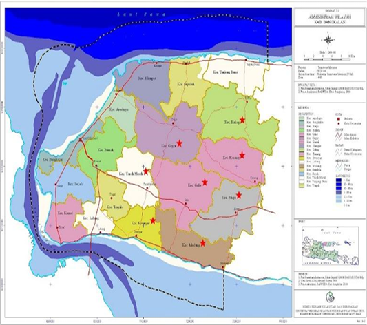
The study was conducted in eight sub-districts with relatively large cattle populations, including two coastal sub-districts (Kwanyar and Modung) and six highland sub-districts (Kokop, Konang, Tanah Merah, Galis, Blega, and Geger). Each sub-district was represented by one selected village. Kwanyar and Modung are located in lowland areas at approximately 25 meters above sea level, whereas the remaining sub-districts are situated in highland areas ranging from approximately 25 to 200 meters above sea level (Figure 1).
Sample collection and fecal examination
In the eight selected sub-districts, cattle populations ranged from approximately 400 to 850 animals per sub-district. Most farmers owned between one and three cattle. To obtain a total sample size of 400 animals, approximately 50 cattle were sampled per sub-district.
When only one animal was present on a farm, that animal was included in the study. When two or more animals were present, individuals were selected using a simple random sampling procedure. Each animal was assigned a temporary identification number based on its stall position or ear tag, and selection was performed using a random number method to minimize selection bias. This approach ensured that each eligible animal had an equal probability of being included.
Sampling was conducted between March and May 2024. Animals were categorized into three age groups: less than 6 months, 6 months to 2 years, and more than 2 years. Both bulls and cows were included.
In addition to fecal sampling, structured interviews and questionnaires were administered to farmers to collect information on farmer characteristics (age, gender, education level, and farming experience) and herd management practices (number of animals, management system, previous treatments, pen structure and materials, environmental conditions, and sanitation practices).
Fresh fecal samples were obtained directly from the rectum, placed into labeled polyethylene zip-lock bags, and preserved in 2.5% potassium dichromate solution. Samples were transported in an ice box to the laboratory for examination.
Laboratory analysis was performed using direct smear (native), sedimentation, and modified Fulleborn flotation techniques ; . Helminth eggs and protozoan cysts or oocysts were identified based on morphological characteristics using standard identification keys . No staining or molecular diagnostic techniques were applied. Therefore, parasite identification was limited to morphologically distinguishable taxa and could not determine molecular subtypes or confirm zoonotic genotypes.
An animal was considered positive if helminth eggs or protozoan cysts/oocysts were detected by any of the examination methods. Prevalence was calculated as: Prevalence (%) = (Number of positive samples / Total number of samples examined) × 100.
Data analysis
Data were analyzed descriptively and inferentially. Prevalence was expressed as percentages with 95% confidence intervals (95% CI). Differences in parasite prevalence according to sex, age group, and sub-district were evaluated using the Chi-square (χ²) test. When expected cell frequencies were less than five, Fisher’s exact test was applied. A p-value of less than 0.05 was considered statistically significant. All statistical analyses were performed using IBM SPSS Statistics for Windows, Version 26.0 (IBM Corp., Armonk, NY, USA).
RESULTS
Overall prevalence of zoonotic intestinal parasites
Out of 400 fecal samples examined from Madura cattle across eight sub-districts in Bangkalan Regency, 116 samples were positive for zoonotic intestinal parasites, resulting in an overall prevalence of 29% (95% CI: 24.6–33.4%) (Table 1). The research area and distribution of sampling locations are presented in Figure 1.
Table 1: Prevalence of zoonotic intestinal parasites in Madura cattle by sub-district.
Sub-district
Number of samples
Positive (n)
Prevalence (%)
Konang
50
9
18
Kokop
50
8
16
Blega
50
20
40
Galis
50
18
36
Geger
50
12
24
Tanah Merah
50
15
30
Modung
50
9
18
Kwanyar
50
25
50
Total
400
116
29.00
The highest prevalence was recorded in Kwanyar (25/50; 50%), whereas the lowest prevalence was observed in Kokop (8/50; 16%) (Table 1). The difference in prevalence among sub-districts was statistically significant (χ² = 24.48; df = 7; p < 0.001), as summarized in Table 2.
Table 2: Summary of chi-square analysis for factors associated with zoonotic intestinal parasite infection in madura cattle.
Variable
Pearson Chi-Square (χ²)
Df
Asymp. Sig. (2-sided)
Interpretation
Sub-district
24.48
7
< 0.001
Significant
Sex
0.04
1
0.839
Not significant
Age group
28.79
2
< 0.001
Significant
N of valid cases
400
Distribution of parasite species
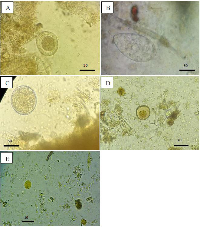
Five zoonotic gastrointestinal parasite species were identified, consisting of two helminths (Toxocara vitulorum and Fasciola gigantica) and three protozoa (Blastocystis hominis, Balantidium coli, and Entamoeba spp.) (Table 3). The microscopic morphology of the identified parasites is shown in Figure 2.
Blastocystis hominis was the most frequently detected species, identified in 78 of 400 samples (19.5%). This was followed by Balantidium coli (15/400; 3.75%), T. vitulorum (6/400; 1.5%), F. gigantica (5/400; 1.25%), and Entamoeba spp.(4/400; 1.0%) (Table 3).
Single-species infections were more common than mixed infections. Co-infection with B. hominis and T. vitulorum was observed in eight samples. One animal from Blega sub-district harbored four parasite species simultaneously (B. hominis, B. coli, F. gigantica, and T. vitulorum) (Table 3).
Table 3: Distribution of zoonotic intestinal parasite species detected in Madura cattle (n = 400).
Parasite species
Positive samples (n)
Prevalence (%)
Blastocystis hominis
78
19.5
Balantidium coli
15
3.75
Toxocara vitulorum
6
1.5
Fasciola gigantica
5
1.25
Entamoeba spp.
4
1.0
Mixed infection (B. hominis + T. vitulorum)
8
2.0
Four-species infection
1
0.25
Regarding distribution by sub-district, only one parasite species was detected in Konang, Galis, and Modung. Two parasite species were identified in Kokop, Geger, Tanah Merah, and Kwanyar, while Blega showed the highest diversity, with four different parasite species detected (Table 3).
Prevalence based on sex
Among 90 bulls examined, 25 were positive (27.77%), whereas 91 of 310 cows were infected (29.35%) (Table 4). Statistical analysis revealed no significant association between sex and infection status (χ² = 0.04; df = 1; p = 0.839), as shown in Table 5.
Table 4: Prevalence of zoonotic intestinal parasites based on sex.
Sex
Number of samples
Positive (n)
Prevalence (%)
Bulls
90
25
27.77
Cows
310
91
29.35
Total
400
116
29.00
Table 5: Chi-square analysis of association between sex and zoonotic intestinal parasite infection.
Variable
χ²
df
p-value
Interpretation
Sex
0.04
1
0.839
Not significant
Chi-square test: χ² = 0.04; df = 1; p = 0.839.
At the sub-district level, the highest prevalence among bulls was observed in Kwanyar (7/9; 77.78%), while the lowest prevalence occurred in Konang, Kokop, and Modung (one positive case each). Among cows, the highest prevalence was also recorded in Kwanyar (18/41; 43.9%), whereas the lowest was found in Kokop (7/40; 17.5%) (Table 4).
Prevalence based on age group
Age group was significantly associated with parasite infection (χ² = 28.79; df = 2; p < 0.001), as presented in Table 7. Cattle aged 6 months to 2 years showed the highest overall prevalence (42.4%), followed by cattle aged <6 months (35.8%), whereas cattle aged >2 years had the lowest prevalence (15.87%) (Table 6).
Cattle younger than 6 months showed an overall infection prevalence of 35.8%. Although T. vitulorum is biologically relevant in this age group due to potential prenatal and lactogenic transmission, the percentage reported above reflects total zoonotic intestinal parasite infection rather than species-specific prevalence (Table 6).
Environmental and management observations
Fecal sampling was conducted in areas with varying altitudes. Modung, Blega, and part of Kwanyar are located in lowland areas (<25 meters above sea level), whereas Konang, Kokop, Galis, Geger, and part of Tanah Merah are situated in highland areas (25–200 meters above sea level). The geographical distribution of these areas is illustrated in Figure 1.
Table 6: Prevalence of zoonotic intestinal parasites based on age group.
Age group
Number of samples
Positive (n)
Prevalence (%)
< 6 months
53
19
35.8
6 months–2 years
158
67
42.4
> 2 years
189
30
15.87
Total
400
116
29.00
Table 7: Chi-square analysis of association between age group and zoonotic intestinal parasite infection.
Variable
χ²
df
p-value
Interpretation
Age group
28.79
2
< 0.001
Significant
Blega, Galis, Tanah Merah, and Kwanyar function as major livestock gathering and temporary holding points for inter-island trade and religious slaughter during Idul Adha. These sub-districts showed relatively higher infection prevalence compared to others (Table 1).
Field observations indicated suboptimal pen sanitation, limited drainage systems, and improper fecal disposal practices. These conditions were commonly observed across study sites and may contribute to environmental contamination and sustained transmission.
DISCUSSION
This study demonstrated that zoonotic intestinal parasite infections remain present in Madura cattle in Bangkalan Regency, with an overall prevalence of 29%. Although lower than several previously reported studies in Indonesia, this finding confirms the continued circulation of gastrointestinal parasites with zoonotic potential in traditional cattle production systems.
The overall prevalence observed in this study was lower than the 71.4% protozoan infection rate previously reported in coastal areas of Bangkalan . Differences in prevalence may reflect variations in diagnostic techniques, ecological settings, seasonal factors, and herd management practices. In contrast, the prevalence in the present study was comparable to findings in Jombang, where gastrointestinal parasites were detected in 35% of beef cattle (Ifqiyah et al., 2021), but lower than reports from dairy cattle in Grati, where protozoan infections reached 85% . These variations highlight the influence of environmental and management conditions on parasite transmission dynamics.
Significant differences in prevalence were observed among sub-districts, with Kwanyar showing the highest infection rate (50%) and Kokop the lowest (16%). Several high-prevalence sub-districts, including Blega, Galis, Tanah Merah, and Kwanyar, function as livestock gathering and temporary holding points for inter-island trade and religious slaughter. Increased animal movement and congregation in these areas may facilitate parasite transmission through environmental contamination and close animal contact. However, because movement data were not quantitatively analyzed in this study, this interpretation should be considered epidemiologically plausible rather than causally confirmed.
Five zoonotic parasite taxa were identified: Toxocara vitulorum, Fasciola gigantica, Blastocystis hominis, Balantidium coli, and Entamoeba spp. Among these, B. hominis was the most frequently detected species. The predominance of Blastocystis spp. in livestock has also been reported in other regions, and high prevalence has been associated with gastrointestinal disturbance and potential host pathology ; . Reports from Sumbawa indicated protozoan infections in Bali cattle reaching 62%, including Blastocystis spp., Balantidium spp., and Entamoeba spp. , supporting the widespread distribution of protozoan parasites in tropical production systems.
The detection of Entamoeba spp. warrants cautious interpretation. Morphological examination does not allow differentiation among species or confirmation of zoonotic genotypes. Although Entamoeba coli is generally considered non-pathogenic in humans, its detection—when present—may indicate fecal contamination and suboptimal hygiene conditions . Because molecular characterization was not performed in this study, the specific zoonotic relevance of the detected Entamoeba spp. cannot be conclusively determined.
Mixed infections were observed in several animals, including one case harboring four parasite species simultaneously. Polyparasitism may negatively affect nutrient absorption and immune function, potentially reducing growth performance. Although clinical outcomes were not assessed in this study, mixed infections are generally associated with cumulative physiological burdens in livestock.
No significant association was found between sex and infection status, suggesting comparable exposure risk among bulls and cows under similar management systems. This contrasts with findings from India, where higher infection rates were reported in females . Differences between studies may reflect variations in herd composition, physiological status, and management conditions.
Age was significantly associated with infection, with the highest prevalence observed in cattle aged 6 months to 2 years. This age-related pattern is consistent with previous findings indicating increased susceptibility in young animals due to incomplete immune development and wider grazing exposure . Cattle younger than six months showed substantial overall infection prevalence; this age group is epidemiologically important for T. vitulorum, which can be transmitted transplacentally or through colostrum. However, species-specific prevalence was not analyzed separately within each age category.
Environmental and management conditions likely contribute to parasite persistence. Traditional extensive grazing systems expose cattle to contaminated fields and communal grazing areas. Parasite eggs and oocysts can survive for extended periods in moist environments, particularly during rainy seasons ; . Previous studies in similar settings have documented soil contamination with helminth eggs and protozoan oocysts around cattle pens and grazing areas ; .
Field observations in this study indicated suboptimal pen sanitation, limited drainage systems, and improper fecal disposal. Such conditions may facilitate environmental contamination and reinfection cycles. In addition, flies such as Musca domestica may act as mechanical vectors of protozoan cysts and oocysts, potentially contributing to parasite dissemination . Similar environmental contamination associated with inadequate sanitation has been reported in livestock farms in Korea .
Although the parasites identified in this study have recognized zoonotic potential, this research did not assess human infection in Bangkalan. Therefore, zoonotic risk should be interpreted in the context of established transmission pathways and environmental exposure rather than confirmed human case data from the study area. Further integrated One Health investigations are needed to evaluate potential cross-species transmission.
Overall, these findings indicate that zoonotic intestinal parasites remain endemic in Madura cattle and are influenced by geographic distribution and age-related susceptibility. Strengthened biosecurity, routine fecal monitoring, improved sanitation, and targeted control measures in livestock aggregation areas may contribute to reducing infection prevalence and limiting potential zoonotic transmission.
CONCLUSION
Zoonotic intestinal parasites remain prevalent in Madura cattle in Bangkalan Regency, with an overall infection rate of 29%. Five parasite taxa were identified, namely Toxocara vitulorum, Fasciola gigantica, Blastocystis hominis, Balantidium coli, and Entamoeba spp., with B. hominis being the most frequently detected. Infection prevalence differed significantly among sub-districts and age groups but was not associated with sex. Cattle aged 6 months to 2 years showed the highest susceptibility. These findings indicate that zoonotic intestinal parasites are maintained within traditional cattle production systems in Bangkalan. Strengthened routine parasitological monitoring, improved sanitation and waste management, enhanced biosecurity practices, and targeted control strategies in high-prevalence and livestock-aggregation areas are recommended to reduce infection pressure and mitigate potential zoonotic risks while improving cattle health and productivity.
ACKNOWLEDGEMENTS
The authors would like to express their sincere gratitude to the Dean of the Faculty of Veterinary Medicine, Universitas Airlangga, for providing financial support for this research through the Faculty’s RKAT fund under Rector’s Decree of Universitas Airlangga No. 886/Unair/2024, dated 27 March 2024. The authors also gratefully acknowledge the support of the Research Group of One Health, Faculty of Veterinary Medicine, Universitas Airlangga. We also thank the Animal Husbandry Office of Bangkalan Regency for granting permission and providing facilities for obtaining fecal samples from Madurese beef cattle raised in smallholder farms.
NOVELTY STATEMENT
To the best of our knowledge, this study provides new information on the identification of five gastrointestinal helminth and protozoan parasites with potential zoonotic significance in fecal samples from Madurese beef cattle raised in smallholder farms. The occurrence of these potentially zoonotic gastrointestinal parasites in Madurese cattle has important implications for both veterinary and public health. The findings provide baseline information that may support relevant veterinary and public health institutions in developing appropriate parasite surveillance and control strategies to reduce the potential risk of zoonotic transmission.
Author’S Contribution
PH designed the study and supervised the research. NDRL & LTS conducted laboratory examinations. M & ZY contributed to data analysis and manuscript preparation. All authors reviewed and approved the final manuscript.
Generative AI and AI assisted technology
statement
The authors declare that no generative AI and AI assisted
technology was used in the creation of this manuscrip
Conflict of interest
The authors declare no conflict of interest.
Figures
Figure 1:.
Research area ().
Figure 2:.
A. Toxocara vitulorum, B. Fasciola gigantica, C. Balantidium coli, D. Blastocystis hominis and E. Entamoeba spp.
References
Badan Pusat Statistik Kabupaten Bangkalan (2023). Hasil pencacahan lengkap sensus pertanian 2023. Badan Pusat Statistik Kabupaten Bangkalan, Bangkalan, Indonesia.
Haajidah J, Sukmanadi M, Kusnoto, Suprihati S, Nangoi L, Hastutiek P (2020). Identification of nematode worms in caecum and colon of sacrificial cattle slaughtered during Eid al-Adha 1439 H in East Surabaya. J. Parasite Sci., 4(1): 25–30. https://doi.org/10.20473/jops.v4i1.20272[CrossRef]
Haidar A, De Jesus O (2023). Entamoeba coli infection. In: StatPearls [Internet]. StatPearls Publishing, Treasure I. (FL).
Hamdani A, Lastuti NDR, Dhamayanti Y, Koesdarto S, Sunarso A, Hastutiek P (2021). Prevalence of gastrointestinal protozoa in bali cattle in Lopok Sub-district, Sumbawa District. J. Parasite Sci., 5(2): 55–60. https://doi.org/10.20473/jops.v5i2.30373[CrossRef]
Hastutiek P, Yuniarti WM, Djaeri M, Lastuti NDR, Suprihati E, Suwanti L (2019). Prevalence and diversity of gastrointestinal protozoa in madura cattle in bangkalan regency, East Java, Indonesia. Vet. World, 12(2): 198–204. https://doi.org/10.14202/vetworld.2019.198-204[CrossRef]
Hastutiek P, Lastuti NDR, Suprihati E, Kurniawati DA, Suwanti LT, Matsubayashi M, Sunarso A (2022). Coproparasitological examinations and molecular determination of eimeria species in madura cattle reared on madura island, Indonesia. Parasitol. Int., 86: 102478. https://doi.org/10.1016/j.parint.2021.102478[CrossRef]
Ifqiyyah MM, Setiawan B, Wijaya A (2021). Prevalence of gastrointestinal parasites in beef cattle in jombang district. J. Parasite Sci., 5(2): 45–50. https://doi.org/10.20473/jops.v5i2.30367[CrossRef]
Ismail HA, Jeon HK, Yu YM, Do C, Lee YH (2010). Intestinal parasite infections in pigs and beef cattle in rural areas of Chungcheongnam-do, Korea. Korean J. Parasitol., 48(4): 347–349. https://doi.org/10.3347/kjp.2010.48.4.347[CrossRef]
Mahartina D, Hastutiek P, Mulyati S, Soeharsono, Hambal M (2020). Identification and distribution of soil-transmitted helminths around sheds and grazing fields of Madura cattle in Geger Sub-district, Bangkalan Regency. J. Parasite Sci., 4(2): 49–54. https://doi.org/10.20473/jops.v4i2.29153[CrossRef]
Margaretha JI, Hastutiek P, Permatasari DA, Sunarso A, Rahardjo D, Suwanti LT (2024). Identification, prevalence, and degree of gastrointestinal protozoan infection in dairy cows at koperasi usaha tani ternak suka makmur, Grati, Pasuruan. J. Parasite Sci., 8(2): 51–54. https://doi.org/10.20473/jops.v8i2.60454[CrossRef]
Marskole P, Verma Y, Dixit AK, Swamy M (2016). Prevalence and burden of gastrointestinal parasites in cattle and buffaloes in Jabalpur, India. Vet. World, 9(11): 1214–1217. https://doi.org/10.14202/vetworld.2016.1214-1217[CrossRef]
Mumpuni S, Subekti S, Koesdarto S, Puspitawati H, Kusnoto (2007). Penuntun praktikum ilmu penyakit helminth veteriner. Fac. Vet. Med., Universitas Airlangga, Surabaya, Indones.
Nurgiartiningsih VMA (2011). Evaluation of performance in madura cattle on madura island, Indonesia. Anim. Prod., 18(3): 125–130. https://doi.org/10.20884/1.anprod.2016.18.3.573[CrossRef]
Popescu CP, Smith WG (1988). A cytogenetic investigation of madura cattle. Reprod. Domestic Anim., 23(3): 145. https://doi.org/10.1111/j.1439-0531.1988.tb01092.x[CrossRef]
Putri, C. H. S., Sarmanu, S., Maslachah, L. (2021). Identification and incident rate of phylum Protozoa around the sad and grazing fields of Madura cattle in Sub-District of Geger, Bangkalan District. Journal of Parasite Science, 5(2), 51–54. https://doi.org/10.20473/jops.v5i2.30368[CrossRef]
Singh E, Kaur P, Singla LD, Bal MS (2017). Prevalence of gastrointestinal parasitism in small ruminants in western Punjab, India. Vet. World, 10(1): 61–66. https://doi.org/10.14202/vetworld.2017.61-66[CrossRef]
Soulsby EJL (1986). Helminths, arthropods and protozoa of domesticated animals. 7th Ed. Baillière Tindall, London.
Stensvold CR, Suresh GK, Tan KS, Thompson RCA, Viscogliosi E (2007). Terminology for blastocystis subtypes – a consensus. Trends Parasitol., 23(3): 93–96. https://doi.org/10.1016/j.pt.2007.01.004[CrossRef]
Sudradjat DS (1991). Epidemiologi dan ekonomi veteriner. Yayasan Agribisnis Indonesia Mandiri, Jakarta, Indonesia.
Susana Y, Suwanti LT, Suprihati E (2019). Identification and prevalence of gastrointestinal parasites in beef cattle in Siak Sri Indrapura, Riau, Indonesia. https://doi.org/10.20473/ijtid.v7i6.10392[CrossRef]
Suwanti LT, Susana Y, Hastutiek P, Suprihati E, Lastuti NDR (2020). Blastocystis spp. subtype 10 infection in beef cattle in kamal and socah, Bangkalan, Madura, Indonesia. Vet. World, 13(2): 231–237. https://doi.org/10.14202/vetworld.2020.231-237[CrossRef]
Szostakowska B, Łozowska WK, Racewicz M, Knight R, Tamang L, Myjak P, Graczyk TK (2004). Cryptosporidium parvum and giardia lamblia recovered from flies on a cattle farm and in a landfill. Appl. Environ. Microbiol., 70(6): 3742–3744. https://doi.org/10.1128/AEM.70.6.3742-3744.2004[CrossRef]
Villeneuve A, Polley L, Jenkins E, Schurer J, Gilleard J, Kutz S, Conboy G, Benoit D, Sewell W, Gagné F (2015). Parasite prevalence in fecal samples from shelter dogs and cats across Canadian provinces. Parasites Vectors, 8: 281. https://doi.org/10.1186/s13071-015-0870-x[CrossRef]
Yoshikawa H, Wu Z, Pandey K, Pandey BD, Sherchand JB, Yanagi T (2009). Molecular characterization of blastocystis isolates from children and rhesus monkeys in Kathmandu, Nepal. Vet. Parasitol., 160(3–4): 295–300. https://doi.org/10.1016/j.vetpar.2008.11.029[CrossRef]
Zalizar L (2017). Gastrointestinal helminthiasis in dairy cattle. J. Ilmu-ilmu Peternakan, 27(2): 1–7. https://doi.org/10.21776/ub.jiip.2017.02s7.02.01[CrossRef]