Research Article · Volume 13, Issue 2, 6 June 2025 · Open Access
Prevalence of Toxocara spp. in Cats and Detection of Intestinal Helminth Infections in Humans
· · · · ·
Affiliations & Notes ⌄
- 1Department of Physiology, College of Veterinary Medicine, Wasit University, Wasit, Iraq
- 2Ibn Sina University of Medical and Pharmaceutical Sciences, College of Medicine, Baghdad, Iraq
- 3Topical Biology Research Unit, College of Science, University of Baghdad, Baghdad, Iraq
- 4Department of Medical Laboratory Technique, Medical Technical College
- 5Al-Farahidi University, Baghdad, Iraq
- 6Microbiology Department, College of Veterinary Medicine, Wasit University, Wasit, Iraq
- 7Department of Internal and Preventive Veterinary Medicine, College of Veterinary Medicine, University of Wasit, Wasit, Iraq. *Correspondence | Alaa Zeyad Kokaz, Department of Physiology, College of Veterinary Medicine, Wasit University, Wasit, Iraq
- 8Email: zaidalani242@yahoo.com
Article Info ⌄
- Journal:
- Journal of Animal Health and Production
- Published:
- 6 June 2025

From the article
- The highest infection rate of 16.3% (18/110) was observed in the 20-29 years age group, with a significant difference (P ≤ 0.01) (Table 2).
- The study concludes that conventional diagnostic methods are effective for detecting specific intestinal helminths in humans, with notable variations observed across age and gender.
- While intestinal helminth infections are relatively uncommon in Baghdad, the infection rate was significantly higher in males than in females and varied across different age groups.
- Additionally, although Toxocara spp. eggs were detected in both domestic and stray cats, the difference in infection rates between the two was not statistically significant.
Selected from the article's own text.
Abstract
INTRODUCTION
Zoonotic helminths—parasitic worms transmissible from animals to humans—pose a serious threat to public health. These include nematodes such as Ascaris lumbricoides, Toxocara canis, and Ancylostoma caninum; cestodes like Echinococcus granulosus; and trematodes such as Schistosoma spp. and Fasciola spp. Socioeconomic factors including poverty, poor sanitation, limited access to healthcare, and low levels of health education contribute to the high burden of these diseases . Various helminth parasites reside in livestock such as cattle, sheep, and goats, with complex life cycles that involve multiple developmental stages within the host. These parasites can contaminate meat and transmit infections to humans, particularly through the consumption of raw or undercooked meat. Poor processing, hygiene, and handling further amplify the risk of transmission ; .
Helminth eggs are highly resistant to environmental factors, surviving for long periods in soil despite exposure to dryness and chemicals. In tropical and subtropical regions, intestinal nematode infections like Strongyloides stercoralis are endemic, affecting over 30 million people ; . These parasites, particularly S. stercoralis and A. lumbricoides, can cause severe disease in immunocompromised individuals due to their ability to reproduce and maintain infection within the host through autoinfection .
To detect intestinal helminths and adult worms, fecal samples are examined using direct smear and flotation methods (NaCl solution and Sheather’s technique) . Toxocara cati, a zoonotic intestinal parasite, is commonly found in domestic and wild cats, especially kittens. Toxocara spp. frequently infect the intestines of companion animals and pose significant public health risks due to their zoonotic potential. Toxocariasis remains a global health concern because of its ability to cause systemic infections in humans .
The objective of this study was to investigate the prevalence of intestinal helminth infections in humans using conventional diagnostic methods, detect Toxocara spp. serologically, and determine its incidence in cats.
MATERIALS AND METHODS
Ethical Approval
The current study was approved in accordance with the Iraqi Human and Animal Welfare Code by Al-Bayan University.
Human Stool Sample Collection
In a comprehensive field investigation, 450 stool samples were collected from various locations around Al-Najaf City. The samples were obtained immediately after defecation and evenly divided between 225 male and 225 female participants. Each sample was carefully placed in a sealed container, accompanied by detailed information regarding the participant’s age and gender. The samples were then promptly transported to the Parasitology Research Laboratory for immediate microscopic examination. Both direct smear and flotation techniques were employed, with the flotation process using Sheather’s sugar solution. This solution was prepared by dissolving 500 grams of sugar in 320 mL of distilled water, with 6.5 grams of phenol added as a preservative, following the methodology described by Alani et al. (2022).
Human Sera Collection and Processing
A field investigation involving 96 human blood samples from various regions of Baghdad Province was conducted. The samples were collected from individuals across different age groups. ELISA testing was performed to detect antibodies against Toxocara spp., with wells designated for controls and test specimens arranged on a plate strip holder . After incubating the plate at room temperature (15–25°C) for 10 minutes, the wells were washed and then enriched with 100 µL of enzyme conjugate. Following a 5-minute incubation at room temperature, the wells were washed again, and 100 µL of chromogenic substrate was added to each well. The mixture was gently mixed by lightly tapping the side of the plate holder for approximately 15 seconds.
Collection of Animal Fecal Samples
Stool samples from 100 cats were recently collected from a private veterinary clinic in Baghdad. Each sample was placed in a clean, clearly labeled container with a secure lid, including relevant data such as the sex of the animal. The samples were then transported in clean containers to the Parasitology Laboratory at the Department of Medical Health Techniques, Al-Bayan University, for immediate examination. The samples were analyzed using standard microscopic methods (40x magnification) following both direct smear and flotation techniques, as described by Lamy and Kawan (2022).
Examination of Fecal Samples
Direct wet method: Upon arrival at the laboratory, each stool sample was promptly processed. The direct smear technique was employed by taking approximately one gram of stool using a wooden stick and mixing it with tap water. The mixture was then applied to a glass slide, followed by the addition of a drop of saline (prepared on 2–3 slides). The sample was carefully spread to form a thin layer and covered with a cover slip for microscopic examination. Observations were made under both 10x and 40x magnifications, following the methodology described by Alani and Kawan (2024). This meticulous direct smear approach enabled thorough examination and facilitated the identification and analysis of any helminth infections present in the samples.
Flotation technique using sheather’s sugar solution: The procedure was implemented as follows: Approximately one gram of stool was mixed with tap water and filtered through gauze. The filtered sample was then transferred into a centrifuge tube, to which 10 mL of Sheather’s sugar flotation solution was added. The tube was filled to the brim with the flotation solution and centrifuged at 1,200 rpm for five minutes. After centrifugation, the tube was carefully removed, and a cover slip was placed on the top of the tube, allowing it to stand undisturbed for 10 minutes. Following this, the cover slip was gently lifted and placed on a glass slide. The entire area under the cover slip was examined microscopically for the presence of parasitic ova or larvae. Identification and confirmation were carried out using both 40x and 100x objective lenses to ensure a thorough analysis. This flotation technique, following the protocol described by Cringoli et al. (2010), significantly enhanced the sensitivity of parasite detection in the processed stool samples.
Statistical Analysis
To assess significant differences between group means, the study employed the Chi-square test. The results were presented as mean, with statistical significance defined as P < 0.05. The data from the experiment were statistically analyzed using the Statistical Analysis System (SAS, version 20.1) .
RESULTS
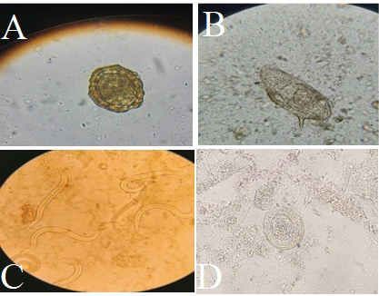
In the current study, fecal samples were collected and examined microscopically to detect the eggs and adult worms of intestinal helminths. The results revealed the presence of eggs of Ascaris lumbricoides, Schistosoma mansoni, and Hymenolepis nana, as well as adult worms of Strongyloides stercoralis (Figure 1). A total of 450 human stool samples were examined, and the infection rate with intestinal helminths was 5.7% (26/450) according to traditional techniques (Table 1). Regarding age groups, no infection was recorded in the 30-39 and ≥ 40 years categories. The highest infection rate of 16.3% (18/110) was observed in the 20-29 years age group, with a significant difference (P ≤ 0.01) (Table 2). In terms of gender, males had the highest infection rate at 7.5% (17/225), while females had a lower infection rate of 4% (9/225), with a significant difference (P ≤ 0.01) (Table 3).
Table 1: Total infection rate of intestinal helminths in humans.
| Parasite | Positive No. | %From total (n=450) samples | % From infected samples |
| Strongyloides stercoralis | 2 | 0.4 | 7.6 |
| Schistosoma mansoni | 3 | 0.6 | 11.5 |
| Hymenolepis nana | 8 | 1.7 | 30 |
| Ascaris lumbricoides | 13 | 2.8 | 50 |
| Total | 26 | 5.7 |
Table 2: Age-wise infection rate of intestinal helminths in humans.
| Age | Examined No. | Positive No. | Percentage |
| < 20yr | 112 | 8 | 7.14 |
| 20yr-29yr | 110 | 18 | 16.3 |
| 30yr-39yr | 111 | 0 | 0 |
| ≥ 40yr | 117 | 0 | 0 |
| Total | 450 | 26 | 5.8 |
| Chi-Square (χ2) | 11.802 | ||
| Significance | P≤0.01 |
Table 3: Gender-wise infection rate of intestinal helminths in humans.
| Gender | No. of examined samples | No. of positive Samples | Percentage |
| Males | 225 | 17 | 7.5 |
| Females | 225 | 9 | 4 |
| Total | 450 | 26 | 5.7 |
| Chi-Square (χ2) | 7.155 | ||
| Significance | P≤0.01 | ||
Toxocara spp. incidence was determined using an indirect IgG ELISA on human blood samples. The study also assessed the impact of various risk factors, including age and gender, on the seroprevalence of Toxocara spp. in Baghdad. The seroprevalence of Toxocara spp. was 10.4%, based on the assessment of IgG antibodies. Highly significant differences (P ≤ 0.01) were observed when comparing age groups. The highest seroprevalence of 24% (6/25) was recorded in the ≥ 40 years age group, followed by 8% (2/25) in the 30-39 years age group, and 18.1% (2/11) in the < 20 years age group, with no infections (0/35) in the 20-29 years age group (Table 4). Regarding gender, the infection was more prevalent in females (15.2%, 7/46) than males (6%, 3/50), with a highly significant difference (P ≤ 0.01) (Table 5).
Table 4: Age-wise seroprevalence of Toxocara spp. in humans detected by indirect ELISA (IgG).
| Age | No. of examined samples | No. of positive samples | Percentage |
| < 20yr | 11 | 2 | 18.1 |
| 20yr-29yr | 35 | 0 | 0 |
| 30yr-39yr | 25 | 2 | 8 |
| ≥ 40yr | 25 | 6 | 24 |
| Total | 96 | 10 | 12.5 |
| Chi-Square (χ2) | 14.302 | ||
| Significance | P≤0.01 |
Table 5: Gender-wise Seroprevalence of Toxocara spp. infection in Humans detected by indirect ELISA (IgG).
| Genders | No. of examined samples | No. of positive Samples | Percentage |
| Males | 50 | 3 | 6 |
| Females | 46 | 7 | 15.2 |
| Total | 96 | 10 | 10.6 |
| Chi-Square (χ2) | 11.535 | ||
| Significance | P≤0.01 | ||
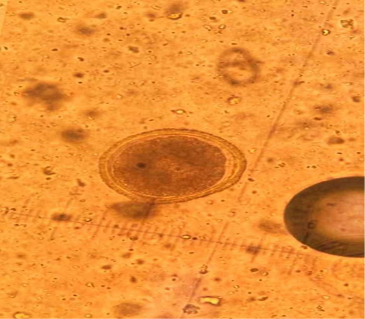
In the current study, eggs of Toxocara spp. were observed in the fecal samples of cats (Figure 2). The overall infection rate among domestic and stray cats was 12% (12/100). Specifically, domestic cats showed an infection rate of 10% (5/50), while stray cats had a higher rate of 14% (7/50), indicating a statistically significant difference (Table 6).
Table 6: Prevalence of Toxocara spp. in domestic and stray cats.
| Ages | Stray cats | Domestic cats | Total | ||||||
| No. of examined samples | Infection | % | No. of examined samples | Infection | % | No. of examined samples | Infection | % | |
| Kittens | 19 | 5 | 26.3 | 15 | 3 | 20 | 34 | 8 | 23.15 |
| Adults | 31 | 2 | 6.4 | 35 | 2 | 5.7 | 66 | 4 | 6 |
| Total | 50 | 7 | 14 | 50 | 5 | 10 | 100 | 12 | 12 |
| Chi-Square (X2) | 7.155 | ||||||||
| Significance | P≤0.01 | ||||||||
DISCUSSION
Intestinal helminths pose a significant public health concern globally, affecting the economic stability of many countries. Contaminated food and water can expose children to parasites both at home and at school ; . These parasites are transmitted through various routes, most commonly via ingestion of infectious eggs due to poor hygiene practices (anus-to-mouth transmission). Inhalation of aerosolized eggs or contact with contaminated surfaces, especially in pinworm infections, also contributes to transmission .
This study highlighted an epidemic of Hymenolepis nana and Ascaris lumbricoides infections in Iraq, likely linked to the use of untreated human and animal waste as fertilizer during crop cultivation. Iraq is among the endemic regions for helminth infections in the Middle East .
According to Saheb et al. (2017), Ascaris infection affected 89 patients, while Hymenolepis nana infected 173 individuals. No infections with Strongyloides stercoralis or Schistosoma mansoni were detected in that study. The highest incidence of Ascaris was recorded in Najaf (78 patients), with the lowest in Muthanna (1 patient). For Hymenolepis nana, Najaf also had the highest prevalence (41 cases), while other regions such as Kirkuk, Diyala, and Missan reported only two cases each.
Barakat et al. (2014) reported that Taenia infections had an incidence of 0.46% in Diyala. From 2003 to 2010, 25 patients were infected with Taenia spp. Various helminths, such as nematodes, cestodes, and trematodes, are zoonotic parasites. In this study, four helminths were recorded in humans: Strongyloides stercoralis (0.4%), Schistosoma mansoni (0.6%), Hymenolepis nana (1.7%), and Ascaris lumbricoides (2.8%). These findings differ from Musa (2017), who reported 30 Ascaris cases in Najaf. Males had a slightly higher infection rate (24 cases) compared to females (20 cases). Strongyloides infections were highest in Sulaymaniyah (4 cases), and Hymenolepis nana was most common in Babylon (22 cases), with no cases in Diyala.
Toxocariasis, caused by the zoonotic nematode Toxocara cati, is a serious concern. Cats can act as reservoirs, and the development of resistance to dewormers complicates control efforts ; . The current study’s results differ from those of Roldán et al. (2009), who reported a 44.92% seroprevalence of human toxocariasis in rural population of Peru. Jin et al. (2013) found a positive predictive value of 78.7%, while Stensvold et al. (2009) reported a lower seroprevalence of 2.4%. However, this study aligns with findings from Iran (9.3% seroprevalence) and Al-Nasiri’s report of 8% in Iraq. Risk factors such as pet ownership, geophagia, and animal contact significantly influence Toxocara transmission .
Our current results regarding prevalence of Toxocara ssp. in cat were differ from Macpherson (2013), who reported 42.5% of feces contained T. cati eggs. Similar data were noted by Dantas-Torres (2020) in Brazil (16.7%) and Martínez-Barbabosa et al. (2003) in Russia (52%). In Iraq, T. cati eggs were found in 25.58% of 90 samples and 34.75% of 125 samples . These eggs are environmentally resilient and can remain infectious for years. Animals infected with adult worms shed eggs through feces, contaminating the environment. Cats burying feces in soil may contribute to wide dispersal. Differences in infection rates may result from variations in sample size, diagnostic methods, and environmental or epidemiological factors.
CONCLUSIONS AND RECOMMENDATIONS
The study concludes that conventional diagnostic methods are effective for detecting specific intestinal helminths in humans, with notable variations observed across age and gender. While intestinal helminth infections are relatively uncommon in Baghdad, the infection rate was significantly higher in males than in females and varied across different age groups. Additionally, although Toxocara spp. eggs were detected in both domestic and stray cats, the difference in infection rates between the two was not statistically significant. The highest seroprevalence in humans was observed among adults.
ACKNOWLEDGMENTS
The authors wish to acknowledge the field technicians who have assisted in research activity and would like to thank the Al-Bayan University and College of Health Medical Techniques.
NOVELTY STATEMENT
This study highlights the link between intestinal parasites transmitted from cats to humans, emphasizing their potential health risk to human populations.
AUTHOR’S CONTRIBUTIONS
All of the trials were designed by Alaa Zeyad Kokaz and Sarah Basheer. Ali Mahdi Salih and Riyam Ahmed Saber conducted all of the tests, gathered the data, and composed the manuscript draft. Zeid Alsadoon and Hasanain A.J. Gharban helped with the data analysis that was done to prepare the work for submission to the journal. The final draft of the work was reviewed and approved by all authors for publication in the Journal of Animal and Health Production.
Authors Declaration
We so attest that every figure in the manuscript belongs to us.
Conflict of Interest
None.
Figures

Eggs of intestinal helminths in human stool samples. A: Egg of Ascaris lumbricoides. B: Egge of Schistosoma mansoni. C: Adult worms of Strongyloides stercoralis D: Egg of Hymenolepis nana.

Egg of Toxocara spp. in cat fecal sample. (40X).
References
- Al-Aredhi HS (2015). Prevalence of gastrointestinal parasites in domestic cats (Felis catus) in Al-Diwaniya province/Iraq. Int. J. Cur. Mic. App. Sci., 4(5): 166-171.
- Al-Rammahi HM, Kareem SM, Hammadi AK (2014). Prevalence of intestinal helminthes in feral cats in Babylon province/Iraq, urban and rural locations. Mir. Res. Vet. Sci. Anim., 3(2): 44-52.
- Agbajelola VI (2025). Addressing Zoonotic Helminths in Nigeria: Bridging Public Health and Veterinary Strategies. World News Nat. Sci., 58: 259-269.
- Aboamer MM, Mohamed HA, Osman YG, Abdel Rahman HE, El Shanawany E (2019). Inactivation of Toxocara vitulorum eggs by Ammonia in combination with solar energy. Egypt. J. Aquat. Biol. Fish., 23(4): 201-214. https://doi.org/10.21608/ejabf.2019.52938[CrossRef]
- Alani ZK, Kawan MH (2024). Prevalence and molecular analysis of Toxocara cati in Baghdad Province. J. Adv. Vet. Anim. Res., 11(2): 392. https://doi.org/10.5455/javar.2024.k788[CrossRef]
- Abdul-Zahra NI, Taher JH, Ismail MA (2021). Level of eosinophil cationic protein in Strongyloides stercoralis infection in Najaf province/Iraq. Nveo-Natural Volatiles and Essential Oils J., Nveo: 4645-4650.
- Alani ZK, Jasim HJ, Barakat H, Al-Yasari JTO, Hameed HM (2022). Prevalence and Molecular Studies of Isospora Spp. in House and Stray Cats at Baghdad Province. Rev. Electron. Vet., 335-343.
- Bisoffi Z, Buonfrate D, Montresor A, Requena-Mendez A, Munoz J, Krolewiecki AJ, Albonico M (2013). Strongyloides stercoralis: a plea for action. PLoS Negl. Trop. Dis., 7(5): e2214. https://doi.org/10.1371/journal.pntd.0002214[CrossRef]
- Barakat R, El Morshedy H, Farghaly A (2014). Human schistosomiasis in the Middle East and North Africa region. Negl. Trop. Dis. Middle East N. Afr., 23-57. https://doi.org/10.1007/978-3-7091-1613-5_2[CrossRef]
- Bowman DD, Lucio-Forster A (2010). Cryptosporidiosis and giardiasis in dogs and cats: veterinary and public health importance. Exp. Parasitol., 124(1): 121-127. https://doi.org/10.1016/j.exppara.2009.01.003[CrossRef]
- Cringoli G, Rinaldi L, Maurelli MP, Utzinger J (2010). FLOTAC: new multivalent techniques for qualitative and quantitative copromicroscopic diagnosis of parasites in animals and humans. Nat. Protoc., 5(3): 503-515. https://doi.org/10.1038/nprot.2009.235[CrossRef]
- Deneke TT, Bekele A, Moore HL, Mamo T, Almaw G, Mekonnen GA, Berg S (2022). Milk and meat consumption patterns and the potential risk of zoonotic disease transmission among urban and peri-urban dairy farmers in Ethiopia. BMC Public Health, 22(1): 222. https://doi.org/10.1186/s12889-022-12665-4[CrossRef]
- Dantas-Torres F (2020). Toxocara prevalence in dogs and cats in Brazil. Adv. Parasitol., 109: 715-741. https://doi.org/10.1016/bs.apar.2020.01.028[CrossRef]
- Fadhil AI, Abed HH, Fadel SR, Al-Zubaidi MTS (2022). Molecular diagnosis of nematode worms Parabronema Skrjabini in camels (camelus dromedaries) in Iraq. Iraqi J. Agric. Sci., 53(3): 584-588. https://doi.org/10.36103/ijas.v53i3.1567[CrossRef]
- Fillaux J, Magnaval JF (2013). Laboratory diagnosis of human toxocariasis. Vet. Parasitol., 193(4): 327-336. https://doi.org/10.1016/j.vetpar.2012.12.028[CrossRef]
- Gazal FM (2010). Prevalence of Enterobiasis in Ninevah Governorate for (2004-2006) Years. TJPS, 15(1).
- Jin Y, Shen C, Huh S, Sohn WM, Choi MH, Hong ST (2013). Serodiagnosis of toxocariasis by ELISA using crude antigen of Toxocara canis larvae. Korean J. Parasitol., 51(4): 433. https://doi.org/10.3347/kjp.2013.51.4.433[CrossRef]
- Lamy SA, Kawan MH (2022). Seroprevalence of toxoplasmosis in quail birds (Coturnix coturnix) in Baghdad City, Iraq. Int. J. Health Sci., (I): 10377-10387. https://doi.org/10.53730/ijhs.v6nS1.7490[CrossRef]
- Lukashev AN, Ruzina MN, Akhmadishina LV (2020). Toxocara prevalence in dogs, cats and the environment in Russia. Adv. Parasitol., 109: 801-817. https://doi.org/10.1016/bs.apar.2020.01.019[CrossRef]
- Mansoor NT, Falih IB, Al-Nasiri FS (2020). Diagnosis of myxobolus bramae (myxosporea: myxobolidae) in the kidneys tissue of carasobarbus luteus and histopathological changes associated with infection. Iraqi J. Agric. Sci., 51(3): 874-884. https://doi.org/10.36103/ijas.v51i3.1042[CrossRef]
- Martı́nez-Barbabosa I, Tsuji OV, Cabello RR, Cárdenas EMG, Chasin OA (2003). The prevalence of Toxocara cati in domestic cats in Mexico City. Vet. Parasitol., 114(1): 43-49. https://doi.org/10.1016/S0304-4017(03)00038-4[CrossRef]
- Macpherson CN (2013). The epidemiology and public health importance of toxocariasis: a zoonosis of global importance. Int. J. Parasitol., 43(12-13): 999-1008. https://doi.org/10.1016/j.ijpara.2013.07.004[CrossRef]
- Musa IS (2017). Incidence of helminthiasis in humans in Iraq. Karbala Int. J. Mod. Sci., 3(4): 267-271. https://doi.org/10.1016/j.kijoms.2017.08.001[CrossRef]
- Mohamed A, Abebe M, Birhanu W, Abdirahman M, Wali MA (2021). Prevalence of Taenia saginata cysticerci in Addis Ababa Abattoir Enterprise, Ethiopia. Food Waterborne Parasitol., 25: e00135. https://doi.org/10.1016/j.fawpar.2021.e00135[CrossRef]
- Njiru J, Muhoho N, Simbauni J, Kabiru E (2016). Effects of soil-transmitted helminths and Schistosoma species on nutritional status of children in Mwea irrigation scheme, Kenya. J. Appl. Life Sci. Int., 5(1): 1-8. https://doi.org/10.9734/JALSI/2016/25053[CrossRef]
- Roldán WH, Espinoza YA, Huapaya PE, Huiza AF, Sevilla CR, Jiménez S (2009). Frequency of human toxocariasis in a rural population from Cajamarca, Peru determined by DOT-ELISA test. Rev. Inst. Med. Trop. São Paulo, 51: 67-71. https://doi.org/10.1590/S0036-46652009000200002[CrossRef]
- Shaalan NN (2015). Epidemiology study and role of anti-helminthes in treatment of dermatitis due to infection with Ancylostoma duodenale parasite. Int. J. Nat. Soc. Sci., 3(8): 2347-4580.
- Saheb EJ, Mahdi SG, Mosa IS, Karim MIA, Khistawi AN (2017). Epidemiology of Some Parasitic Helminthes in Iraq from 2011 until 2015. Iraqi J. Sci., 789-796.
- Stensvold CR, Skov J, Møller LN, Jensen PM, Kapel CM, Petersen E, Nielsen HV (2009). Seroprevalence of human toxocariasis in Denmark. Clin. Vaccine Immunol., 16(9): 1372-1373. https://doi.org/10.1128/CVI.00234-09[CrossRef]
- Virta M, Huitu O, Heikkinen J, Holmala K, Jokelainen P (2022). High Toxocara cati prevalence in wild, free-ranging Eurasian lynx (Lynx lynx) in Finland, 1999–2015. Int. J. Parasitol. Parasites Wildl., 17: 205-210. https://doi.org/10.1016/j.ijppaw.2022.02.004[CrossRef]
Services
Article usage statistics
Loading…