Special Issue: Emerging and Re-Emerging Animal Health Challenges in Low and Middle-Income Countries · Volume 13, Issue 1, 10 July 2025 · Open Access
Phylogenetic Analysis and Molecular Identification of Staphylococcus aureus Isolated from Skin Lesions of Cats
·
Affiliations & Notes ⌄
- 1Department of Internal and Preventive Veterinary Medicine, College of Veterinary Medicine, University of Baghdad, Baghdad, Iraq. *Correspondence | Mawj Firas Abdulameer, Department of Internal and Preventive Veterinary Medicine, College of Veterinary Medicine, University of Baghdad, Baghdad, Iraq
- 2Email: mawj.abd2201.m@covm.uobaghdad.edu.iq
Article Info ⌄
- Journal:
- Journal of Animal Health and Production
- Published:
- 10 July 2025

From the article
- This study concluded that there are limitations to conventional methods for identifying S. aureus.
- Phylogenetic analysis of the 23s rRNA gene of S. aureus showed Iraqi isolates formed a distinct cluster, suggesting that there was local adaptation to feline hosts in Iraq, while the Iranian isolate was genetically distant.
- Epidemiologically, young cats were more susceptible to S. aureus infection.
- Merging the conventional and molecular diagnostics tools is an important consideration for accurate identification of S. aureus in veterinary practices.
Selected from the article's own text.
Abstract
INTRODUCTION
Feline dermatology includes many skin disorders that effect the health of cats (Noli et al., 2023). Staphylococcus aureus is an opportunistic pathogen in cats ; . Also, Staphylococcus species have important role in feline skin disease. Staphylococcus species can convert to pathogenic when host defense mechanisms are compromised, causing conditions such as pyoderma and abscesses .
Staphylococcus aureus is Gram-positive bacteria, which may cause various types of infections that depends on virulence factors. Therefore, S. aureus can be isolated from different sources of samples . The development of antimicrobial resistance and virulence genes is an emerging concern around the globe . Staphylococcus aureus, as MRSA, is documented as a major pathogen in human skin infections . However, MRSA strains have been commonly isolated from healthy cats and may act as reservoirs of Staphylococcus spp . It has previously been identified that silver nanoparticles and gentamicin are promising candidates in controlling the antimicrobial resistance of S. aureus in mastitis in animals .
Polymerase chain reaction (PCR) offers quick and accurate methods for detecting S. aureus in veterinary medicine. Owing to its high-sensitivity and specificity, it offers confirmatory and gold standard for molecular testing . A study conducted on feline otitis externa confirmed infection of Staphylococcus aureus. This study revealed the infection rate to be 5.8%according to the results of 23S rRNA of S. aureus ; . Radhy (2023) has reported that S. aureus was isolated from 10% of eye infections in cats. Ahmed and Yousif (2021a) have isolated S. aureus from mastitis in ewes and molecular identification was performed using 23srRNA genes and investigated antimicrobial resistance genes (acc-aph) frequency, On the other hands, Ahmed and Yousif (2021b) have reported the presence mecA gene in S. aureus with a rate 16.32%. The present study aims to identify and characterize S. aureus infections in cats and offer to contribute in investigating the Staphylococcus spp. and related antimicrobials resistance genes in the variable cases of skin lesions in the cats.
MATERIAL AND METHODS
Samples Collection
A total of 100 skin lesions swab samples were collected from 100 individual cats from Baghdad province during November 2023 to June 2024. These cats were examined clinically for detecting skin lesions. There were a total of 59 females and 41 males, whereas 43 cats were less than one year and 57 cats were equal to or more than one year in age. The distribution of breed include: 68 Crossbreed, 13 Persians, 9 Scot, and 10 Himalayans. The skin swabs were collected by sterile cotton swab and transported with icebox to the bacterial laboratory of high education of the Clinical Pathology of the Department of Internal and Preventive Veterinary Medicine, College of Veterinary Medicine, University of Baghdad.
Bacteriological Identification of S. Aureus
All swabs were streaked firstly on mannitol salt agar and incubated at 37°C for 24-48 hours. Then cultured on 5% sheep blood agar with an incubation period of 24-48 hours at 37°C, to detect a hemolysis around the colony. The selective media (STAPh110) were used for staphylococcus identification at 37°C for 24 hours (Markey et al., 2014). The Gram stain was performed according to the instructions from the commercial kit. The biochemical tests were performed in the present study as per earlier reports , including catalase, oxidase, and coagulase tests. To confirm identification of Staphylococcus aureus, VITEK® system was used. The Staphylococcal isolates were stored at -20° brain heart infusion broth with 10% glycerin .
Molecular Diagnosis of S. Aureus
The DNA was extracted from the bacterial suspensions at concentrations up to 1x10⁹ CFU/ml after cultivation on nutrient broth with an incubation period of 24 hours at 37°C according to the instructions of the manufacturer of the kit (Presto mini gDNA bacteria kit, Geneaid, Taiwan), and the final elute of the DNA was stored at -20 °C for conducting molecular assays. The target area of the 23s rRNA gene of S. aureus, about 350 bp, was identified by two primers, F-TCGGAATCTGGGAGGACCAT and R-AATCGTAAGTCGGTTCGGTCC .
The total volume of the PCR reaction was 25 μL, including 12.5 μL of master mix (50 units/mL of Taq DNA polymerase at pH 8.5, 3 mM MgCl2, 400 μM each of dNTP), and 1 μL for each forward and reverse primer (10 picomol), and completed the volume into 25 μL by Nuclease-free water according to the instructions of Promega (USA). The protocol of thermocycles is documented in the Table 1. The PCR products were loaded on electrophoresis with red-safe stain (ABM, Canada), then visualized under a gel documentation system (ACCURIS/ USA).
Table 1: The protocol of thermocycles for amplifying 23s rRNA gene of S. aureus.
| Steps | Temperature (C°) | Time | Cycles |
| Initial denaturation | 94 | 10 min. | 1 |
| Denaturation | 94 | 45 sec. | 35 |
| Annealing | 58 | 45 sec. | |
| Extension | 72 | 45 sec. | |
| Final extension | 72 | 7 min. | 1 |
| Hold | 4 | 24 hr. | 1 |
The sequence data of the 23s rRNA gene of S. aureus were submitted to National Centre for Biotechnology Information (NCBI) for three samples under accession numbers PQ476659.1, PQ476660.1, and PQ476661.1. The sequences of S. aureus were aligned with sequence data that were available in the NCBI of the same species. The phylogenetic tree was constructed using MEGA6 software and depended on the evolutionary distances and number of base substitutions per site .
Statistical Analysis
A One-way ANOVA test was used for detecting the significant difference of the mean of the values in the study, while the odds ratio confidence interval (CI) was used for estimating the risk factors of the data in the study. Chi-square and two-proportion Z-Test were used to detect differences between datasets. Differences were considered significant at (P < 0.05) (SPSS, version 20).
RESULT AND DISCUSSION
Bacteriological Identification
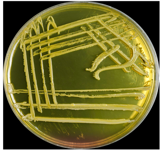
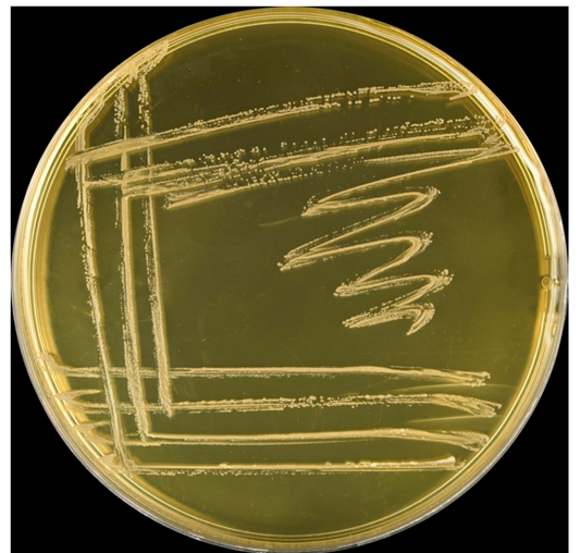
The main aim of the study was the recovery of S. aureus isolates from feline skin infections cases, which was conducted by conventional methods such as culture, morphological appearances under microscope, biochemical tests. Analysis of the cultures indicated Staphylococcus aureus produced yellow colonies, on the Mannitol Salt Agar (MSA) indicating mannitol fermentation (Figure 1). While Staphylococcus aureus on blood agar produced colonies that were surrounded by beta-hemolysis, which is a clear hemolytic zone, S. aureus on STAPH110 media appeared as yellow golden colonies (Figure 2). Gram- stained smears of Staphylococcus aureus were conducted and revealed that Gram-positive single or in grouped cocci.
The Staphylococcus aureus biochemical profile indicated that results were positive for catalase and coagulase tests and negative for oxidase test, as expected. Beta hemolysis was observed on blood agar, and mannitol fermentation in MSA media. The total positive culture isolates were 52 out of 100 skin lesions identified as S. aureus by the conventional culture method. To confirm identification of Staphylococcus aureus, VITEK® system was used. A total of 12 Staphylococcus aureus cultures out of 52 isolates were confirmed by VITEK examination.
Table 2: Infection rate of Staphylococcus aureus according to category of diagnosis.
| Category of diagnosis | Infection rate | Statistical value | |
| Confirmation by Vitek | 12/52 (23%) | Chi-square | 47.98 |
| Confirmation by PCR | 47/52 (90.3%)* | p-value | 0.0043 |
Molecular Detection of S. aureus
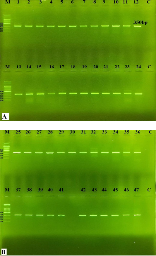
All bacterial isolates (52 out of 100 skin lesion samples) were identified by using conventional methods. However, only 47 out of 52 (90.3%) isolates were confirmed by conventional PCR as S. aureus (Table 2), which produced a specific targeting fragment of 350 bp of the 23s rRNA gene (Figure 3). When the diagnosis was confirmed by the Vitek system, only 12 out of 52 isolates (23%) were detected, indicating low sensitivity of Vitek method in S. aureus diagnosis of skin samples and high sensitivity of PCR method for detecting S. aureus (Table 2).
A larger proportion of Staphylococcus aureus was detected using conventional methods. However, using the VITEK® system, only a total of 12 S. aureus isolates were confirmed. These results display the importance of molecular methods for bacterial identification, but conventional methods remain widely used because of low cost, especially in non-specialist laboratories . However, these methods must be required to distinguish S. aureus from other Staphylococcus species in animals through biochemical profiling . The VITEK® system can produce inaccurate results of some bacteria identification because of the use of low range of biochemical panel through the tests, and increase the risk of contamination . Identification of only 12 out of the 52 isolates suggests that there is relatively low significance to identify S. aureus using classical methods. This also indicate that the necessity of used method in diagnostic bacteriology for accuracy, especially with isolates from animal sources. Moreover, misdiagnosis of Staphylococcus species had negative impact on clinical practice, treatment, and antimicrobial susceptibility testing . Therefore, combination of conventional and molecular methods can provide a highly reliable result and diagnostic confidence to staphylococcal identification.
Clinical and Physical Examination
The present study analyzed clinical, physical examination and vital parameters between non-infected and infected cats with S. aureus. The body temperature and respiratory rate in the infected cats were significantly increased compared to the non-infected cats. In contrast, the heart rate was not significant differences between the infected and non-infected cats (Table 3).
Table 3: Vital signs of non-infected and infected cats with S. aureus (confirmed based on PCR).
| Parameters | Non infected (53) | Infected cats (47) | P value |
| Temperature °C/minute | 38.65 ± 0.05 | 39.29 ± 0.05* | 0.00001 |
| Respiratory rate/minute | 25.02 ± 0.40 | 30.21 ± 0.41* | 0.00001 |
| Heart rate / minute | 176.13 ± 2.97 | 179.68 ± 2.71 | 0.3839 |
Range and M+ SE, *significant differences at (P<0.05).
The most common and documented clinical lesions of Staphylococcus skin infections in cats were pus formation in wounds or post-operative sites and hair loss and redness on the abdomen. While less frequently observed lesions included ophthalmia and periocular hair loss, omphalitis in kittens, and external ear crusting. Statistical analysis revealed that there are no significant differences between lesion type and Staphylococcus infection, indicating that the presence of a specific skin lesion does not significantly predict infection status 0f Staphylococcus (Table 4).
Table 4: Infection rate of Staphylococcus aureus confirmed based on PCR and according to category of skin lesions.
| Lesion Type | Total Number | Infected cats | Infection Rate (%) |
| hair loss and redness on the abdomen | 28 | 13 | 46.43% |
| Crusting of external ear | 3 | 1 | 33.33% |
| Ophthalmitis and hair loss around the eyes | 4 | 2 | 50.0% |
| Omphalitis in kitten | 8 | 4 | 50.0% |
| Pus formation in wound and post-operation | 57 | 27 | 47.37% |
*Chi-square= 0.275; p-value= 0.991. No statistically significant association between skin lesion type.
The fever in the infection significantly occurred due to stimulation in the hypothalamic thermoregulatory center, and by mediated endogenous pyrogens . The elevated respiratory rate could be attributed to the systemic inflammatory response and the associated metabolic demand in small animals with bacterial dermatitis (Faccin et al., 2023). However, the heart rate was not significantly different between groups of cats. This may suggest that the compensatory cardiovascular response was variable in cats, and needed to adaptation of the cats . The skin lesions were variable in this study. Pus formation indicating that suppurative dermatitis with S. aureus due to its enzymes and toxins productions . Similarly, hair loss and erythema were related to folliculitis or dermal invasion . Also, ear lesion in cat can be associated to multifactorial etiology of includes bacteria, mites, fungi, and environmental allergens .
Distribution of the Infection According to the Gender and Age
Infection rates of Staphylococcus confirmed based on the PCR results, was classified to gender and age of cats. Analysis indicated female cats were 39.02%, male cats were 52.54%. Cats with less than one year were 65.12%, and aged one year or older were 33.33%. Although the infection rate was higher in males, no statistically significant association was established. However, the difference was significant in less than one-year cats. The infection rate of Staphylococcus according to PCR results was not statistically and significantly higher in male than female cats, this finding is consistent with Backel and Cain (2017), where authors have identified that male cats may be more susceptible to the skin infections because of their history of fight. More notably, the infection rate was significantly higher in young cats and susceptibility to Staphylococcus infection because of young cats may have immature immune systems, and high susceptible to opportunistic pathogens, and increasing risk of bacterial colonization and infection . The season and physiological statues in cats were significantly affected and will require further investigations .
Phylogenetic Study of 23S RRNA of S. Aureus
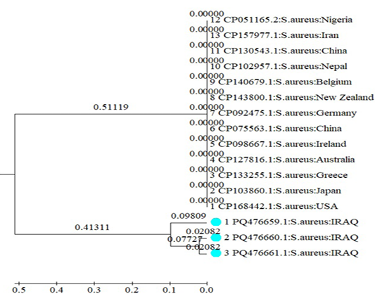
The phylogenetic analysis of the 23s rRNA gene of Staphylococcus aureus isolated from skin infection was conducted with reference strains from other geographical countries, including USA, China, Iran, Germany, Japan, Nigeria and others (Figure 4). The isolates characterized in this study (PQ476659.1, PQ476660.1, PQ476661.1) formed as separate clade, because of distinct genetic relationship, and were separated from other reference strains including Iranian strains (CP157977.1). The nearest world clade had zero genetic distance between them.
The difference between the Vitek system and PCR in detection of bacterial infection revealed limitations of conventional diagnostic methods. These results are consistent with findings of Luteijn et al. (2011) who have noted higher sensitivity and specificity of PCR methods in the bacterial identification. Vitek systems depended on metabolic profiles and phenotypic characteristics, which can be produced intervening between coagulase-positive and coagulase-negative Staphylococcus spp., leading to false negative or even misidentification . In contrast, the PCR targets specific sequences of specific gene of bacteria, allowing rapid and accurate identification . In veterinary skin infection, where samples can contaminate, or chronic infections may impair bacterial culture. These support that the molecular diagnostics can be used in routine identification of S. aureus in skin infections in cats for accurate identification and antimicrobial therapy use to control he spread of this pathogen.
The current study was conducted on skin lesions of 100 cats, with an infection rate of 47% of S. aureus. The analysis of 23S rRNA of S. aureus from ears of cats revealed the infection rate to be 5.8% and association with feline otitis ; . Radhy (2023) has reported that S. aureus was isolated from 10% of eye infections in cats whereas Ahmed and Yousif (2021a) have isolated S. aureus from mastitis in ewes and molecularly characterize them with 23s rRNA genes. Moreover, Hayyawi (2012) has studied many bacterial isolates from external ears in sheep and the most pathogens were S. aureus with a rate of about 30.76 %. Saher et al. (2009) have identified S. aureus with a rate of 21.8 % from dogs with tumor tissue’s samples. The culturing of the samples was conducted from nose, throat and ear of human patients and they have identified S. aureus as the most common pathogen (81.6%) . The differences in the percentage between the present study and other studies in Baghdad may be attributed to selection of one organ of cats in the current study. All previous studied have highlighted the ability of 23srRNA genes for investigating S. aureus in animals and human’s samples. The isolates reported in this study clustered within a distinct clade, and close genetic relationship may indicate a localized adaptation of certain S. aureus in the feline host. The host-specific evolutionary patterns and limited diversity of S. aureus has been demonstrated . However, Iranian strain did not group with the Iraqi cluster due to host specific factors and due to the fact that the Iranian isolates was isolated from urine of human.
CONCLUSIONS AND REcOMMENDATIONS
This study concluded that there are limitations to conventional methods for identifying S. aureus. Phylogenetic analysis of the 23s rRNA gene of S. aureus showed Iraqi isolates formed a distinct cluster, suggesting that there was local adaptation to feline hosts in Iraq, while the Iranian isolate was genetically distant. Epidemiologically, young cats were more susceptible to S. aureus infection. Merging the conventional and molecular diagnostics tools is an important consideration for accurate identification of S. aureus in veterinary practices.
ACKNOWLEDGMENTS
The authors sincerely thank for all staff members of the Department of Internal and Preventive Veterinary Medicine of the College of Veterinary Medicine, University of Baghdad, and the staff of the Veterinary Hospital in Baghdad province for their valued support and cooperation for conducting of this study.
NOVELTY STATEMENTS
The novelty of this study is included the molecular detection and phylogenetic analysis of 23s rRNA gene of S. aureus, which isolated from skin lesions in cats.
AUTHOR’S CONTRIBUTIONS
Mawj Firas Abdulameer collected and cultured the samples and completed other diagnostic tests. Ayman Barzan Abdulgafor designed the study, interpreted the results, and reviewed the manuscript.
Conflict of Interest
The authors have no conflict of interest to disclose.
Figures

The colonies of Staphylococcus aureus on mannitol salt agar (yellow in color), after incubation 37°C for 24-48 hours.

The colonies of Staphylococcus aureus on STAPH 110 (golden yellow), after incubation 37°C for 24-48 hours.

Amplification of 350bp fragment of 23srRNA of Staphylococcus aureus (Agarose gel electrophoresis 1.2%); showing. M: ladder 100 sizers, 47 lanes positive cases. Lane C negative control under blue light documentation.

Phylogenetic tree of Staphylococcus aureus (23s rRNA gene) characterized in this study.
References
- Abbas LA, Rady AM (2023a). Isolation and molecular detection of Staphylococcus aureus from feline otitis in Baghdad. Iraqi J. Vet. Sci., 37(Suppl I–IV): 141–146. https://doi.org/10.33899/ijvs.2023.1375560.2696[CrossRef]
- Abbas LA, Rady AM (2023b). Molecular and phylogenetic analysis of Staphylococcus aureus isolated from feline otitis externa. J. Surv. Fish. Sci., 10(3S): 723–727. https://sifisheriessciences.com/journal/index.php/journal/article/view/78/72
- Abbott JA (2005). Heart rate and heart rate variability of healthy cats in home and hospital environments. J. Feline Med. Surg., 7(3): 195–202. https://doi.org/10.1016/j.jfms.2004.09.005[CrossRef]
- Al-Dujaily AH, Mahmood AK (2022). Evaluation of antibacterial and antibiofilm activity of biogenic silver nanoparticles and gentamicin against Staphylococcus aureus isolated from caprine mastitis. Iraqi J. Vet. Med., 46(1): 10–16. https://doi.org/10.30539/ijvm.v46i1.1309[CrossRef]
- Ahmed ZA, Yousif AA (2021b). Molecular and phylogenetic analysis of methicillin-resistant Staphylococcus aureus isolated from subclinical mastitis in lactating ewes. Iraqi J. Vet. Sci., 35(Suppl III): 121–126. https://doi.org/10.33899/ijvs.2021.131854.2013[CrossRef]
- AL-Ani ANA, Al-Badrawi TYG, Faraj BR, Badawi NM (2019). Seasonal and physiological changes on renal function parameters in Iraqi cats. Indian J. Ecol., 46(Spl Issue 8): 237–241. https://www.researchgate.net/publication/339362869
- Ahmed ZA, Yousif AA (2021a). Isolation, antimicrobial susceptibility and molecular detection of Staphylococcus aureus from clinical mastitis in ewes. Teikyo Med. J., 44(6): 03875547.
- Al-Hamadany WS (2013). Isolation and identification of Gram-positive cocci bacterial pathogens from suppurative otitis media infections. Iraqi J. Vet. Med., 37(1): 129–133. https://doi.org/10.30539/iraqijvm.v37i1.345[CrossRef]
- Al-Rasheed AA, Sana’a SA, Al-Jashamy KA, Garba B (2022). Immunopathological responses to the bovine mastitis associated with Staphylococcus species infection. Iraqi J. Vet. Med., 46(2): 7–11. https://doi.org/10.30539/ijvm.v46i2.1398[CrossRef]
- Backel K, Cain C (2017). Skin as a marker of general feline health: cutaneous manifestations of infectious disease. J. Feline Med. Surg., 19(11): 1149–1165. https://doi.org/10.1177/1098612X17735764[CrossRef]
- Bannoehr J, Guardabassi L (2012). Staphylococcus pseudintermedius in the dog: taxonomy, diagnostics, ecology, epidemiology and pathogenicity. Vet. Dermatol., 23(4): 253–e52. https://doi.org/10.1111/j.1365-3164.2012.01046.x[CrossRef]
- Clifford RJ, Milillo M, Prestwood J, Quintero R, Zurawski DV, Kwak YI, McGann P (2012). Detection of bacterial 16S rRNA and identification of four clinically important bacteria by real-time PCR. PLoS One, 7(11): e48558. https://doi.org/10.1371/journal.pone.0048558[CrossRef]
- Del Giudice P (2020). Skin infections caused by Staphylococcus aureus. Acta Derm. Venereol., 100(9): adv00110. https://doi.org/10.2340/00015555-3466[CrossRef]
- Dinarello CA (2004). Infection, fever, and exogenous and endogenous pyrogens: some concepts have changed. J. Endotoxin Res., 10(4): 201–222. https://doi.org/10.1179/096805104225006129[CrossRef]
- Faccin M, Wiener DJ, Rech RR, Santoro D, Rodrigues Hoffmann A (2023). Common superficial and deep cutaneous bacterial infections in domestic animals: a review. Vet. Pathol., 60(6): 796–811. https://doi.org/10.1177/03009858231176558[CrossRef]
- Faires MC, Tater KC, Weese JS (2009). An investigation of methicillin-resistant Staphylococcus aureus colonization in people and pets in the same household with an infected person or infected pet. J. Am. Vet. Med. Assoc., 235(5): 540–543. https://doi.org/10.2460/javma.235.5.540[CrossRef]
- Funke G, Funke Kissling P (2005). Performance of the new VITEK 2 GP card for identification of medically relevant gram-positive cocci in a routine clinical laboratory. J. Clin. Microbiol., 43(1): 84–88. https://doi.org/10.1128/JCM.43.1.84-88.2005[CrossRef]
- Gatta MN, Al Graibawi MAA (2021). Phenotypic and genotypic detection of biofilm producing methicillin resistant Staphylococcus aureus (MRSA) isolated from human. Ann. Rom. Soc. Cell Biol., 25(6): 19047–19058. https://annalsofrscb.ro/index.php/journal/article/view/9481
- Hanselman BA, Kruth SA, Rousseau J, Weese JS (2009). Coagulase-positive staphylococcal colonization of humans and their household pets. Can. Vet. J., 50(9): 954–958. https://pmc.ncbi.nlm.nih.gov/articles/PMC2726022
- Hayyawi SM (2012). Comparison of microbial isolates isolated from external ear canal of sheep and their susceptibility to antibiotics. Iraqi J. Vet. Med., 36(0E): 41–48. https://doi.org/10.30539/iraqijvm.v36i0E.379[CrossRef]
- Jafaar FA, Kandala NJ (2025). Molecular detection of adhesive matrix molecules for Staphylococcus aureus isolated from different samples. Iraqi J. Agric. Sci., 56(2): 784–798. https://doi.org/10.36103/ngr0nm29[CrossRef]
- Karmakar A, Dua P, Ghosh C (2016). Biochemical and molecular analysis of Staphylococcus aureus clinical isolates from hospitalized patients. Can. J. Infect. Dis. Med. Microbiol., 2016: 9041636. https://doi.org/10.1155/2016/9041636[CrossRef]
- Krapf M, Müller E, Reissig A, Slickers P, Braun SD, Müller E, Monecke S (2019). Molecular characterisation of methicillin-resistant Staphylococcus pseudintermedius from dogs and the description of their SCCmec elements. Vet. Microbiol., 233: 196–203. https://doi.org/10.1016/j.vetmic.2019.04.002[CrossRef]
- Luteijn JM, Hubben GAA, Pechlivanoglou P, Bonten MJ, Postma MJ (2011). Diagnostic accuracy of culture-based and PCR-based detection tests for methicillin-resistant Staphylococcus aureus: a meta-analysis. Clin. Microbiol. Infect., 17(2): 146–154. https://doi.org/10.1111/j.1469-0691.2010.03202.x[CrossRef]
- Markey BK, Leonard FC, Archambault M, Cullinane A, Maguire D (2014). Clinical Veterinary Microbiology. 2nd ed. Mosby Elsevier, 105–120.
- Monecke S, Coombs G, Shore AC, Coleman DC, Akpaka P, Borg M, Ehricht R (2011). A field guide to pandemic, epidemic and sporadic clones of methicillin-resistant Staphylococcus aureus. PLoS One, 6(4): e17936. https://doi.org/10.1371/journal.pone.0017936[CrossRef]
- Mustapha M, Bukar Kolo YM, Geidam YA, Gulani IA (2014). Review on methicillin-resistant Staphylococcus aureus (MRSA) in dogs and cats. Int. J. Anim. Vet. Adv., 6(2): 61–73. https://doi.org/10.1111/j.1748-5827.2004.tb00180.x[CrossRef]
- Nardoni S, Ebani VV, Fratini F, Mannella R, Pinferi G, Mancianti F, Perrucci S (2014). Malassezia, mites and bacteria in the external ear canal of dogs and cats with otitis externa. Slovenian Vet. Res., 51(3): 113–118. https://core.ac.uk/download/pdf/80256382.pdf
- Noli C, Borio S, Varina A, Schievano C (2016). Development and validation of a questionnaire to evaluate the quality of life of cats with skin disease and their owners, and its use in 185 cats with skin disease. Vet. Dermatol., 27(4): 247–e58. https://doi.org/10.1111/vde.12341[CrossRef]
- Phumthanakorn N, Prapasarakul N, Yindee J, Gronsang D (2022). Frequency, distribution, and antimicrobial resistance of coagulase-negative staphylococci isolated from clinical samples in dogs and cats. Microb. Drug Resist., 28(2): 236–243. https://doi.org/10.1089/mdr.2020.0586[CrossRef]
- Rajapaksha P, Elbourne A, Gangadoo S, Brown R, Cozzolino D, Chapman J (2019). A review of methods for the detection of pathogenic microorganisms. Analyst, 144(2): 396–411. https://pubs.rsc.org/en/content/articlelanding/2019/an/c8an01488d
- Ryman VE, Kautz FM, Nickerson SC (2021). Case study: misdiagnosis of nonhemolytic Staphylococcus aureus isolates from cases of bovine mastitis as coagulase-negative staphylococci. Animals, 11(2): 252. https://doi.org/10.2460/javma.235.5.540[CrossRef]
- Saher MH, Abdulrazak MA, Aboud AS, Al Khaban JM, Iesa AM (2009). Study on bacterial isolation from dogs affected with malignant tumor. Iraqi J. Vet. Med., 33(1): 120–131. https://doi.org/10.30539/iraqijvm.v33i1.725[CrossRef]
- Saraiva MM, De Leon CM, Santos SC, Stipp DT, Souza MM, Santos Filho L, Oliveira CJ (2018). Accuracy of PCR targeting different markers for Staphylococcus aureus identification: a comparative study using matrix-assisted laser desorption/ionization time-of-flight mass spectrometry as the gold standard. J. Vet. Diagn. Invest., 30(2): 252–255. https://doi.org/10.1177/1040638717732370[CrossRef]
- Tamura K, Stecher G, Peterson D, Filipski A, Kumar S (2013). MEGA6: molecular evolutionary genetics analysis version 6.0. Mol. Biol. Evol., 30(12): 2725–2729. https://doi.org/10.1093/molbev/mst197[CrossRef]
- Vinshia JJ, Padmavathy K, Sathyapriya B (2024). Comparison of the VITEK® 2 system with conventional methods for species identification and antimicrobial susceptibility pattern of staphylococcal carrier isolates. Int. Res. J. Multi. Scope, 5(2): 940–948. https://doi.org/10.47857/irjms.2024.v05i02.0689[CrossRef]
- Woudstra S, Wente N, Zhang Y, Leimbach S, Kirkeby C, Gussmann MK, Krömker V (2023). Reservoirs of Staphylococcus spp. and Streptococcus spp. associated with intramammary infections of dairy cows. Pathogens, 12(5): 699. https://doi.org/10.3390/pathogens12050699[CrossRef]
Services
Article usage statistics
Loading…