
Autosomal recessive primary microcephaly (MCPH) is a rare neurodevelopmental disorder characterized by severe reduction in the brain volume that affects especially the neocortex, and intellectual disability. To date 17 genes have been linked with this disease. Biallelic mutations in the abnormal spindle-like, microcephaly associated gene ASPM causes MCPH type 5, the most common MCPH subtype. In the current study we recruited two families from Sialkot region of Pakistan which were assessed by whole exome sequencing and cosegregation analysis. We identified a previously described mutation c.4802C>G (p.S1601*) in the ASPM gene in both families. This study further underlines that mutations in the ASPM gene are a common cause for microcephaly in the Pakistani population and updates our knowledge regarding the frequent involvement of ASPM in microcephaly.
Short Communication · Volume 50, Issue 5, 25 August 2018 · Open Access
Homozygous Nonsense Mutation in the ASPM Gene Causes MCPH in Consanguineous Pakistani Families
· · · · · · · · · ·
Affiliations & Notes ⌄
- 1Department of Zoology, Mirpur University of Science and Technology, Mirpur 10250, Pakistan 2Institute of Neuroanatomy and Cell Biology, Charité-Universitätsmedizin Berlin, Berlin, Germany 3Berlin Institute of Health, Anna-Louisa-Karsch Strasse 2, 10178, Berlin, Germany 4Center for Chronically Sick Children (Sozialpädiatrisches Zentrum, SPZ), Charité-Universitätsmedizin Berlin, Berlin, Germany 5Department of Zoology, University of Azad Jammu and Kashmir, P.O. Box 13100, Muzaffarabad, Pakistan 6De
Article Info ⌄
- Journal:
- Pakistan Journal of Zoology
- Published:
- 25 August 2018

From the article
- We have identified via WES a previously reported nonsense mutation c.4802C>G located in exon 22 of the ASPM gene transcript 1 (NM_018136) in both families.
- The homozygous nonsense mutation (c.4802C>G) in the ASPM gene leads to a truncated protein, or nonsense mediated mRNA decay, resulting in the complete loss of the ASPM protein.
- We report two Pakistani families with a homozygous nonsense mutation in the ASPM gene and expand the knowledge about ASPM related microcephaly cases in the Pakistani population.
- This updates our knowledge regarding the frequent involvement of ASPM in microcephaly.
Selected from the article's own text.
Abstract
Materials and methods
The study was reviewed and approved by the local ethical committee and the ethical committee of the Charité University of Medicine, Berlin Germany (EA1/212/08). Informed written consent was obtained from the patients and guardians of the families. Patients were recruited from Sialkot region of Pakistan by field survey. All the patients were examined by medical experts for clinical assessment. Blood samples were taken from all available individuals of affected pedigrees, and DNA was extracted using standard laboratory protocols.
For whole exome sequencing (WES), genomic DNA libraries were prepared through an Agilent Sure SelectXT Human All Exon V6 kit following the manufacturer’s protocol. The sequencing libraries were sequenced on an Illumina HiSeq 2000 platform. The preliminary whole exome data analysis was performed through BWA and GATK software to generate a BAM and a VCF file, respectively. Annotation of the VCF files were carried out through the wANNOVAR software and the data was manually analyzed for the presence of candidate pathogenic variant.
For segregation analysis, identified mutations in the ASPM gene (NM_018136) were verified through Sanger sequencing and segregation analysis. Sanger sequencing was performed on all available patients and one parent was used as control. Primer3web (version 4.0.0) tool was used to design the primers in the flanking region of pathogenic variants. Sequencing primers F5´- GTGCTTATGAGTTATTCTACCGGC -3´, R5´- TTTGCACCAAGTGGAGTGGG -3´, were used for Sanger sequencing.
Table I.- Clinical and morphological features of affected patients from Family A and B with ASPM gene mutation.
| Family | Pedigree ID | Gender | Age | Phenotype | OFC | Weight (kg) | Height (cm) |
| A | IV.2 | Male | 55 years | Microcephaly, Aggressiveness | -6.4 SD | 40 | 161.5 |
| IV.3 | Male | 45 years | Microcephaly, Cognitive delay | -5.5 SD | 55 | 152.4 | |
| B | IV.3 | Male | 12 years | Microcephaly, Speech delay, Motor delay | -8.8 SD | 25 | 145 |
| IV.4 | Male | 12 years | Microcephaly, Speech delay, Motor delay | -8.8 SD | 24 | 143 |
Results
All affected individuals of family A had primary microcephaly with occipital frontal head circumference of 45.72 cm and 46.99 cm (IV.2 at 55 years; -6.4 SD and IV.3 at 45 years; -5.5 SD) and moderate ID, while patient IV.2 also showed aggressive behavior (Fig. 1A). In family B both patients had primary microcephaly with occipital frontal head circumferences of 40.64.cm (IV.3 at 12 years; -8.8 SD) and 40.64cm (IV.4 at 12 years; -8.8SD) and mild ID (Fig. 2A). In addition, both patients displayed a speech and motor delay. Affected members in both families were born at complete term with normal delivery. Clinical features of patients are summarized in (Table I).
In both families affected members showed small head with frontal narrowing. However, patients in family A with ASPM mutations had facial features in the form of prominent glabella, arched, thin eyebrows, and prominent eyes in infancy. With age this facial phenotype became subtle. Both the families had no relationship with each other. The forehead became sloping and the prominent eyes became less apparent. Both the families were recruited from two different regions and had no previous history of common ancestry.
We have identified via WES a previously reported nonsense mutation c.4802C>G located in exon 22 of the ASPM gene transcript 1 (NM_018136) in both families. The mutation was confirmed via Sanger sequencing and cosegregates within the pedigrees (Figs. 1B, 2B). The reported variant is not present in ExAC, 1000 genomes browser, or exome variant server, but reported in NCBI variation viewer and dbSNP as known disease mutation (rs199422189) and only pathogenic for primary autosomal recessive microcephaly type 5. The homozygous nonsense mutation (c.4802C>G) in the ASPM gene leads to a truncated protein, or nonsense mediated mRNA decay, resulting in the complete loss of the ASPM protein.
Discussion
We identified a previously reported nonsense mutation c.4802C>G (p.S1601*; rs199422189) in the ASPM gene in two Pakistani families showing clinical signs of primary microcephaly and ID. The mutation cosegregates with the disease phenotype in both families.
The ASPM gene is mapped on chromosome 1q31.3 and encodes for a 10887 bp transcript (NM_018136) containing 28 exons that is translated into a 3477 amino acid protein (Fig. 3). Fetal expression pattern of ASPM mRNA in mouse brain revealed an expression at sites of active neurogenesis in the neuroepithelium . Thereby, the ASPM protein is involved in cleavage plane orientation, regulating the mitotic spindle function in neuronal progenitors. The loss of ASPM leads to a premature switch from symmetric to asymmetric cell division, decreasing the number of postmitotic neurons in the developing cortex, reflected in a reduced cortical surface area and a simplified gyral pattern. Therefore, the ASPM gene is involved in determining cerebral cortical size during development and linked to primary autosomal recessive microcephaly type 5 (MCPH5; (#605481)). To date 171 mutations in the ASPM gene have been identified comprising 73 nonsense, 4 missense and 14 splice site mutations along with 68 microdeletions 8 duplications and 1 insertion leading to frame shift mutations . Moreover one complex rearrangement along with two additional microdeletions has also been reported . In this study, we identified a homozygous nonsense mutation p.S1601* located in exon 22 that encodes for the calmodulin-binding IQ domains that serves as binding site for EF-hand proteins like calmodulin and PKC phosphorylation site . In a previous study we have reported mutation in CRB1 gene causing blindness in Kashmiri families . Calmodulin is known to regulate cell proliferation via the interaction with ASPM to regulate spindle assembly (Berchthold and Villalobo, 2014). We hypothesize that our nonsense mutation leads to a truncated protein or nonsense-mediated mRNA decay, resulting in the complete loss of the ASPM protein. Several publications support this notion by showing that no correlation between the severity of the disorder and the length of the truncated protein exist (Bond et al., 2003) and that the lack of the C-terminal domain of ASPM, which mediates mid body localization, seems to be sufficient to cause microcephaly in humans .
Conclusion
We report two Pakistani families with a homozygous nonsense mutation in the ASPM gene and expand the knowledge about ASPM related microcephaly cases in the Pakistani population. This updates our knowledge regarding the frequent involvement of ASPM in microcephaly. Thus, ASPM should be included in an NGS panel to study microcephaly cases in a Pakistani cohort.
Acknowledgements
This study was supported by the German Research Foundation (SFB665) and the Berlin Institute of Health (CRG1). We are thankful to the patients and their families for their participation in the present study.
Statement of conflict of interest
Authors have declared no conflict of interest.
Figures
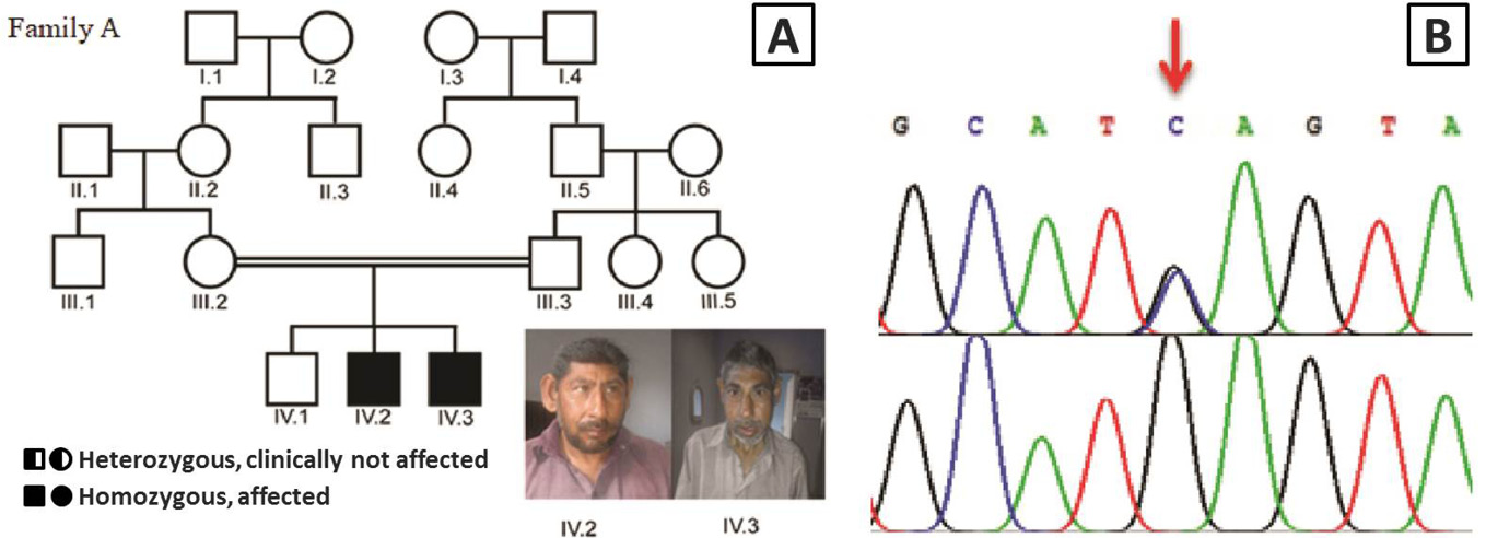
A, Pedigree of the consanguineous family A of Pakistani descent with two patients affected by microcephaly (□, male; ○, female; ═, consanguineous marriage); B, Sanger sequencing results of patient IV.2 from family A showing a homozygous mutation c.4802C>G in the ASPM gene that leads to a nonsense mutation p.S1601* (red arrow). The parental control III.2 is heterozygous for the ASPM mutation.
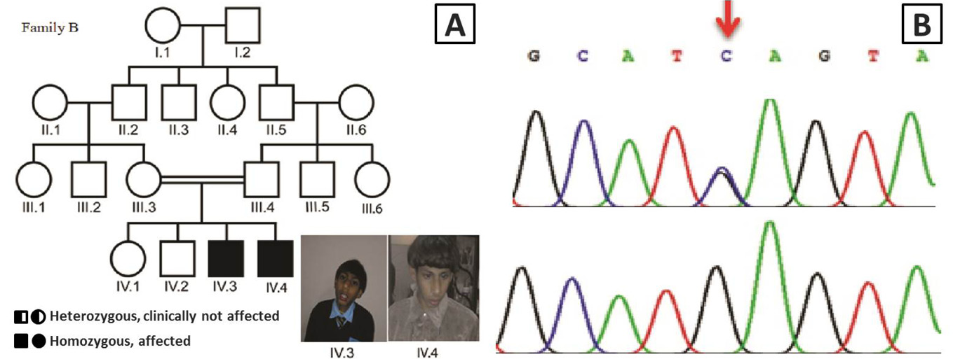
A, Pedigree of the consanguineous family B of Pakistani descent with two patients affected by microcephaly, speech delay and motor delay (□, male; ○, female; ═, consanguineous marriage); B, Sanger sequencing results of patient IV.2 from family B showing a homozygous mutation c.4802C>G in the ASPM gene that leads to a nonsense mutation p.S1601* (red arrow). The parental control III.3 is heterozygous for the ASPM mutation.
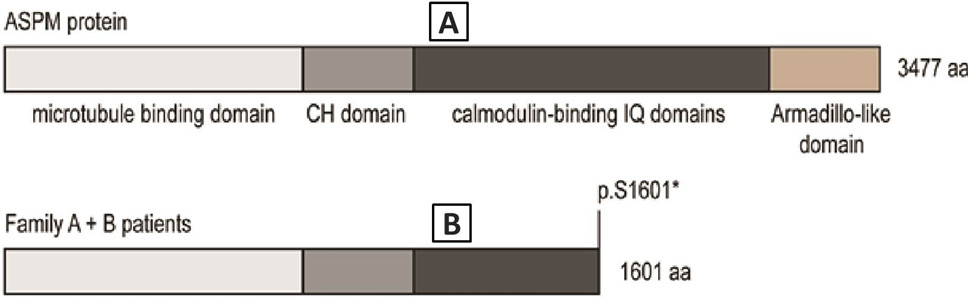
A, the ASPM gene codes for a 3477 aa protein with four protein domains: microtubule binding domain (light grey), CH domain (grey), calmodulin-binding IQ domains (dark grey) and the Armadillo-like domain (brown); B, the nonsense mutation p.S1601* in patients of family A and B lead to a truncated protein, which is missing the Armadillo-like domain and part of the calmodulin-binding IQ domain.
References
- Abdel-Hamid, M.S., Ismail, M.F., Darwish, H.A., Effat, L.K., Zaki, M.S. and Abdel-Salam, G.M., 2016. Am. J. Med. Genet. Part A, 170: 2133-2140. https://doi.org/10.1002/ajmg.a.37724[CrossRef]
- Baudier, J., Deloulme, J.C., van Dorsselaer, A., Black, D. and Matthes, H.W., 1991. J. Biol. Chem., 266: 1229-1237.
- Berchtold, M.W. and Antonio, V., 2014. Mol. Cell. Res., 2: 398-435. https://doi.org/10.1016/j.bbamcr.2013.10.021[CrossRef]
- Bilgüvar, K., Öztürk, A.K., Louvi, A., Kwan, K.Y., Choi, M., Tatli, B., Yalnizoğlu, D., Tüysüz, B., Çağlayan, A.O., Gökben, S. and Kaymakçalan, H., 2010. Nature, 467: 207. https://doi.org/10.1038/nature09327[CrossRef]
- Bond, J., Roberts, E., Mochida, G.H., Hampshire, D.J., Scott, S., Askham, J.M., Springell, K., Mahadevan, M., Crow, Y.J., Markham, A.F. and Walsh, C.A., 2002. Nat. Genet., 32: 316. https://doi.org/10.1038/ng995[CrossRef]
- Bramham, J., Hodgkinson, J.L., Smith, B.O., Uhrı́n, D., Barlow, P.N. and Winder, S.J., 2002. Structure, 10: 249-258. https://doi.org/10.1016/S0969-2126(02)00703-7[CrossRef]
- Faheem, M., Naseer, M.I., Rasool, M., Chaudhary, A.G., Kumosani, T.A., Ilyas, A.M., Pushparaj, P.N., Ahmed, F., Algahtani, H.A., Al-Qahtani, M.H. and Jamal, H.S., 2015. BMC Med. Genom., 8: 4. https://doi.org/10.1186/1755-8794-8-S1-S4[CrossRef]
- Fish, J.L., Kosodo, Y., Enard, W., Pääbo, S. and Huttner, W.B., 2006. Proc. natl. Acad. Sci., 103: 10438-10443. https://doi.org/10.1073/pnas.0604066103[CrossRef]
- Jouan, L., Daoud, H., Dionne-Laporte, A., Dobrzeniecka, S., Spiegelman, D., Rochefort, D., Hince, P., Szuto, A., Lassonde, M., Barbelanne, M. and Tsang, W.Y., 2016. Eur. J. Human Genet., 24: 607-610. https://doi.org/10.1038/ejhg.2015.156[CrossRef]
- Khan, A., Wang, R., Han, S., Ahmad, W. and Zhang, X., 2018. Genet. Test Mol. Biomark., 22: 159-164. https://doi.org/10.1089/gtmb.2017.0229[CrossRef]
- Kraemer, N., Picker-Minh, S., Abbasi, A.A., Fröhler, S., Ninnemann, O., Khan, M.N., Ali, G., Chen, W. and Kaindl, A.M., 2016. Clin. Genet., 89: 744-745. https://doi.org/10.1111/cge.12685[CrossRef]
- Latif, Z., Kathrin, B., Tufail, H.T., Muhammad, N.K., Ghazanfar, A., Ansar, A.A., Abdul, R., Hao, H. and Angela, M.K., 2017. Pakistan J. Zool., 6: 2313-2317. http://doi.org/10.17582/j.pjz/2017.49.6.2313.2317[CrossRef]
- Leipe, D.D., Koonin, E.V. and Aravind, L., 2004. J. mol. Biol., 343: 1-28. https://doi.org/10.1016/j.jmb.2004.08.023[CrossRef]
- Nicholas, A.K., Swanson, E.A., Cox, J.J., Karbani, G., Malik, S., Springell, K., Hampshire, D., Ahmed, M., Bond, J., Di Benedetto, D. and Fichera, M., 2009. J. med. Genet., 46: 249-253. https://doi.org/10.1136/jmg.2008.062380[CrossRef]
- Paramasivam, M., Chang, Y.J. and Lo-Turcou, J.J., 2007. Cell. Cycle, 6:1605-1612. https://doi.org/10.4161/cc.6.13.4356[CrossRef]
- Passemard, S., Titomanlio, L., Elmaleh, M., Afenjar, A., Alessandri, J.L., Andria, G., de Villemeur, T.B., Boespflug-Tanguy, O., Burglen, L., Del Giudice, E. and Guimiot, F., 2009. Neurology, 73: 962-969. https://doi.org/10.1212/WNL.0b013e3181b8799a[CrossRef]
- Tan, C.A., Topper, S., Melver, C.W., Stein, J., Reeder, A., Arndt, K. and Das, S., 2014. Brain Develop., 36: 351-355.
- Zaqout, S., Morris-Rosendahl, D. and Kaindl, A.M., 2017. Neuropediatrics, 48: 135-142. https://doi.org/10.1055/s-0037-1601448[CrossRef]
Services
Article usage statistics
Loading…