
Cotugnia sp. is a parasitic tapeworm belonging to the class Cestoda and found in the gastrointestinal tract of various birds worldwide. The GIT tract is where food is digested, and parasites are normally observed due to a favourable environment for food and shelter. The present study aims to investigate the histopathological changes in the intestine of Melopsittacus undulatus naturally infected with cestode Cotugnia karachiensis. Infected tissues were fixed in formalin, and 5-7μm thick paraffin sections were made, stained and mounted to study histopathology. This study revealed that mucosa was thickened, necrosis was observed in serosa, villus hypertrophy, vacuole formation, crypt gland destruction and channel formation in the intestinal layer were observed. The present study indicated a state of grave problems facing a parrot (Melopsittacus undulates) when infected with cestode Cotugnia karachiensis.
Short Communication · Volume 58, Issue 1, 2 January 2026 · Open Access
Histopathological Changes in the Intestine of a Parrot (Melopsittacus undulatus) Naturally Infected with Cestode, Cotugnia karachiensis, from Karachi, Sindh, Pakistan
· ·
Affiliations & Notes ⌄
- 1Department of Zoology, University of Karachi, Karachi-75270, Pakistan. * Corresponding author: rubabmalik943@gmail.com
Article Info ⌄
- Journal:
- Pakistan Journal of Zoology
- Published:
- 2 January 2026

From the article
- During the study of infected tissues of the large intestine, serosa, mucosa and muscularis mucosa degeneration were observed.
- The degeneration of villi and haemorrhage in blood capillaries is observed due to infection of Cotugnia karachiensis (Fig. 1A).
- Changes in the size of intestinal glands and haemorrhages have been noticed (Fig. 1C).
- Alterations in villi and length of epithelium, crypt depth and thickness of tunica muscularis are all because of the gastro-intestine of indigenous chicken in Nepal.
Selected from the article's own text.
Abstract
Materials and methods
The dead parrots were brought into the Parasitology Laboratory at the University of Karachi. They were dissected, and intestines were examined in 0.8% physiological saline for the parasitic helminth infection. The damaged sections were separated and preserved in cold 70% ethanol for further histological procedures. Infected intestine tissues were placed in Bouin’s solution for twenty-four hours. Fixation is essential to maintain the tissue’s molecular and structural composition and enhance the absorbance during staining. After fixation, tissue is processed through dehydrated ethanol series, cleared in xylene, and maintained in wax for an hour in a 60°C oven with two baths containing melted paraffin wax at a 1:1 ratio. The wax infiltrates the tissue’s structure, increasing optical differentiation, hardening the tissue, and easier tissue sectioning. Tissue was placed into cavity blocks with somewhat hardened wax, and then melted paraffin wax was applied to the tissue. 6–8 µm thick strips were produced using standard histology methods on slides. To extend the strips, the slides were placed on a hot plate set to 40°C. Staining was utilised to contrast the tissue slice’s component sections. Then, in each grade, went through a descending series of alcohol (100%-30%) for 8 min, followed by an ascending series of alcohol (30%-100 %) for 10 min. To eliminate all traces of alcohol, waited for 15 min and mounted with Canada balsam. Microphotographs of permanently mounted tissue slices were obtained with a Nikon photomicroscope.
Results and discussion
During the study of infected tissues of the large intestine, serosa, mucosa and muscularis mucosa degeneration were observed. The degeneration of villi and haemorrhage in blood capillaries is observed due to infection of Cotugnia karachiensis (Fig. 1A). The villi are blunt-ended and more expanded than their original size (Fig. 1B). Changes in the size of intestinal glands and haemorrhages have been noticed (Fig. 1C). The hypertrophic condition in villi has been observed (Fig. 1D). The invasion of scolex revealed deep blockage towards the mucosal linings (Fig. 1E).
Crucial deformation of epithelial cells with degenerative and small-size villi has been observed (Fig. 1F). In Figure 1G, enlarged villi with unpacking crypts were observed. In Figure 1H, a cyst with defensive cells has been observed (although the author is not sure about the cyst). It was obvious that there would be a severe infection because of erupted villi from the mucous layer, resulting in a prime disruption causing unrecognisable intestinal structures.
The layers of the intestine in psittacine birds are more like the warm-blooded animal intestine that possess mucosa, sub-mucosa, muscularis and serosa . Abdel-Rahman et al. (2019) observed that acute histopathological alteration in the intestines of birds can be seen because of the presence of helminths observed in the present research. Infection in birds GIT can lead to abnormality in the immune response in the host, leading to lethal effects (Hassan et al., 2019). The findings align with Samad et al. (1986) and Pinto et al. (1997), who observed atrophied villi enteritis with cellular insinuation and granulomas origination. Moreover, in the intestine, minor haemorrhages were noticed . Uddin et al. (2010) observed several nodules in the intestinal mucosa. Bhowmik et al. (1982) noted that obstruction, gross lesions and sores were noticed in the intestine due to cestode invasion.
Ayaz et al. (2003) observed multiple cestode infestations in the intestines of broiler chickens, leading to poor function. Moreover, Nisar et al. (2015) suggested that a portion of parasites sometimes remain in the muscular layer. In the current study, some scolices were penetrated in intestinal layers. Anwar et al. (2000) observed
hypertrophy and atrophy in villi due to the presence of Raillietina echinobothrida in the domestic fowl intestine. Adang et al. (2014) stated that Aves can be affected by parasitic infection. The current investigation shows that macrophages and lymphocytes are quite active at the site of infection. Alterations in villi and length of epithelium, crypt depth and thickness of tunica muscularis are all because of the gastro-intestine of indigenous chicken in Nepal.
Declarations
Acknowledgements
The first author is thankful to her supervisor for her guidance and support.
Funding
The study received no external funding.
Statement of conflict of interest
The authors have declared no conflict of interest.
Figures
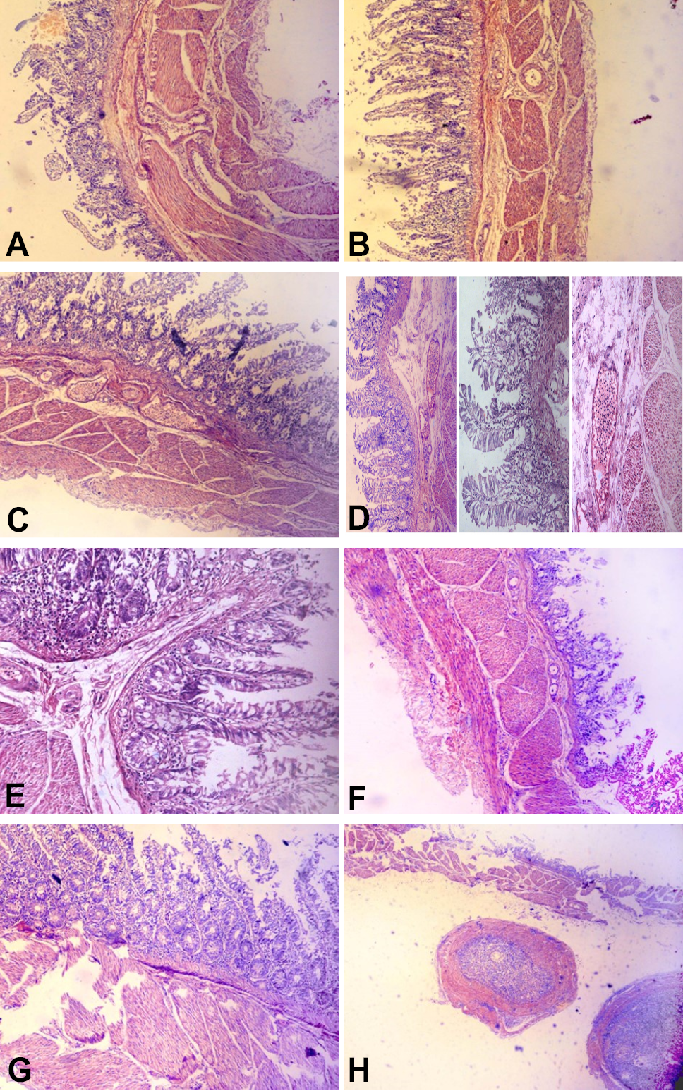
Effect of cestode infection on histological structure of small intestine of parrot. A shows massive degeneration of serosa and muscularis mucosa with heavy cellular infiltration. Mucosal layer and the villi and intestinal glands show destructive changes. B shows abnormally elongated blunt-ended villi and necrotic patches. C shows haemorrhages with broken blood capillaries; change in the size and shape of intestinal glands can be seen. D shows empty intestinal glands and severe congestion in the submucosal layer (b) villi showing hypertrophic condition (20x) (c) Photomicrograph showing heavy cellular infiltrate. E shows the penetration of cestode parasite with heavy infiltration of cells along with abnormally elongated villi. F shows an intense villi epithelial desquamation, an evident degeneration of villi and a smaller sized villi known as dwarf villi and an increased cellular infiltrate. G shows hypertrophy and degeneration of mucosal layer with bleeding between the glands while emptied glands of Lieberkühn and a visible increase in lymphocytic cells can be seen. H shows a cyst (cestode) with defensive cells. The serosa and muscularis mucosa can be seen demolished. Magnification: A, B, C, D, F, G, H 10X; E, 20X. Stain: Hematoxylin & Eosine.
References
- Abdel-Rahman, M.M.I.A., Tolba, H.M.N. and Abdel-Ghany, H.M., 2019. Adv. Anim. Vet. Sci., 7: 66-72. https://doi.org/10.17582/journal.aavs/2019/7.2.66.72[Full text][CrossRef]
- Adang, K.L., Asher, R. and Abba, R., 2014. Asian J. Poult. Sci., 8: 32-40. https://doi.org/10.3923/ajpsaj.2014.32.40[CrossRef]
- Anwar, A.H., Rana, S.H., Shah, A.H., Khan, M.N. and Akhtar, M.Z., 2000. Pathol. J. Agric. Sci., 37: 1-2.
- Ayaz, M.M., Akhtar, M., Hayat, C.S., Hafeez, M.A. and Haq, A., 2003. Pak. Vet. J., 23: 51-52.
- Bhowmik, M.K., Sasmal, N.K. and Chakraborty, A.K., 1982. Indian Vet. med. J., 6: 100-102.
- Hassan, T.A., El-Gawady, H.M., El-Gayer, A.K. and Sallam, N.H., 2023. J. Adv. Vet. Res., 13: 352-359.
- Malik, R., Khatoon, N. and Waheed, S., 2022. Pak. J. Nematol., 40: 120-126. https://doi.org/10.17582/journal.pjn/2022/40.2.120.126[Full text][CrossRef]
- Nisar, S., Khan, F.M., Khan, A.M., Arshad, A., Mehboob, U. and Raza, A., 2015. Int. J. Fauna Biol. Stud., 2: 36-39.
- Pinto, R.M., Vicente, J.J. and Noronha, D., 1997. Mem. Inst. Oswaldo Cruz, 92: 209-214. https://doi.org/10.1590/S0074-02761997000200013[CrossRef]
- Reavill, D.R. and Dorrestein, G.M., 2010. Vet. Clin. Exot. Anim. Pract., 13: 135-150. https://doi.org/10.1016/j.cvex.2009.12.001[CrossRef]
- Samad, M.A., Alam, M.M. and Bari, A.S.M., 1986. Vet. Parasitol., 21: 279-284. https://doi.org/10.1016/0304-4017(86)90055-5[CrossRef]
- Slaoui, M. and Fiette, L., 2011. Histopathology procedures from tissue sampling to histopathological evaluation. Methods Mol. Biol. Protocols, 691: 69-82. https://doi.org/10.1007/978-1-60761-849-2_4[CrossRef]
- Uddin, M.M., Khan, M.Z.I., Islam, K.N., Kibria, A.S.M.G., Adhikary, G.N., Parvez, M.N.H., Basu, J., Uddin, M.B. and Rahman, M.M., 2010. Pak. Vet. J., 30: 67-71.
Services
Article usage statistics
Loading…