Research Article · Volume 14, Issue 7, 20 July 2026 · Open Access
Antibiotic Resistance of Escherichia coli Strains Isolated From Pet Parrots in Hanoi, Vietnam
· · · · · · ·
Affiliations & Notes ⌄
1Faculty of Veterinary Medicine, Vietnam National University of Agriculture, Gia Lam, Hanoi, Vietnam
2Undergraduate student, Faculty of Veterinary Medicine, Vietnam National University of Agriculture, Gia Lam, Hanoi, Vietnam. *Correspondence | Truong Ha Thai, Faculty of Veterinary Medicine, Vietnam National University of Agriculture, Gia Lam, Hanoi, Vietnam
We found that 52 of 53 isolates (98.1%) were resistant to at least one antibiotic, and the most common resistance was identified as amoxicillin (94.3%).
Overall, the E. coli strains we isolated were resistant to many antibiotics, and many were MDR.
This suggests that pet birds could be reservoirs of antibiotic-resistant bacteria, which is a concern for both human and animal health.
Although we did not study transmission to humans, the presence of MDR E. coli in pet birds is a potential public health concern that warrants further investigation.
Selected from the article's own text.
Abstract
Pet birds can be birds can carry and spread various bacteria that cause illness in humans, including E. coli. We investigate the prevalence and antibiotic resistance of E. coli strains isolated from pet parrots raised in Hanoi, Vietnam. Overall, 53 (59.6%) out of 89 fecal samples from pet parrots contained E. coli. Red-breasted parakeet samples had the highest rate (75.0%, 9/12 samples), followed by Monk parakeet (70.0%, 7/10 samples), Macaw parrot (66.7%, 8/12 samples). Meanwhile, fecal samples from Lovebirds, Sun parakeet, and African grey parrot had E. coli positive rates ranging from 50.0-58.8%. The isolated E. coli strains exhibited resistance to all 10 antibiotics belonging to 7 different antibiotic classes. Resistance was highest to amoxicillin (94.3%), followed by trimethoprim/sulfamethoxazole (50.9%) and streptomycin (47.2%). Resistance to norfloxacin, levofloxacin, and nalidixic acid was 35.8%, 41.5% and 45.3%, respectively. Cefotaxime, kanamycin and neomycin were resistant at rates ranging from 30.2-34.0%. Notably, three strains (5.7%) were resistant to meropenem, a last-resort antibiotic for serious human infections. Only one isolate out of the 53 isolates was susceptible to all the antibiotics tested. The other 52 strains (98.1%) were resistant to at least one antibiotic and exhibited 31 different resistance phenotypes. The resistance rates for 1 and 2 antibiotic classes were 15.1% and 20.8%, respectively. Meanwhile, the resistance rates for 3-6 antibiotic classes ranged from 13.2% to 17.0%. Only one strain (1.9%) was resistant to 7 antibiotic classes. Of these, 33 (62.3%) were resistant to three or more different classes of antibiotics (multidrug-resistant). This suggests that pet birds could be reservoirs of antibiotic-resistant bacteria, which is a concern for both human and animal health.
Birds are the third most popular pets, after dogs and cats, and are close companions to humans, and their numbers are rising worldwide and they play an important role in human life . The majority of captive birds belong to two orders: Passeriformes, which includes canaries and sparrows, and Psittaciformes, which includes parrots, parakeets, and lovebirds . However, many recent studies have shown that companion animals can carry and spread zoonotic bacteria, including E. coli ; . E. coli is also one of the most common pathogens, responsible for a range of diseases such as colibacillosis, respiratory infections and septicemia in birds , as well as urinary tract infections, diarrhea and blood infections in humans ; , and is considered a source of bacteria containing genes associated with antibiotic resistance ; . E. coli can easily adapt to different environments, which helps it develop many drug resistance mechanisms, and even harmless E. coli can spread antibiotic resistance genes in bacteria .
Antibiotic resistance is a One Health problem, affecting humans, animals, and the environment . Antibiotic resistance keeps getting worse and, unfortunately, has serious health and economic effects around the world. Numerous studies have shown that drug-resistant bacteria and/or bacteria carrying antibiotic resistance genes can be transmitted from animals to humans and vice versa, through contaminated food, the environment, or through contact ; ; Pompa et al., 2017). Close contact between people and their pet birds creates many chances for bacteria to spread, including of antibiotic-resistant bacteria ; ; . Furthermore, owners often treat sick birds themselves, without veterinary advice, which can promote antibiotic resistance .
In recent years, keeping pet birds, including parrots, has become quite popular in Vietnam. However, few studies have looked at diseases in pet birds or the risk of them spreading diseases to people. Specifically, to our knowledge, there are currently no studies have examined antibiotic resistance in bacteria from pet birds in Vietnam. Therefore, investigating the prevalence and antibiotic resistance of E. coli strains isolated from pet parrots in this study will provide additional data on the antibiotic resistance situation in livestock in Vietnam.
MaTERIALS AND METHODS
Sampling
In this study, we collected 89 fecal samples from healthy parrots kept as pets in people’s homes in Hanoi, Vietnam, from June 2025 to April 2026. The samples were collected following Vietnam’s national technical regulation on animal diseases - General requirements for sample collection, storage and shipment of the Ministry of Agriculture and Rural Development (2011). Briefly, fresh fecal samples from the cages were carefully collected by using sterile spoons, each sample in its own sterile bag, labeled, stored in a cooler with ice, and taken immediately to the laboratory of Department of Veterinary microbiology and Infectious diseases, Faculty of Veterinary Medicine, Vietnam National University of Agriculture for analysis within 24 hours.
Isolation of E. coli
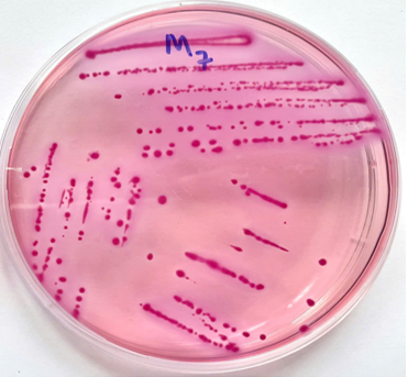
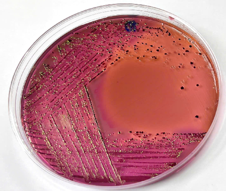
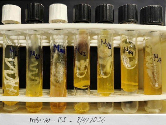
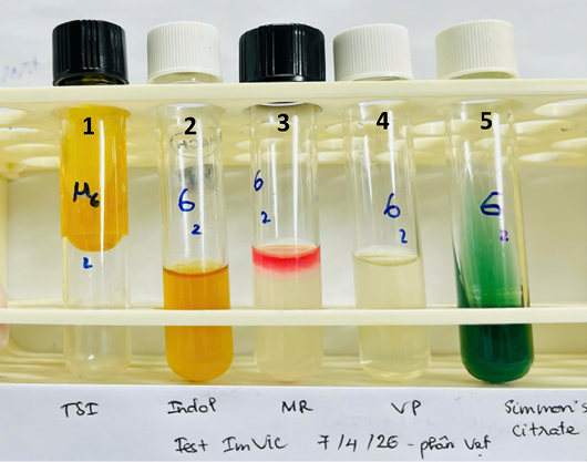
At the laboratory, about one gram of each fecal sample was mixed with buffered peptone water (BPW, Merck, Germany) at a 1:9 ratio. Next, a loopful of the mixed culture was streaked onto MacConkey agar (Merck, Germany) and incubated at 37°C for 24 hours. Then, the pink colonies (Figure 1) were cultured onto eosin methylene blue agar (EMB, Merck, Germany) and continuously incubated at 37°C for 24 hours. Only one colony with the typical metallic green sheen on EMB agar (Figure 2) were streaked into triple sugar iron agar (TSI, Merck, Germany) tube and incubated at 37°C for 24 hours. Colonies exhibiting a typical TSI profile, including glucose and lactose fermentation with gas production and no H2S (Figure 3), were identified as E. coli by Gram staining and biochemical tests, such as indole production, methyl red, Voges-Proskauer, and citrate utilisation reactions, with the expected IMViC result being “++--” (Figure 4). We stored all isolates in brain heart infusion broth with 50% glycerol at -20°C for subsequent experiments.
Confirmation of E. coli
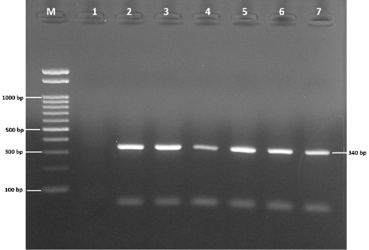
DNA was extracted using the TopPURE® Genomic DNA Extraction Kit (ABT, Vietnam) according to the manufacturer’s instructions. We used specific primers with the following sequences based on the 16S gene for E. coli (5’-GTTAATACCTTTGCTCATTGA-3’ and 5’-ACCAGGGTATCTAATCCTGTT-3’) with an expected PCR product of 340 bp were used (Figure 5). PCR cycling conditions were initial denaturation at 94 ºC for 5 min followed by 35 cycles of denaturation at 94 ºC for 30 sec, annealing at 60 ºC for 30 sec and extension at 72 ºC for 45 sec followed by final extension at 72 ºC for 10 min. The reaction components included 12.5 µl of GoTaq® Green Master Mix (Promega, USA), 1 µl each of the forward and reverse primers (10 µM), 8.5 µl of purified water, and 2 µl of template DNA. The PCR products were electrophoresed on 1.5% agarose gel supplemented with RedSafe™ nucleic acid staining solution (Intron, Korea).
Antimicrobial susceptibility test
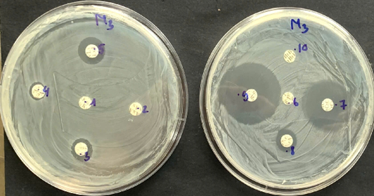
Antibiotic susceptibility test was examined according to the guidelines of the Clinical and Laboratory Standards Institute . Agar diffusion method was performed on Mueller-Hinton agar (MHA, Merck, Germany) following Bauer et al. (1966) and 10 different antibiotic agents (Nam Khoa, Vietnam) belonging to seven classes were used (Figure 6), including penicillins (amoxicillin, 10 µg), carbapenems (meropenem, 10 µg), cephalosporins (cefotaxime, 30 µg), aminoglycosides (kanamycin, 30 µg; neomycin, 30 µg; streptomycin, 10 µg), quinolones (nalidixic acid, 30 µg), fluoroquinolones (levofloxacin, 5 µg; norfloxacin, 10 µg), sulfonamides (trimethoprim/sulfamethoxazole, 1.25/23.75 µg). The inhibition zone diameters of the ten antibiotics for E. coli are shown in Table 1. Escherichia coli ATCC 25922 strain was used for quality control. An isolate was determined to be antibiotic-resistant or multidrug-resistant (MDR) based on the definition of Magiorakos et al. (2012).
Table 1: CLSI breakpoints (inhibition zone diameters) for ten antibiotic tested.
Order
Antibiotic classes
Kind of
antibiotic
Zone inhibition diameters (mm)
Susceptible
Intermediate
Resistance
1
Penicillin
Amoxicillin
≥ 17
14-16
13 ≤
2
Cephalosporin
Cefotaxime
≥ 26
23-25
22 ≤
3
Carbapenem
Meropenem
≥ 23
20-22
19 ≤
4
Aminoglycosides
Kanamycin
≥ 18
14-17
13 ≤
Neomycin
≥ 18
14-17
13 ≤
Streptomycin
≥ 15
12-14
11 ≤
5
Quinolone
Nalidixic acid
≥ 19
14-18
13 ≤
6
Fluoroquinolone
Levofloxacin
≥ 21
17-20
16 ≤
Norfloxacin
≥ 17
13-16
12 ≤
7
Sulfonamide
Trimethoprim/ Sulfamethoxazole
≥ 16
11-15
10 ≤
Data analysis
The isolation and antibiotic resistance rates of E. coli strains were recorded and calculated using Microsoft Excel 2016. The 95% confidence intervals (95% CI) for the proportions were estimated using the Clopper-Pearson binomial distribution (Exact Binomial Test) with R software.
RESULTS
Isolation of bacteria
Overall, the rate of E. coli isolation from fecal samples was 59.6% (Table 2). Of these, the fecal samples from Red-breasted parakeet samples had the highest rate (75.0%), followed by Monk parakeet (70.0%), Macaw parrot (66.7%). Isolation rates for other species ranged from 50.0% to 58.8% in fecal samples from other parrot species such as Lovebirds, Sun parakeet, and African grey parrot.
Antibiotic susceptibility
The isolates showed resistance to each of the 10 antibiotics belonging to 7 different antibiotic classes, but at different rates (Table 3). Resistance was highest to amoxicillin (94.3%), followed by trimethoprim/sulfamethoxazole (50.9%) and streptomycin (47.2%). Resistance to norfloxacin, levofloxacin, and nalidixic acid was 35.8%, 41.5%, and 45.3%, respectively. Cefotaxime, kanamycin and neomycin were resistant at rates ranging from 30.2-34.0%. Notably, three (5.7%) strains were resistant to meropenem, a last-resort antibiotic for serious human infections.
Table 2: Isolation rates of E. coli from fecal samples collected from pet parrots (n = 89).
Order
Species
No. of samples
Positive n (%)
Negative n (%)
95% confidence interval
1
African grey parrot (Psittacus erithacus)
17
10 (58.8)
7 (41.2)
(32.9%, 81.6%)
2
Love-bird (Agapornis fischeri)
10
5 (50.0)
5 (50.0)
(18.7%, 81.3%)
3
Macaw parrot (Ara chloropterus)
12
8 (66.7)
4 (33.3)
(34.9%, 90.1%)
4
Monk parakeet (Myiopsitta monachus)
10
7 (70.0)
3 (30.0)
(34.8%, 93.3%)
5
Red-breasted parakeet (Psittacula alexandri)
12
9 (75.0)
3 (25.0)
(42.8%, 94.5%)
6
Sun parakeet/Sun conure (Aratinga solstitialis)
9
5 (55.6)
4 (44.4)
(21.2%, 86.3%)
7
Other species
19
9 (47.4)
10 (52.6)
(24.4%, 71.1%)
Total
89
53 (59.6)
36 (40.4)
(48.6%, 69.8%)
* Note: Other species include Alexandrine parakeet (Psittacula eupatria, n = 4); Amazon parrot (Amazona ochrocephala, n=5); Budgie (Melopsittacus undulatus, n=6); Cockatiel (Nymphicus hollandicus, n =4).
Table 3: Antibiotic susceptibility of E. coli strains isolated from pet parrot fecal samples (n = 53).
Order
Antibiotic classes
Kind of antibiotic
Resistance n (%)
Intermediate n (%)
Susceptible n (%)
1
Penicillin
Amoxicillin
50 (94.3)
3 (5.7)
0 (0.0)
2
Cephalosporin
Cefotaxime
16 (30.2)
6 (11.3)
31 (58.5)
3
Carbapenem
Meropenem
3 (5.7)
4 (7.5)
46 (86.8)
4
Aminoglycosides
Kanamycin
16 (30.2)
8 (15.1)
29 (54.7)
Neomycin
18 (34.0)
23 (43.4)
12 (22.6)
Streptomycin
25 (47.2)
5 (9.4)
23 (43.4)
5
Quinolone
Nalidixic acid
24 (45.3)
4 (7.5)
25 (47.2)
6
Fluoroquinolone
Levofloxacin
22 (41.5)
4 (7.5)
27 (50.9)
Norfloxacin
19 (35.8)
1 (1.9)
33 (62.3)
7
Sulfonamide
Trimethoprim/ Sulfamethoxazole
27 (50.9)
2 (3.8)
24 (45.3)
Table 4: Antibiotic resistance phenotypes of the isolated E. coli strains (n = 53).
Only one of the 53 isolates was susceptible to all the antibiotics tested (Table 4). The remaining 52 (98.1%) strains were resistant to at least one antibiotic and exhibited 31 different resistance phenotypes. The most common pattern was resistance to amoxicillin alone (10 strains, 18.9%), followed by AMX-CTX-KAN-NEO-STM-NAL-LEV-NOR-SXT resistance was found in 5 (9.43%) strains isolated from African grey parrots and Red-breasted parrots. The resistance rates for 1 and 2 antibiotic classes were 15.1% and 20.8%, respectively (Table 5). Meanwhile, the resistance rates for 3-6 antibiotic classes ranged from 13.2% to 17.0%. Only one strain (1.9%) was resistant to seven classes. Of these, 33 isolates (62.3%) were resistant to three or more antibiotic classes, meeting the definition of multidrug-resistant (MDR).
Table 5: Resistance to different antibiotic classes of isolated E. coli strains (n= 53).
*Note: “No resistance” means susceptible to all 10 antibiotics tested. The number of strains resistant to three or more antibiotic classes is 33 (62.3%).
DISCUSSION
Previous studies conducted in Bangladesh and Turkey reported isolation rates of 48.7% in 150 and 52.3% in 172 parrots and parakeets samples, respectively. However, lower isolation rates, ranging from 18.3% to 36.0%, were observed in studies conducted in Bangladesh , Italy , Brazil , and Egypt . These differences could be due to geography, sampling methods, or lab techniques. Additionally, sanitary conditions in the cages, along with the hot and humid climate in northern Vietnam, may be related to the high rate of E. coli isolates in fecal samples from parrot species.
We found that 52 of 53 isolates (98.1%) were resistant to at least one antibiotic, and the most common resistance was identified as amoxicillin (94.3%). High amoxicillin resistance (81-100%) has also been seen in other studies in Brazil , Italy and Bangladesh . Cefotaxime is a critically important antibiotic for human medicine (WHO, 2017), but resistance was observed in 30.2% of the E. coli strains in present study. One study in Egypt found even higher resistance (66.7%) in parrots with respiratory illness . Meropenem is a last-resort antibiotic for serious human infections (WHO, 2017). Similar studies conducted in Brazil , Egypt , and Bangladesh all showed that all isolated E. coli strains were susceptible to this antibiotic. Although only three strains (5.7%) were resistant to meropenem in this study, this is concerning because meropenem is not approved for use in animals.
Trimethoprim/sulfamethoxazole is an oral broad-spectrum antibiotic often used in birds, and it is particularly proper used for therapy of birds . Resistance to trimethoprim/sulfamethoxazole were 44.4% and 46.0% in studies conducted in Egypt and Turkey . However, this antibiotic resistance rate reached 85.0% and 100% in studies from Bangladesh and Italy . Notably, parrot species-derived E. coli strains exhibited trimethoprim/sulfamethoxazole resistance at rates ranging from 12.1-33.0% in a study conducted in Brazil ; ; . The rates of resistance to aminoglycosides such as kanamycin, neomycin, and streptomycin were 30.2%, 34.0%, and 47.2%, respectively. Aminoglycosides are important therapeutic agents in clinical practice, often used to treat infections, especially severe infections caused by Gram-negative bacteria, but they are used less often in birds because they can be toxic . However, some previous studies have documented aminoglycoside resistance in E. coli strains isolated from parrots, such as resistance rates of 25.0% and 34.0% for kanamycin and streptomycin in Turkey ; and resistance rates of 29.8% and 42.6% for kanamycin and neomycin in Bangladesh . In Brazil, E. coli strains isolated from parrots showed streptomycin resistance ranging from 2.0% to 37.2% and 74.0% ; ; . Different lab methods and study populations may explain the varying resistance rates.
The rates of resistance to quinolone and fluoroquinolone classes such as norfloxacin, levofloxacin, and nalidixic acid ranged from 35.8% to 45.3%. Lower resistance rates for these antibiotics, from 13.0% to 17.0%, were observed in E. coli strains isolated from parrots in Turkey . In Brazil, parrot-derived E. coli strains showed resistance to nalidixic acid at levels of 6.1% - 25.6% - 30.0% ; ; . Levofloxacin resistance of E. coli strains in a similar study conducted in Bangladesh was 19.1% . Notably, parrot-derived E. coli strains with respiratory tract infections in Egypt showed resistance to norfloxacin at a high level of 51.9% . Differences in antibiotic resistance rates and phenotypes may be related to differences in antibiotic use practices, veterinary surveillance, and differences in hygiene and biosecurity measures between countries.
We found 31 different resistance patterns among the isolates, of which 33 (62.3%) were identified as multidrug-resistant. This variety may come from sampling different parrot species kept under different conditions of care, disease prevention, and treatment. Household pets are recognised as significant risks for zoonotic diseases and considered as a source of multidrug-resistant (MDR) bacteria ; , which can pose serious risks. The rates of MDR E. coli were consistent with the 59.0% and 67.0% observed in E. coli strains isolated from parrots in Brazil and Turkey . However, in previous studies conducted in Italy and Brazil , the rates of multidrug-resistant strains were identified as 40.1% and 43.2%, respectively. Notably, MDR rates were reported as high as 100% in studies in Bangladesh . The rise of MDR bacteria calls for tighter control of antibiotic use to minimize the spread of MDR pathogens in livestock, including pet birds, and avoid significant risks to animal and community health ; .
Limitations
This study was only conducted with a small sample size on healthy parrots raised in Hanoi, and specific drug resistance genes such as ESBL, carbapenemase (blaNDM, blaKPC, blaOXA-48, blaIMP, blaVIM…) have yet to be identified. However, this study has provided basic data on the infection rates and antibiotic resistance of E. coli isolates from pet birds, including many introduced species. Therefore, larger-scale studies on more bird species are needed, as well as the use of molecular methods to detect antibiotic resistance genes, in order to determine the relationship between drug resistance genes, including plasmids, and multidrug-resistant phenotypes. In fact, finding out exactly how these bacteria become resistant would help us understand their role in the One Health context.
CONCLUSION
Overall, the E. coli strains we isolated were resistant to many antibiotics, and many were MDR. This suggests that pet birds could be reservoirs of antibiotic-resistant bacteria, which is a concern for both human and animal health. Although we did not study transmission to humans, the presence of MDR E. coli in pet birds is a potential public health concern that warrants further investigation. Future studies should examine ESBL and carbapenemase genes using PCR, and birds should be sampled at different times to see if resistance changes over time.
ACKNOWLEDGEMENTS
The authors would like to thank the students for transporting samples and the pet owners for their cooperation and assistance in the sampling process.
NOVELTY STATEMENT
This preliminary study documented the presence of antibiotic-resistant E. coli strains in pet parrots in Hanoi, Vietnam. The study results suggest that pet parrots may be carriers of multidrug-resistant E. coli and contribute to the spread of antibiotic resistance in the environment. This underscores the need for monitoring and controlling antibiotic use in both animal husbandry and veterinary medicine to mitigate this problem in Vietnam.
AUTHOR’S CONTRIBUTION
CTTH: Study design, investigation and methodology, and manuscript writing. HTH, NTT, NMH, LBN, NTTM: Sample collection, experimentation and data analysis. TLO: Laboratory research design, sample testing supervision. THT: Conceptualization, supervision, writing, review and editing, project management.
Funding information
The present study received no external funding. The authors contributed to this study independently.
Ethical approval
The present study was conducted by collecting samples in accordance with the guidelines outlined in the animal welfare and safety procedures of the Committee on Animal Research and Ethics (CARE), Faculty of Veterinary Medicine, Vietnam National University of Agriculture, Vietnam (Approval No. CARE-2025/08).
Generative AI and AI assisted technology statement
AI-generated tools were not used to create any scientific content. Any AI assistance was limited to minor language editing, and all ideas, interpretations, and conclusions remain entirely the authors.
Statement of conflict of interest
The authors have declared no conflict of interest.
Figures
Figure 1:.
E. coli colonies isolated from parrot feces exhibited pink colour on MacConkey agar.
Figure 2:.
E. coli colonies isolated from parrot feces exhibited green metallic sheen on EMB agar.
Figure 3:.
E. coli strains isolated from parrot on TSI agar. The yellow butts and yellow slants, accompanied by gas bubbles due to the production of acid and CO2 during the fermentation of sugars such as glucose and lactose/sucrose.
Figure 4:.
The IMViC tests for E. coli confirmation (from left). Tube 1: Sugar fermentation and gas production on TSI agar by E. coli; Tube 2: Indole (+); Tube 3: Methyl red (+); Tube 4: Voges-Proskauer (-); Tube 5: Simmons citrate (-).
Figure 5:.
Representative electrophoresis of the 16S gene for E. coli isolates (M: marker, 100 bp; lane 1: negative control; lane 2: positive control; lane: 3, 4, 5, 6, 7 positive 16S genes).
Figure 6:.
Antibiotic susceptibility testing of 10 antibiotics against E. coli strains isolated from parrot feces. (1): Kanamycin; (2): Amoxicillin; (3): Streptomycin; (4): Neomycin; (5): Levofloxacin; (6): Nalidixic acid; (7): Cefotaxime; (8): Norfloxacin; (9): Meropenem; (10): Trimethoprim/Sulfamethoxazole
References
Argudín MA, Deplano A, Meghraoui A, Dodémont M, Heinrichs A, Denis O, Nonhoff C, Roisin S (2017). Bacteria from animals as a pool of antimicrobial resistance genes. Antibiotics (Basel), 6(2): 12. https://doi.org/10.3390/antibiotics6020012[CrossRef]
Bauer AW, Kirby WM, Sherris JC, Turck M (1966). Antibiotic susceptibility testing by a standardized single disk method. Am. J. Clin. Pathol., 45(4): 493-496. Available at: https://academic.oup.com/ajcp/article-abstract/45/4_ts/493/4821085?redirectedFrom=fulltext. https://doi.org/10.1093/ajcp/45.4_ts.493 https://doi.org/10.1093/ajcp/45.4_ts.493[CrossRef]
Boroomand Z and Faryabi S (2020). Bird Zoonotic Diseases., Journal of Zoonotic Diseases., 4(3):20-33.
Boseret G, Losson B, Mainil JG, Thiry E, Saegerman C (2013). Zoonoses in pet birds: Review and perspectives. Vet. Res., 44(1): 36. https://doi.org/10.1186/1297-9716-44-36[CrossRef]
CLSI (2020). Performance standards for antimicrobial susceptibility testing, 30th Edition. CLSI supplement M100. Wayne, PA, USA: Clinical and Laboratory Standards Institute; pp. 32-41. Available at: https://www.nih.org.pk/wp-content/uploads/2021/02/CLSI-2020.pdf
Damborg P, Broens EM, Chomel BB, Guenther S, Pasmans F, Wagenaar JA, Weese JS, Wieler LH, Windahl U, Vanrompay D, Guardabassi L (2016). Bacterial zoonoses transmitted by household pets: State of the art and future perspectives for targeted research and policy actions. J. Comp. Pathol., 155(Suppl 1): S27-40. https://doi.org/10.1016/j.jcpa.2015.03.004[CrossRef]
Day MJ (2016). Pet-Related Infections. Am. Fam. Phys., 94(10): 794-802; Available via https://www.aafp.org/pubs/afp/issues/2016/1115/p794.html
Diren Sigirci B, Baran C, Barış H, Mehmet A, Ilgın K, Kemal M, Serkan I, Funda B, Naciye O, Seyyal A, Beren BK (2020). Antimicrobial resistance profiles of Escherichia coli isolated from companion birds. J. King Saud Univ. Sci., 32: 1069-1073. https://doi.org/10.1016/j.jksus.2019.09.014[CrossRef]
Diren Sigirci B, Celik B, Kahraman BB, Bagcigil AF, Ak S (2019). Tetracycline resistance of Enterobacteriaceae isolated from feces of synanthropic birds. J. Exot. Pet Med., 28: 13–18. https://doi.org/10.1053/j.jepm.2017.12.003[CrossRef]
Flammer, K., 2006. Antibiotic drug selection in companion birds. J. Exot. Pet Med., 15(3): 166–176. https://doi.org/10.1053/j.jepm.2006.06.003[CrossRef]
Giacopello C, Foti M, Fisichella V, Piccolo FL (2015). Antibiotic-resistance patterns of Gram-negative bacterial isolates from breeder canaries (Serinus canaria domestica) with clinical disease. J. Exot. Pet Med., 24 (1): 84–91. https://doi.org/10.1053/j.jepm.2014.12.009[CrossRef]
Hasib FMY, Bayzid M, Das T, Rana EA, Kamal T, Hossain MA, Alim MA (2025). Multidrug-resistant extended-spectrum beta-lactamase (ESBL) producing Escherichia coli in pet birds of Bangladesh. Vet. Med. Sci., 11(5):e70509. http:///doi:10.1002/vms3.70509[CrossRef]
Hosseinian SA (2022). Zoonotic diseases associated with pet birds. J. Zoonotic. Dis., 6(3): 91-112.
Karpman D, Ståhl AL (2014). Enterohemorrhagic Escherichia coli pathogenesis and the host response. Microbiol. Spectr. 2(5). https://doi.org/10.1128/microbiolspec.EHEC-0009-2013[CrossRef]
Lopes ES, Maciel WC, Albuquerque AH, Machado DN, Amorim Bezerra WG, Vasconcelos RH, Castro Teixeira RS (2015). Prevalence and antimicrobial resistance profile of enterobacteria isolated from psittaciformes of illegal wildlife trade. Acta Sci. Vet., 43 (1): 1–9; Available via https://www.cabidigitallibrary.org/doi/pdf/10.5555/20163045093
Machado DN, Lopes ES, Albuquerque AH, Horn RV, Bezerra WGA, Siqueira RAS, Cardoso WM (2018). Isolation and antimicrobial resistance profiles of Enterobacteria from nestling grey-breasted parakeets (Pyrrhura griseipectus). Braz. J. Poult. Sci., 20(1): 103–110. https://doi.org/10.1590/1806-9061-2017-0551[CrossRef]
Magiorakos AP, Srinivasan A, Carey RB, Carmeli Y, Falagas ME, Giske CG, Harbarth S, Hindler JF, Kahlmeter G, OlssonLiljequist B, Paterson DL, Rice LB, Stelling J, Struelens MJ, Vatopoulos A, Weber JT, Monnet DL (2012). Multidrugresistant, extensively drug-resistant and pandrug-resistant bacteria: An international expert proposal for interim standard definitions for acquired resistance. Clin. Microbiol. Infec., 18(3): 268-281. https://doi.org/10.1111/j.1469-0691.2011.03570.x[CrossRef]
Malinen E, Kassinen A, Rinttilä T, Palva A (2003). Comparison of real-time PCR with SYB Green I or 5’-nuclease assays and dot-blot hybridization with rDNA-targeted oligonucleotide probes in quantification of selected faecal bacteria. Microbiology (Reading), 149(Pt1): 269-277. https://doi.org/10.1099/mic.0.25975-0[CrossRef]
Marques AR, Pascoal-Filho NM, Teixeira RSC, Silva ING, Melo LS, Lima BP, Campos EMSQ, Maciel WC (2024). Investigation of enterobacteria with zoonotic and multi-resistant potential in exotic parrots kept in a domestic environment. Pesq. Vet. Bras., 44: e07387. https://doi.org/10.1590/1678-5150-pvb-7387[CrossRef]
Ministry of Agriculture and Rural Development (MARD), (2011). National technical regulation on animal diseases - General requirements for sample collection, storage and shipment. QCVN 01-83:2011/BNNPTNT, pp: 5–10. Promulgated under Circular No. 71/2011/TT-BNNPTNT of MARD. Available via https://sgtvt.bacninh.gov.vn/documents/154127/14279228/QCVN+01-83-2011-BNNPTNT.pdf/2a692d85-15d0-445c-969e-8964f5dadcad
Monteiro HIG, Silva V, de Sousa T, Calouro R, Saraiva S, Igrejas G, Poeta P (2025). Antimicrobial resistance in European companion animals practice: A one health approach. Animals (Basel), 5(12): 1708. https://doi.org/10.3390/ani15121708[CrossRef]
Nupur MN, Afroz F, Hossain MK, Harun-ur-Rashid SM, Rahman MG, Kamruzzaman M, Ferdous KA, Haque MA (2023). Prevalence of potential zoonotic bacterial pathogens isolated from household pet birds and their antimicrobial profile in northern Bangladesh. Agrobiol. Rec., 11: 28-38. https://doi.org/10.47278/journal.abr/2023.005[CrossRef]
Ohene Larbi R, Ofori LA, Sylverken AA, Ayim-Akonor M, Obiri-Danso K (2021). Antimicrobial Resistance of Escherichia coli from broilers, pigs, and cattle in the greater Kumasi Metropolis, Ghana. Int. J. Microbiol., 2021: 5158185. https://doi.org/10.1155/2021/5158185[CrossRef]
Pakbin B, Brück WM, Rossen JWA (2021). Virulence factors of enteric pathogenic Escherichia coli: A review. Int. J. Mol. Sci., 22(18): 9922. https://doi.org/10.3390/ijms22189922[CrossRef]
Pomba C, Rantala M, Greko C, Baptiste KE, Catry B, van Duijkeren E, Mateus A, Moreno MA, Pyörälä S, Ružauskas M, Sanders P, Teale C, Threlfall EJ, Kunsagi Z, Torren-Edo J, Jukes H, Törneke K (2017). Public health risk of antimicrobial resistance transfer from companion animals. J. Antimicrob. Chemother., 72(4): 957-968. https://doi.org/10.1093/jac/dkw481[CrossRef]
Pontes PS, Coutinho SDA, Iovine RO, Cunha MPV, Knöbl T, Carvalho VM (2018). Survey on pathogenic Escherichia coli and Salmonella spp. in captive cockatiels (Nymphicus hollandicus). Braz. J. Microbiol., 49 (Suppl 1): 76-82. https://doi.org/10.1016/j.bjm.2018.05.003[CrossRef]
Prestinaci F, Pezzotti P, Pantosti A (2015). Antimicrobial resistance: A global multifaceted phenomenon. Pathog. Glob. Health., 109: 309-318. https://doi.org/10.1179/2047773215Y.0000000030[CrossRef]
Samir A, Mosallam T, Aboul-Ella H, Ali A, Samir O, Hegab M, Erian M, Youssef F, Zaher H (2025). Zoonotic relevance of multidrug-resistant bacteria in parrots with respiratory illness. Vet. Res. Commun., 49(4): 194. https://doi.org/10.1007/s11259-025-10752-6[CrossRef]
Szmolka A, Nagy B (2013). Multidrug resistant commensal Escherichia coli in animals and its impact for public health. Front. Microbiol., 4: 258. https://doi.org/10.3389/fmicb.2013.00258[CrossRef]
Varriale L, Dipineto L, Russo TP, Borrelli L, Romano V, D’Orazio S, Pace A, Menna LF, Fioretti A, Santaniello A (2020). Antimicrobial resistance of Escherichia coli and Pseudomonas aeruginosa from companion birds. Antibiotics (Basel), 9(11): 780. https://doi.org/10.3390/antibiotics9110780[CrossRef]
WHO (2017). Critically important antimicrobials for human medicine. Geneva, Switzerland: WHO. Available via http://apps.who.int/iris/bitstream/10665/255027/1/9789241512220-eng.pdf?ua=1 (Accessed 24 August 2020).