Research Article · Volume 11, Issue 6, 23 May 2023 · Open Access
Histomorphometrical and Histochemical Study of Caecum in Adult Muscovy Ducks (Cairina moschata)
·
Affiliations & Notes ⌄
1Department of Anatomy, College of Veterinary Medicine, University of Mosul, Mosul, Iraq. *Correspondence | Omar Younis Altaey, Department of Anatomy, College of Veterinary Medicine, University of Mosul, Mosul, Iraq
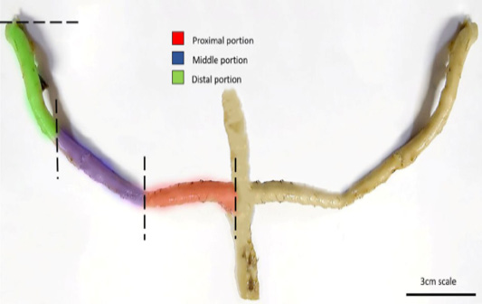
Gross morphological characteristics of the caecum indicated that it was demarcated into three distinct portions according to the thickness of wall, namely the proximal, middle, and distal portions, each of which exhibits significant differences in length.
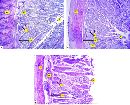
Villi were found to be significantly longer in the proximal portion, and the depth of caecal crypts was higher in this region compared to the middle and distal (Figure 3, Table 3).
The lymphatic nodules were observed to have a greater diameter in the proximal portion compared to the middle and distal portions, with a higher number of nodules in the distal region compared to the rest of the caecum (Table 3).
The villi of the cecal middle portion showed intense staining activity, suggesting a greater amount of acidic mucin compared to the proximal and distal portions.
Selected from the article's own text.
Abstract
Muscovy ducks are becoming an increasingly important source of food worldwide. Caecal microbial digestion is crucial for the growth and maintenance of the health in these birds. The main objective of this study is to understand the anatomy, histology, histochemistry, and distribution of lymphoid tissue in the caeca of Muscovy ducks. Samples from eight healthy adult Muscovy ducks (Cairina moschata) were fixed, processed, sectioned, and stained with Hematoxylin and Eosin as well as Periodic Acid Schiff: Alcian blue pH 2.5 and toluidine blue stains for histochemical analysis. Dissecting microscope measurements and histological measures were taken, and immunohistochemical analysis was performed to evaluate B lymphocyte distribution. The study found that in Muscovy ducks, the left caecum was longer and heavier than the right caecum, and the proximal portion of both caeca was wider and had thicker walls compared to the middle and distal portions. Histologically, the length of villi and depth of crypts were greater in the proximal portion compared to the middle and distal portions. Histochemical analysis showed that acidic glycoprotein was dominant in the villi and crypts, with higher density in the middle portion compared to the proximal and distal portions. The density of glycosaminoglycans was also higher in goblet cells at the basal crypts compared to longitudinal crypts. Immunohistochemical analysis revealed higher levels of lymph follicles and B lymphocytes in the proximal portion compared to the middle and distal portions. The complex architecture suggests that caeca in Muscovy duck play a significant role in microbial digestion and immunological functionality and Insight into caecal morphology enhance bird’s productivity and health.
The poultry industry is the foundation of global food security as it presents a dependable source of meat and eggs to support the nutritional demands, besides the short productivity duration, and the effortless management which recently increase interest in the breeding of birds like ducks and geese in many Iraqi villages . Over the last two decades, duck meat production has experienced significant growth worldwide, rising from 2.6 million tons in 1998 to 4.5 million tons in 2018 according to the statistical database of food and agriculture organization of the united nations The Muscovy duck (Cairina moschata) holds a distinct place in terms of domestication, this species was one of the earliest animals to be domesticated in South America and played a crucial role as a source of food and trade . it was used in poultry farming as a result of its considerable size and desirable qualities like leanness, tenderness, and taste .
The digestive system is essential for the growth and health of birds. It is responsible for breaking down food into smaller absorbable molecules that can be easily utilized by the body to provide energy and maintain overall health .
The large intestine of birds is composed of a pair of caeca and a short, smooth rectum, each caecum is comprised of three portions: proximal, middle, and distal, the caecal wall is primarily composed of lymphoid tissues located at its base, referred to as the caecal tonsils. Studies on ducks have revealed that ducks possess two highly developed caeca that are shaped like long, blind sacs. These caeca are positioned in the dorsocoleomic cavity and originated from the lateral wall of the rectum close to the junction with the ilium ; .
Research has revealed that these structures is a key role in various processes, such as the absorption of water, digestion, and absorption of cholesterol, degradation of nitrogenous compounds, and microbial degradation of some carbohydrates, it is also believed that the ceca are an essential in protecting the body against infections .
The caecal microbiota of Muscovy ducks is an important factor in the regulation of metabolism, and fat deposition. The authors emphasized the significant influence of the caecal microbiota on the physiological processes of growth .
many scientific papers have documented the morphology and histology of the digestive system in certain bird species ; ; ; . Despite efforts, limited data is available on the structure of the Muscovy duck’s caeca and the histochemistry and distribution architecture of lymphoid tissue within this structure. accordingly, the current study aims to expand the existing knowledge regarding the morphology, histology, histochemistry of the caecal carbohydrate and the distribution of lymphoid tissue, using immunohistochemical techniques against anti-Mouse clone CD40 ligand (CD40L) in the Muscovy duck’s caeca.
Materials and Methods
Animals preparation
Eight healthy adult Muscovy ducks (Cairina moschata) of either sex were used in this study weighing 3.5 to 4 kg obtained from certified birds’ markets. The ducks were raised under ad libitum feeding and watering conditions, the study was achieved at the Department of Anatomy in the College of Veterinary Medicine University of Mosul, Mosul, Iraq. Samples were collected carefully from the coelomic cavity, washed with physiological saline to remove debris and blood then kept in plastic container filled with Davidsons’ fixative prepared according to for 12 hours later transferred to buffered formalin 10%.
Morphometrical analysis
the total length, and weight were measured using the digital Vernier (LOUISWARE, China) and weighing balance (EK-EW, Australia) further measurements performed using the dissecting microscope (Huma scope stereo 14900/5, Germany) included the diameter of caecum, lumen, and thickness of wall.
Histological and histochemical analysis
Samples underwent a series of processing steps for histological and histochemical evaluation. First, they were washed with distilled water, trimmed, and dehydrated in ascending grades of ethyl alcohol concentration (70%, 80%, 90%, 100%), then cleared with xylene solution (2 changes) for 30 minutes, followed by infiltration with melted paraffin at 58ºC, and cast into blocks. Thin sections (6 micrometers) were cut from the blocks with a rotary microtome (OS-315, UK), then mounted on glass slides with DPX media, and stained with Harris hematoxylin and eosin according to protocol, further slides were stained with Periodic acid-Schiff and Alcian blue PAS/AB pH 2.5 stain as well as toluidine blue stains (Thermo Fisher Scientific, USA) for detection of glycoproteins and glycosaminoglycans in tissue sections. It highlights the neutral glycoproteins (mucins) with a red or magenta color and acidic glycoproteins with a blue color. While toluidine blue stain used for demonstration of glycosaminoglycans by pink reaction metachromasia . The stained sections were examined with a light microscope (Olympus-Dm-CBAD, Japan) for histological and histomorphometric assessments using an 18.0 MP OMAX digital camera equipped with image processing software. 20 histological sections were randomly selected and the parameters were measured in 10 microscopic fields included the length of caecal villi, depth of caecal crypts, diameter of lymphatic nodules and thickness of the muscular layer.
Immunohistochemistry analysis
After the deparaffination and rehydration of the sections and performing antigen retrieval by heating EDTA buffer. The primary antibody against CD40 ligand (CD40L) anti-Mouse clone was applied and incubated overnight, followed by incubation with a secondary antibody conjugated to horseradish peroxidase HRP. The reaction was visualized using DABi (diaminobenzidine) substrate and counterstained with Gill’s hematoxylin, and the sections were then evaluated under a microscope to observe CD40-positive B lymphocytes .
Statistical analysis
The gross and histological data obtained from the caeca were analyzed to quantify the mean and standard error of the measurements. The statistical analysis was performed using IBM SPSS V.25 software, and included a one-way analysis of variance (ANOVA) followed by a Duncan post-hoc test . The purpose of this analysis was to evaluate the significant differences among the different caecal portions, with a significance level set at P ≤ 0.05.
Results and Discussion
Upon detailed macroscopic examination, it was ascertained that the caeca of the Muscovy duck constitute elongated, tubular, and blind structures that emerge bilaterally from the rectum at the ileorectal junction. The examination further revealed that the left caecum measured 14.51 cm in length and weighed 0.53 g, while the right caecum length was 13.66 cm and weighed 0.49 g. Gross morphological characteristics of the caecum indicated that it was demarcated into three distinct portions according to the thickness of wall, namely the proximal, middle, and distal portions, each of which exhibits significant differences in length. particularly, the middle portion was found to be the longest in both the right and left caecum (Figure 1, Table 1).
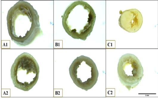
The caecal wall was slightly thicker and the diameter was greater at the proximal portion compared to those of the middle and distal portions. However, there were no significant differences in the luminal length among the three portions (Figure 2, Table 2).
Histological analysis revealed that the caecal wall is composed of four distinctive layers, namely, mucosa, submucosa, muscularis, and serosa. The mucosal layer was characterized by well-developed villi, separated by longitudinal crypts, which extended along the thickness of the mucosal layer. Moreover, the basal portion of the mucosal layer was observed to contain small circular crypts and disseminated lymphatic tissue. Significant differences were observed in the length of villi and depth of caecal crypts across the proximal, middle, and distal regions of the caecum. Villi were found to be significantly longer in the proximal portion, and the depth of caecal crypts was higher in this region compared to the middle and distal (Figure 3, Table 3).
Table 1: The length measurements of the Muscovy duck caecum.
Direction
Total length
M±SEM
Proximal portion
M±SEM (%)
Middle portion
M±SEM (%)
Distal portion
M±SEM (%)
Left caecum (cm)
14.51±0.09
3.48±0.04(24)a
6.24±0.08(43)b
4.79±0.06(33)c
Right caecum (cm)
13.66±0.07
3.28±0.05(23)a
5.87±0.09(43)b
4.51±0.07(34)c
M: mean, SEM: standard error of mean, %: percentages. Different letters among columns indicate statistical significant differences at P≤0.05.
Table 2: The gross measurements of the Muscovy duck caecum.
Variables
Proximal portion
M±SEM
Middle portion
M±SEM
Distal portion
M±SEM
Diameter of caecum (mm)
5.12±0.37 a
4.07±0.02 b
3.78±0.21 a
Lumen (mm)
2.34±0.71 a
3.19±0.12 a
2.82±0.17 a
Wall thickness (mm)
0.91±0.01 a
0.86±0.01 ab
0.48±0.12 b
M: mean, SEM: standard error of mean, %: percentages. Different letters among columns indicate statistical significant differences at P≤0.05.
Table 3: The histological measurements of the Muscovy duck caecum.
Variables
Proximal portion
M±SEM
Middle portion
M±SEM
Distal portion
M±SEM
Length of villus (µm)
766.65±3.05 ab
612.21±4.03 a
537.86±2.53 b
Depth of crypts (µm)
513.40±3.53 a
227.40±4.42 b
454.71±1.74 b
Diameter of lymph follicles (µm)
79.78±0.19 a
69.27±1.44 b
67.91±1.32 b
Thickness of muscular layer (µm)
127.07±3.40 a
87.48±0.75 b
45.23±1.37 c
M: mean, SEM: standard error of mean, %: percentages. Different letters among columns indicate statistical significant differences at P≤0.05.
The lamina propria of the caecal wall was predominantly composed of bundles of collagen fibers and fibrocytes, with diffused lymphatic tissue and lymphatic nodules. The lymphatic nodules were observed to have a greater diameter in the proximal portion compared to the middle and distal portions, with a higher number of nodules in the distal region compared to the rest of the caecum (Table 3).
The thickness of the muscular layer varied across the different regions of the caecum, the proximal and middle portions being relatively thicker than the distal portion. Additionally, the width of the caecal lumen was found to be wider in the middle region compared to the proximal and distal parts (Table 3).
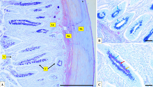
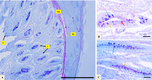
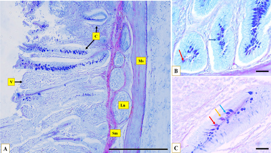
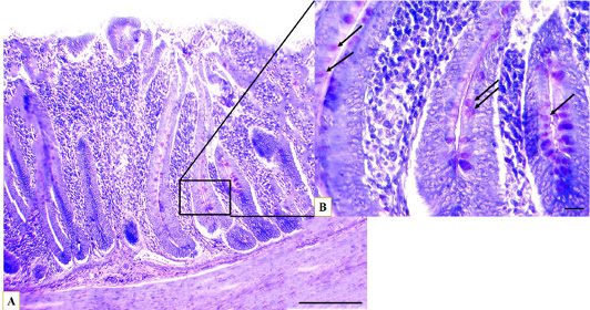
The carbohydrate histochemical analysis of the Muscovy duck’s caeca revealed the presence of a significant amount of glycoprotein (mucin), specifically. Both acidic and natural types of glycoprotein were found distributed throughout most of the caecal tissue. The Alcian blue staining indicated that the dominant type of glycoprotein was acidic. The villi of the cecal middle portion showed intense staining activity, suggesting a greater amount of acidic mucin compared to the proximal and distal portions. The goblet cells in the cecal crypts were also strongly stained with Alcian blue, particularly in the proximal and distal portions in comparison to the middle portion. The amount of acidic glycoprotein was less in the middle portion. On the other hand, the neutral glycoprotein content was much lower than the acidic type, with only weak positive staining activity observed in the villi of the middle cecal portion using the periodic acid-Schiff reagent. Negative staining activity was observed at the proximal and distal portions (Figures 4, 5, 6) (Table 4).
Table 4: The histochemical reaction of the Muscovy duck caecum to PAS, AB and, T.B stains.
Stain
Variables
Proximal portion
Middle portion
Distal portion
PAS
villi
-
+
-
crypts
+
+
+
AB 2.5
villi
++
+++
++
crypts
+++
++
+++
T. B
Longitudinal crypts
+++
++
+
Basal crypts
+
++
+
+++: very strong; ++: strong; +: moderate; -: no reaction.
The histochemical analysis also revealed positive toluidine blue staining activity in the goblet cells in the longitudinal and basal cecal crypts. However, no positive staining was observed in the villi of most caecal regions. The positive findings represented the staining reaction of glycosaminoglycans (GAGs).
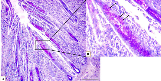
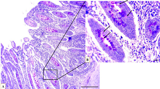
The quantity and distribution of GAGs varied among the different regions of the caeca and between the longitudinal and basal crypts. At the proximal part of the caecum, the basal crypts exhibited poor positive staining activity due to lower quantities of GAGs. In contrast, the longitudinal crypts showed intense positive staining activity. Additionally, the middle part of the caecum showed moderate positive intensity to toluidine blue in both caecal crypts, indicating an average amount of GAGs. While in the distal part of the caecum, both the longitudinal and basal crypts displayed poor positive staining activity (Figures 7, 8, 9)(Table 4).
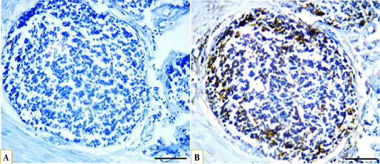
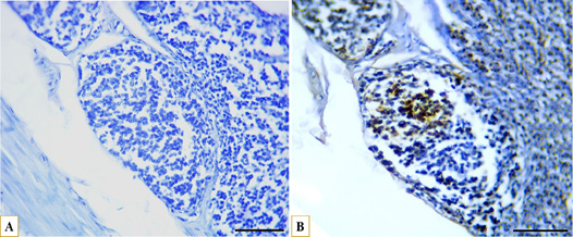
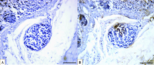
The results of the immunohistochemical analysis revealed the pattern of distribution and concentration of B lymphocytes in lymphatic nodules across different segments of the caecum. Additionally, positive expression of CD40 markers was observed throughout the lymphatic tissue of the mucosal lamina propria and submucosa layers.
In the proximal segment of the caecum, there were fewer lymphatic nodules that were large in size and had a higher density of B lymphocytes, primarily distributed at the edge and center of the nodules. Additionally, the middle segment had a greater number of lymphatic nodules, which were large also in size and had a moderate density of B lymphocytes. In the distal segment, the number of lymphatic nodules increased and the size of the nodules decreased progressively. The density of B lymphocytes was lower, distributed at the edge of lymphatic nodules. Notably, positive marker reaction was observed in the diffused lymphoid tissue, specifically around the basal crypts, in the middle and distal segments of the caecum (Figures 10, 11, 12).
Birds exhibit inter-species variations in caecal anatomy, including the variations in shape, length, thickness of the caecal wall, and symmetry. In addition, differences in histological layers, villi morphology, and lymphatic tissue distribution and organization .
This study found that the caeca of Muscovy ducks is composed of two elongated, blind asymmetrical structures, with the left caecum being longer and heavier than the right and each caecum composed of three portions (proximal, middle, and distal). These findings are consistent with those reported by in long-tailed ducks, where the ceca described as elongated and the left caecum being longer and heavier than the right one. Furthermore, several studies like ; ; conducted in chickens and turkeys have also described the ceca as a pair of blind structures located in the left coelomic cavity, consisting of three distinct parts proximal, middle, and distal. In chickens, the ceca exhibit nearly equal lengths on both sides.
The data from ; ; studies revealed that the length of chicken’s left caecum was 13.14 cm while that of the right caecum was 13.15 cm. In contrast, the left caecum of the long-tailed duck was 9.07 cm and the right caecum was 7.84 cm. while, in turkeys, the left and right caecum length was 24.25 cm and 23.75 cm, respectively. Notably, these measurements were close to those found in the ceca of Muscovy ducks.
The caeca in pigeon according to study was very small, rudimentary bud like and was less than 0.36 cm, while in goose was very long fill the ventral coelomic cavity of bird’s body these result differ from caecal structure in Muscovy duck. The variations observed in the anatomy of the caeca among different species are attributed to differences in their dietary habits (food nature), flying capability and the variations in the size of the digestive systems.
Histologically, the caecal wall in Muscovy ducks had four layers: mucosa, submucosa, muscularis, and serosa. The mucosal layer contained numerous villi and crypts that varied across different regions, with longer villi and higher crypt depth in the proximal portion. Moreover, the lamina propria was mostly composed of fibrous connective tissue, with larger and more numerous lymph nodules in the proximal and middle regions. Additionally, the thickness of the muscular layer varied and was thicker in the proximal and middle portions. Various authors have documented comparable observations in ducks, chickens, turkeys, One such observation by (Abd el-wahab et al., 2017) noted that the mucosa layer of ducks consists of multiple villi and crypts which were long and lumen nearly obstructed in proximal portion of caecum became blunted in middle portion and shorter in distal portion and the depth of crypts were high in proximal portion and numerous in distal portion compared to the proximal and middle portions. while, found that chickens exhibit longer well-developed villi in the proximal portion, with deep basal and longitudinal crypts than in the middle and distal portions. Similarly, turkeys according to displayed caecal histological layers characterized by a narrow lumen and thick muscular layer in the proximal part, compared to a wider lumen and thinner muscular wall in the middle and distal portions. Notably, the observations in this study differ from those reported by in quails, who noted that the villi in the proximal part are very short and absent in some regions of the distal part and the thickness of the caecal wall thicker in proximal part compared to the middle and distal one.
The histochemical findings of the Muscovy duck caeca were found to contain a high amount of glycoprotein, mainly in the form of acidic glycoprotein. The neutral glycoprotein content was significantly lower than the acidic type. The Alcian blue staining technique revealed that the villi of the caecal middle portion contained a greater amount of acidic glycoprotein than the proximal and distal portions. Glycosaminoglycans were positively stained in the goblet cells of the longitudinal and basal caecal crypts and absent in the villi. The quantity and distribution of GAGs were found to differ between the regions of the caecum. The proximal part had lower quantities of GAGs, while the middle and distal parts exhibited an average amount. Several studies, including ; ; , described how the intensity of acidic glycoprotein was greater in the caeca of adult Uttara fowls and quails, particularly at the caecal crypts and surface epithelium of villi. However, neutral glycoprotein quantities were moderate in small age birds and weak in adult caecal lamina propria. Duangnumsawang et al. (2021) reported that acidic mucin, were predominant in the caeca of chickens, particularly at villi and intestinal glands, and that the intensity of acidic mucin increased progressively from the duodenum toward the caeca. Additionally, found that goblet cells were mainly located in the proximal part of the quail caeca, and their number decreasing in the middle part of the caecum and almost disappearing in the distal part.
According to the glycoprotein predominantly was of the acidic type, rather than the neutral type in chickens. Additionally, it appears that acidic glycoprotein was primarily produced prior to hatching, while neutral glycoprotein become more prevalent after hatching. However, the intestinal mucin performs vital functions in preserving the delicate balance of the intestinal microbiome, promoting efficient nutrient transport, and preventing pathogens invasion.
The current study found that B lymphocytes were distributed differently across the caecal segments. The lymphatic nodules in the proximal segment were large with a high density of B lymphocytes, while the middle segment had smaller nodules with an average density of B lymphocytes. As for the distal segment, the nodules were smaller with fewer B lymphocytes. In a comparative study, observed similar outcomes in chickens, where the major lymphatic nodules concentrated in the proximal section of the caecum. They noted that approximately 45% of the lymphatic tissue was situated in this region, which is constantly exposed to bacterial or nonbacterial antigens of extra-caecal origin. Similarly, reported that in ducks, lymphatic nodules were densely distributed in the proximal portion compared to the middle and distal segments of the caecum. In their investigation of pigeon ceca, found that the caecal wall exhibited a significant infiltration of diffuse lymphatic tissue and lymphatic nodules that occupied the entire thickness of the caecal wall. The current research has a restricted focus, primarily at the cellular-level, and does not encompass the application of additional biochemical markers and lake of developmental data.
Conclusions and Recommendations
this study sheds light on the unknown morphology, histochemistry, and lymphatic tissue distribution of the caeca in Muscovy ducks. The caeca were found to be a complex structure consisting of three segments with varying lengths, diameters, and wall thicknesses. The distribution of acidic and neutral glycoprotein and glycosaminoglycans were also found to differ among different caecal regions, and the distribution of lymphatic nodules and lymph cells varied throughout the caeca. These results suggest that the well-developed caecum of Muscovy ducks plays an important role in maintaining their microbial digestion and immunological functionality as herbivorous and insectivore, non-flying birds. Insight into caecal morphology have significant implications for improving bird’s productivity by enhancing immunity and gut health, in order to improve the comprehension of the overall caecal structure, it is advisable that future analysis expand to include a range of morphological parameters and biochemical markers, as well as incorporate the examination of diverse age groups. To understanding and refining the current body of knowledge on the subject matter.
Acknowledgements
The authors are thankful to the Department of Anatomy - College of Veterinary Medicine, University of Mosul, for the encouragement and assistance.
Novelty Statement
The current study introduces innovative approaches to investigate the caecum of Muscovy ducks by utilizing histochemical examination and immunohistochemistry. This combination of techniques allows for an examination of the distribution of carbohydrates and B lymphocytes in different segments of the caecum, providing unique insights into its physiological and immunological aspects.
Author’s Contribution
The research was conceptualized by TS and also conduct the anatomy of the animals, the histological and histochemical examination. OY conducted the microscopic measurements of caeca, the data analysis and writing the initial draft of the research. All authors contributed to the reading, reviewing and revising the manuscript and approved the final version.
Ethical approval
This study was conducted in compliance with the rules and regulations of the Faculty of Veterinary Medicine, Mosul University, Iraq (Approval no. UM.VET, 202١.٠٧٢). Birds euthanized with cervical dislocation in accordance with the AVMA Recommendations and adhered to the guidelines of the institutional animal care and use committee and current legislation on research and ethical approval of the Faculty of Veterinary, Mosul University, Iraq.
Data availability
The authors unanimously concur that the data presented in this study are openly accessible through the Advances in Animal and Veterinary Sciences journal platform, without any limitations or restrictions.
List of abbreviations
CD40L, Cluster of Differentiation 40 Ligand; PAS/AB, Periodic Acid-Schiff and Alcian Blue; AVMA, American Veterinary Medical Association; DPX, Dibutylphthalate Polystyrene Xylene; DABi, (3,3′-Diaminobenzidine); GAGs, glycosaminoglycans.
Conflict of interest
The authors have declared no conflict of interest.
Figures
Figure 1:.
Photograph illustrates caecum of Muscovy duck with it is distinct portions, scale bar= 3cm.
Figure 2:.
Photograph illustrates the lumen and wall of Muscovy duck’s caecum at anterior end of proximal portion (A1), posterior end of proximal portion (A2), anterior end of middle portion (B1), posterior end of middle portion (B2), anterior end of distal portion (C1), posterior end of distal portion. Scale bar =3mm
Figure 3:.
Microphotographs show the layers of Muscovy duck’s caecum, the muscular layer (Ms), the submucosa (Sm), caecal crypts (C), caecal villi (V), the lymph nodules (Ln) at proximal portion (A), middle portion (B), distal portion (C), Hematoxylin and eosin stain, Scale bar = 200µm.
Figure 4:.
Microphotographs show the glycoprotein distribution through the caecum, the muscular layer (Ms), the submucosa (Sm), caecal crypts (C), caecal villi (V), the lymph nodules (Ln) at proximal portion (A), at the basal crypt (B), and at the longitudinal crypt (C), acidic reaction (blue arrow) neutral reaction (red arrow), and mixed reaction (yellow arrow), AB/PAS stain, (A) Scale bar=200 µm, (B)(C) Scale bar=20 µm.
Figure 5:.
Microphotographs show the glycoprotein distribution through the caecum, the muscular layer (Ms), the submucosa (Sm), caecal crypts (C), caecal villi (V), at middle portion (A), at the basal crypt (B) , and at the longitudinal crypt (C), acidic reaction (blue arrow) neutral reaction (red arrow), and mixed reaction (yellow arrow), AB/PAS stain, (A) Scale bar=200 µm, (B)(C) Scale bar=20 µm.
Figure 6:.
Microphotographs show the glycoprotein distribution through the caecum, the muscular layer (Ms), the submucosa (Sm), caecal crypts (C), caecal villi (V), the lymph nodules (Ln), at distal portion (A), at the basal crypt (B), and at the longitudinal crypt (C), acidic reaction (blue arrow), neutral reaction (red arrow), and mixed reaction (yellow arrow), AB/PAS stain, (A) Scale bar=200 µm, (B)(C) Scale bar=20 µm.
Figure 7:.
Microphotographs show the glycosaminoglycans distribution through the caecum, at proximal portion (A), the longitudinal crypt (B) GAGs containing goblet cells (arrows), toluidine blue stain, (A) Scale bar=200 µm, (B) Scale bar=20 µm.
Figure 8:.
Microphotograph show the glycosaminoglycans distribution through the caecum, at middle portion (A), the longitudinal crypt (B) GAGs containing goblet cells (arrows), toluidine blue stain, (A) Scale bar=200 µm, (B) Scale bar=20 µm.
Figure 9:.
Microphotograph show the glycosaminoglycans distribution through the caecum, at distal portion (A), the longitudinal crypt (B) GAGs containing goblet cells (arrows), toluidine blue stain, (A) Scale bar=200 µm, (B) Scale bar=20 µm.
Figure 10:.
Microphotographs showing immunohistochemical expression of CD40 in B lymphocytes in the proximal caecal portion and distribution of B lymphocytes (A) control with negative CD40 expression, (B) strong positive expression of CD40 (brown) localized to the cell membrane of B lymphocytes (blue), IHC, scale bar = 50 µm.
Figure 11:.
Microphotographs showing immunohistochemical expression of CD40 in B lymphocytes in the middle caecal portion and distribution of B lymphocytes (A) control with negative CD40 expression, (B) strong positive expression of CD40 (brown) localized to the cell membrane of B lymphocytes (blue), IHC, scale bar = 50 µm.
Figure 12:.
Microphotographs showing immunohistochemical expression of CD40 in B lymphocytes in the distal caecal portion and distribution of B lymphocytes (A) control with negative CD40 expression, (B) strong positive expression of CD40 (brown) localized to the cell membrane of B lymphocytes (blue), IHC, scale bar = 50 µm.
References
Abd-el-wahab S, Farrag A, El Deeb R, Eltatawy S (2017). Comparative histological and ultrastructural studies on the rectal caeca of three birds. Middle East. J. Appl. Sci., 7(2): 250-261. https://www.curresweb.com/mejas/mejas/2017/250-261.pdf
Arias-Sosa L, Rojas AL (2021). A review on the productive potential of the Muscovy duck. World’s Poult. Sci. J., 77(3): 565-588. https://doi.org/10.1080/00439339.2021.1921668[CrossRef]
Baygeldi S, Güzel B, Ilgün R, Özkan E (2023). Macroanatomy of cecum in German mast geese (Anser Anser) and investigation with scanning electron microscope (Sem). Braz. J. Poult. Sci., 25. https://doi.org/10.1590/1806-9061-2021-1588[CrossRef]
Casotti G (2001). Luminal morphology of the avian lower intestine: evidence supporting the importance of retrograde peristalsis for water conservation. Anat. Rec. Off. Publ. Am. Assoc. Anat., 263(3): 289-296. https://doi.org/10.1002/ar.1104[CrossRef]
Duangnumsawang Y, Zentek J, Goodarzi BF (2021). Development and functional properties of intestinal mucus layer in poultry. Front. Immunol., 12: 745849. https://doi.org/10.3389/fimmu.2021.745849[CrossRef]
Dziala-Szczepanczyk E (2006). Morphometric characteristic of the caecum in long-tailed ducks Clangula hyemalis wintering on the Polish Baltic coast. Electron. J. Pol. Agric. Univ. Ser. Biol., 9(4).
FAOSTAT (2020). Livestock primary. http://www.fao.org/faostat/en/#data/QL. Fundación para la innovación Agraria. Resultadosy Lecciones En Producción de Pato. Muscovy Para Carne. Ministerio de Agricultura Chile.
Hamoda H, Farag A (2018). Histological characterizations of the gut associated lymphatic tissue in pigeon. Alex. J. Vet. Sci., 59(2). https://doi.org/10.5455/ajvs.16178[CrossRef]
Hena SA, Sonfada ML, Danmaigoro A, Bello A, Umar AA (2012). Some comparative gross and morphometrical studies on the gastrointestinal tract in pigeon (Columbia livia) and Japanese quail (Coturnix japonica). Sci. J. Vet. Adv., 1(2): 57-64.
Jones ML (2020). Histotechnology a self instructional text: Carson, FL, Cappellano, C, Chicago, ASCP Press, 2020, 400 pp., 475 images. Price: List 199.29ASCPmember 159.29 (hardcover), ISBN: 978-089189-6760. In: Taylor & Francis.
Kitagawa H, Hiratsuka Y, Imagawa T, Uehara M (1998). Distribution of lymphoid tissue in the caecal mucosa of chickens. J. Anat., 192(2): 293-298. https://doi.org/10.1046/j.1469-7580.1998.19220293.x[CrossRef]
Kshash B, Oda H (2019). Constraints facing poultry producers in Iraq. J. Agric. Exten., 23(2): 90-100. https://doi.org/10.4314/jae.v23i2.10[CrossRef]
Kumary S, Venkatesan S, Geetha R (2009). Microanatomical studies on the caecum of Japanese quail. Indian J. Anim. Sci., 79(10): 1011-1014. https://www.cabdirect.org/cabdirect/abstract/20093304424
Latendresse JR, Warbrittion AR, Jonassen H, Creasy DM (2002). Fixation of testes and eyes using a modified Davidson’s fluid: Comparison with Bouin’s fluid and conventional Davidson’s fluid. Toxicol. Pathol., 30(4): 524-533. https://doi.org/10.1080/01926230290105721[CrossRef]
Lavin SR, Karasov WH, Ives AR, Middleton KM, Garland JT (2008). Morphometrics of the avian small intestine compared with that of nonflying mammals: A phylogenetic approach. Physiol. Biochem. Zool., 81(5): 526-550. https://doi.org/10.1086/590395[CrossRef]
Lee BO, Moyron-Quiroz J, Rangel-Moreno J, Kusser KL, Hartson L, Sprague F, Lund FE, Randall TD (2003). CD40, but not CD154, expression on B cells is necessary for optimal primary B cell responses. J. Immunol., 171(11): 5707-5717. https://doi.org/10.4049/jimmunol.171.11.5707[CrossRef]
Lyu W, Liu X, Lu L, Dai B, Wang W, Yang H, Xiao Y (2021). Cecal microbiota modulates fat deposition in muscovy ducks. Front. Vet. Sci., 8: 609348. https://doi.org/10.3389/fvets.2021.609348[CrossRef]
Majeed M, Al-Asadi F, Nassir AA, Rahi E (2009). The morphological and histological study of the caecum in broiler chicken. Bas. J. Vet. Res., 8(1). https://doi.org/10.33762/bvetr.2009.55203[CrossRef]
Mobini B (2011). Age-dependent morphometric changes of different parts of small and large intestines in the Ross broilers. Int. J. Agro Vet. Med. Sci., 5(5): 456-463. https://www.cabdirect.org/cabdirect/abstract/20123319489, https://doi.org/10.5455/ijavms.20111108123845[CrossRef]
Nnadozie O, Ikpegbu E, Nlebedum UC, Agbakwuru I (2019). Assessment of the morphological development of the caecal tonsil in Turkey (Meleagris gallopavo). Anat. J. Afr., 8(1): 1431-1437. https://doi.org/10.4314/aja.v8i1.182623[CrossRef]
Pandit K, Dhote B, Mahanta D, Sathapathy S, Tamilselvan S, Mrigesh M, Mishra S (2018). Histological, histomorphometrical and histochemical studies on the large intestine of Uttara Fowl. Int. J .Curr. Microbiol. Appl. Sci., 7(3): 1477-1491. https://doi.org/10.20546/ijcmas.2018.703.176[CrossRef]
Petrie A, Watson P (2013). Statistics for veterinary and animal science: John Wiley & Sons.
Pourrhaji Motab J, Hashemi SR (2019). Anatomical, morphometrical and histological study of large intestine in muscovy duck (Cairina moschata). J. Anim. Physiol. Dev., (Quart. J. Biol. Sci.), 12(#T0052 3(46). Retrieved from https://www.sid.ir/en/journal/ViewPaper.aspx?ID=686556
Ravindran V, Abdollahi MR (2021). Nutrition and digestive physiology of the broiler chick: State of the art and outlook. Anim., 11(10): 2795. https://doi.org/10.3390/ani11102795[CrossRef]
Rezaian M, and Hamedi S (2007). Histological study of the caecal tonsil in the cecum of 4–6 months old white leghorn chicks. Am. J. Anim. Vet. Sci., 2: 50-54. https://thescipub.com/abstract/ajavsp.2007.50.54, https://doi.org/10.3844/ajavsp.2007.50.54[CrossRef]
Saleh TF, Altaey OY, Ahmed NS (2022). Avian caecal tonsils Anatomy and Histology, A species comparison in five Iraqi domesticated birds. Al-Qadisiyah J. Vet. Med. Sci., 2(21): 19-30. https://www.iasj.net/iasj/article/257278
Stahl PW (2008). Animal domestication in South America. The handbook of South American archaeology, pp. 121-130. https://doi.org/10.1007/978-0-387-74907-5_8[CrossRef]
Suvarna SK, Layton C, Bancroft JD (2019). Tissue processing. In: Suvarna SK, Layton C, and Bancroft JD (Eds.), Bancroft’s Theory and Practice of Histological Techniques, Elsevier. pp. 73-83.
Svihus B, Choct M, Classen HL (2013). Function and nutritional roles of the avian caeca: A review. World Poult. Sci. J., 69(2): 249-264. https://doi.org/10.1017/S0043933913000287[CrossRef]
Underwood W, Anthony R (2020). AVMA guidelines for the euthanasia of animals: 2020 edition. Retrieved on March, 2013(30).
Uni Z, Smirnov A, Sklan D (2003). Pre-and posthatch development of goblet cells in the broiler small intestine: effect of delayed access to feed. Poult. Sci., 82(2): 320-327. https://doi.org/10.1093/ps/82.2.320[CrossRef]
Ushakumary S, Ramesh G, Vijayaraghavan C (2002). Lymphoid aggregation in caecum and colon of Japanese Quail. Indian J. Vet. Anat., 14: 16-21.
Wang J, Peng K (2008). Developmental morphology of the small intestine of African ostrich chicks. Poult. Sci., 87(12): 2629-2635. https://doi.org/10.3382/ps.2008-00163[CrossRef]
Yildiz M, Aydemir I, Kum S, Eren U (2019). Histological and immunohistochemical studies of the proximal caecum and caecal tonsils of quail (Coturnix coturnix japonica). Anat. Histol. Embryol., 48(5): 476-485. https://doi.org/10.1111/ahe.12469[CrossRef]