
Traditional medicines are used for therapeutic purposes all over the world. Many endemic plants all over the world have magical therapeutic potential. These could be explored further for medicinal purposes and hence can be preserved for their proper propagation. Thermposis turcica is endemic to Turkey. It’s general anti-oxidant and anti-cancerous activities are explored, but no study has been observed on liver cancerous cell line in term of its genotoxicty. So, genotoxic evaluation was carried out for the alcoholic and hexane extracts of T. turcica. Methanol extracts showed the highest DNA damage (20±1) at 200 µg/ml concentration and 11.67±2.52 at 50 µg/ml. Ethanol extracts showed the 2nd highest DNA damage (19±2)at 200 µg/ml concentration and 11 ±1at 50 µg/ml. While least was observed in the hexane extract (10.33±1.15; 6±2) at both concentrations, respectively. All groups were significantly different (P<0.05) from the control group at both concentrations. A current study concluded that T. turcica had the genotoxic effects on the liver cancerous cell line and alcoholic extracts showed the more DNA damage on HepG2 cells.
Short Communication · Volume 51, Issue 1, 16 January 2019 · Open Access
Genotoxic Evaluation of an Endemic Plant Thermopsis turcica Extracts on Liver Cancer Cell Line
·
Affiliations & Notes ⌄
- 1Department of Molecular Biology and Genetics, Faculty of Science and Literature, Afyonkocatepe University, Afyonkarahisar 03200, Turkey. 2Institute of Biochemistry and Biotechnology, University of Veterinary and Animal Sciences, Lahore, Pakistan , * Corresponding author: cigerci@aku.edu.tr
Article Info ⌄
- Journal:
- Pakistan Journal of Zoology
- Published:
- 16 January 2019

Abstract
Materials and methods
Thermposis turcica was collected from Afyon, Sultandagi, near Aksehir Lake in Turkey during the flowering period in May. The collected plants was dried under shade, grounded into powder and extracted with 400 mL of ethanol, methanol, and n-hexane at ambient temperature for 24 h. Dimethyl sulfoxide (DMSO; 1%), was used as solvent to dissolve the all plant extracts .
Frozen liver cancer cells (HepG2) were obtained from Anadolu University, thawed quickly at 37°C water bath. This procedure was done not for more than a minute. Quickly pipetted out into a flask, added the appropriate amount of medium, centrifuged it at 1000 rpm for 5 min. Supernatant discarded and pellet was resuspended with 2 ml medium. The cells were transfered from tube to the 25 cm2 flasks. 4 ml medium added into a flask and placed in incubator (5% CO2). The HepG2 was grown in RPMI 1640 medium, containing supplemented with 15% fetal bovine serum, 1% Penicillin-Streptomycin (10,000 U/mL). After 24 h cells were attached. The culture medium was changed to remove non-adherent cells and replenish nutrients. Again cells were incubated until 80% confluence. Medium was changed at every 3 day.
When cells reached up to 80% confluence then Cells were passaged in to 75 cm2 flask. Removed medium directly by aspiration. Trypsin-EDTA (0.02 to 0.03 ml per cm2 ) for 4-6 min. Then detached cell with trypsin-EDTA added in to Fresh medium. Medium quantity was equal to the quantity of trypsin EDTA. This mixture was centrifuged at 1000 rpm for 5 min. Supernatant was discarded. Pellet was mixed with 2-3 ml fresh medium. Then it is converted into 75 cm2 flask and kept in an incubator. Again medium was changed at every 3rd day and passaging was done until 80 % confluence.
Genotoxic evaluation was carried out by the alkaline comet assay. Approximately 1 x 10⁵ cells were cultured in each flask 25 cm2, and kept for 24 h for stabilization. T. turcica extracts were exposed to 50 µg/ml and 200 µg/ml. Negative solvent control (DMSO) was also taken. After 24 h exposure, cell were washed with PBS, scrapped with cell scraper and 10 µl cells were mixed with LMP agarose (100 µL). Mixture of cells and LMP agarose was then spread over the slide already coated with the NMP agar. Slides were kept at the cold slabs for 2-3 min after covering with long coverslips. Then, coverslips were removed and slides were kept in an alkaline lysis solution for one and half hour at 4oC. Next, the slides were incubated in electrophoresis buffer for 20 min and then electrophoresis was carried out for 20 min at 25V/300 mA. Slides were washed with cold distilled water and stained with ethidium bromide (200 µg/ml). The DNA damage score was calculated as described by the Cigerci et al. (2015, 2016). Briefly, score was given on the basis of tail length from 0 to 4 as shown in Figure 1 (0= no damage to 4= highest damage).
The data from the comet assay was subjected to variance analysis (ANOVA), (p < 0.05).
Results and discussion
Although it is categorized as critically endangered by the Red Data Book , the investigation of its biological activities might increase importance of T. turcica conservation in nature.
Results of genotoxicity caused by the alcoholic and hexane extracts are shown in Table I. Methanol extracts showed the highest DNA damage (20±1) at 200 µg/ml concentration and 11.67±2.52 at 50 µg/ml. Ethanol extracts showed the 2nd highest DNA damage 19±2 at 200 µg/ml concentration and 11 ±1 at 50 µg/ml. While least was observed in the hexane extract (10.33±1.15; 6±2) at both concentrations, respectively. All groups were significantly differ from the control group at 200 µg/ml concentration and 50 µg/ml. While hexane extract was significantly different (P<0.05) from the control group at the highest dose. There was no difference between methanol and ethanol (P<0.05).
Table I.- DNA damage by the alcoholic and hexane extracts of the Thermopsis turcica on HepG2 cells.
| Extracts | DNA damage (Mean±SD)* | |
| 200 µg/ml | 50 µg/ml | |
| Ethanol | 19±2ab | 11±1a |
| Methanol | 20±1ab | 11.67±2.52a |
| Hexane | 10.33±1.15a | 6±2b |
| Control | 2±1.0b | 1±1b |
* Means with the same letter do not differ statistically at the level of P<0.05. SD, standard deviation.
Comet assay was used to assess the genotoxic effects of T. turcica extracts on hepG2. The comet assay, is a very cheap and sensitive test for estimation of DNA damage and provides direct determination of DNA strand breaks in individual cells. DNA strand breaks in individual cells . This test has already been used to evaluate the in vitro/in vivo genotoxicity/antigenotoxicity of several agents and chemicals with various cell lines .
Genotoxic effects of various T. turcica extracts were observed in the present study. More genotoxicity was observed at the highest concentrations. Alcoholic extracts showed the more DNA damage effects on HepG2 cells. Similarly, genotoxic effects of T. turcica extract had also been observed on the root nuclei of onion bulbs in a dose dependent manner . It is already shown that genotoxic components of plant origin can be used as a chemotherapeutic agent, Like, podophyllotoxin, catarantine and scopolamine have the toxic properties and these have been used into therapy .
Thermopsis species have been constantly investigated in several areas. Total flavonoid contents like formononetin, chrysoeriol, apigenin, luteoln, thermopsoside and cynaroside could be the reason of its medicinal properties . The various extcracts had already been investigated to find its cytotoxic, antimicrobial and anti-oxidant properties ; reported that acetone and methanol extracts of T. turcica had radical scavenging effects at 50, 100 and 200 μg/mL concentrations and methanol extract showed the antioxidant results . An ethanolic extract prepared from T. rhombifolia was cytotoxic to HT-29 (colon) and SH-SY5Y (brain) cancer cell lines (Kerneis et al., 2014).
Although some studies have been practiced to T. turcica, but no study has been found on the genotoxic effects of this endemic plant on liver cancerous line by the alcoholic and hexane extracts. This study suggests that alcoholic extracts had the genotoxic effects on the liver cancerous cell line. This study provides the preliminary data on the DNA damage effects on HepG2 cells. Further investigations could be made to find its anticancerous effects on various cell lines at the molecular level.
Acknowledgements
The author is grateful to the TUBITAK 2215 program, for providing the opportunity to pursue Ph.D. in Afyon Kocatepe University, Afyonkarahisar Turkey.
Statement of conflict of interest
Authors have declared no conflict of interest.
Figures
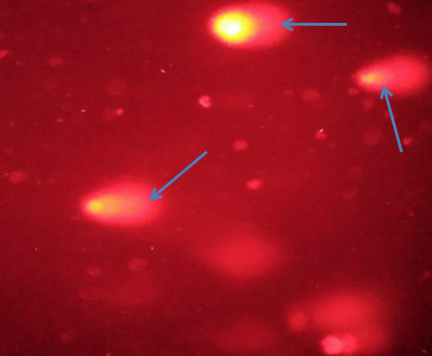
DNA damage is shown in the form of tail. Arrows pointing to the tails of a damaged DNA.
References
- Aksoy, L., Kolay, E., Agilonu, Y., Aslan, Z. and Kargioglu, M., 2013. Saudi J. biol. Sci., 20: 235-239. https://doi.org/10.1016/j.sjbs.2013.02.003[CrossRef]
- Ali, M.M. and Cigerci, I.H., 2017. Br. J. pharmaceut. Res., 16: 1-5. https://doi.org/10.9734/BJPR/2017/33437[CrossRef]
- Bali, E.B., Acik, L., Akca, G., Sarper, M., Elci, M.P., Avcu, F. and Vural, M., 2014. Asian-Pac. J. trop. Biomed., 4: 505-514. https://doi.org/10.12980/APJTB.4.2014B1168[CrossRef]
- Basgel, S. and Erdemoglu, S.B., 2006. Sci. Total Environ., 359: 82-89. https://doi.org/10.1016/j.scitotenv.2005.04.016[CrossRef]
- Blaauboer, B.J., Balls, M., Barratt, M., Casati, S., Coecke, S., Mohamed, M.K., Moore, J., Rall, D., Smith, K.R., Tennant, R., Schwetz, B.A., Stokes, W.S. and Younes, M., 1998. Environ. Hlth. Perspect., 106: 413-418. https://doi.org/10.1289/ehp.98106413[CrossRef]
- Cigerci, I.H., Liman, R., Ozgul, E. and Konuk, M., 2015. Cytotechnology., 67: 157-163. https://doi.org/10.1007/s10616-013-9673-0[CrossRef]
- Cigerci, I.H., Cenkci, S., Kargioglu, M. and Konuk, M., 2016. Cytotechnology., 68: 829-838. https://doi.org/10.1007/s10616-014-9835-8[CrossRef]
- Ekim, T., Koyuncu, M., Vural, M., Duman, H., Aytaç, Z. and Adigüzel, N., 2000. Red data book of Turkish plants (pteridophyta and spermatophyta). TDKA and Van Centennial University Press, Ankara.
- Irfan, A. and Dileep, N.L., 2006. Surgery, 25: 34-41.
- Keeler, R.F. and Baker, D.C., 1990. J. Comp. Pathol., 103: 141-146. https://doi.org/10.1016/S0021-9975(08)80173-9[CrossRef]
- Kerneis, S., Swift, L.H., Lewis, C.W., Bruyere, C., Oumata, N., Colas, P., Ruchaud, S., Bain, J. and Golsteyn, R.M., 2015. Nat. Prod. Res., 29: 1026-1034. https://doi.org/10.1080/14786419.2014.979423[CrossRef]
- Korcan, S.E., Cigerci, I.H., Dilek, M., Kargioglu, M., Cenkci, S. and Konuk, M., 2009. Kuwait J. Sci. Engin., 36: 101-112.
- Kotenko, L.D., Mamatkhanov, A.U., Turakhozhaev, M.T. and Yuldashev, M.P., 2001. Chem. Nat. Comp., 37: 137-139. https://doi.org/10.1023/A:1012322718171[CrossRef]
- Kültür, S., 2007. J. Ethnopharmacol., 111: 341-364. https://doi.org/10.1016/j.jep.2006.11.035[CrossRef]
- McGrath-Hill, C.A and Vicas, I.M., 1997. J. Toxicol. clin. Toxicol., 35: 659-665. https://doi.org/10.3109/15563659709001251[CrossRef]
- Sohail, M., Khan, M.N., Qureshi, N.A. and Chaudhry, A.S., 2017. Pakistan J. Zool., 49: 305-311. http://dx.doi.org/10.17582/journal.pjz/2017.49.1.305.311[CrossRef]
- Valentin-Severin, I., Hegarat, L.L., Lhuguenot, C., Anne-Marie, L.L. and Marie-Christine Chagnon, M.C., 2003. Mutat. Res., 536: 79-90. https://doi.org/10.1016/S1383-5718(03)00031-7[CrossRef]
Services
Article usage statistics
Loading…