
Newcastle disease, caused by avian orthoavulavirus serotype 1, is a highly contagious and economically devastating viral infection affecting both wild and domestic birds. The disease is characterized by severe respiratory, gastrointestinal, and neurological symptoms, often leading to high mortality and substantial production losses, particularly in commercial poultry. In Bangladesh, multiple virulent and avirulent ND virus (NDV) genotypes and subgenotypes continue to circulate despite ongoing vaccination programs. This review presents a comprehensive analysis of the genetic landscape of prevalent NDV strains in Bangladesh between 2010 and 2024 using full-length coding sequences of the fusion (F) gene. Specifically, we focused on genotypes, subgenotypes, host species, and annual distribution patterns in the country. We further investigated phylogenetic relationships, evolutionary divergence, and amino acid substitutions in the full-length F protein. Comparative analyses of amino acid variability and sequence similarity between currently used vaccine strains and prevalent Bangladeshi field isolates were also scrutinized. We have unraveled considerable genetic divergence, and highlighted mismatches in genotypic coverage. These findings underscore the urgent need to develop genotype-matched NDV vaccines to improve disease control strategies in Bangladesh’s poultry sector.
Articles · Volume 8, Issue 3, 4 September 2026 · Open Access
Genetic Diversity and Evolutionary Insights into Newcastle Disease Virus Circulating in Bangladesh
· · · · · · ·
Affiliations & Notes ⌄
- 1Department of Microbiology and Hygiene, Bangladesh Agricultural University, Mymensingh 2202, Bangladesh
- 2Department of Microbiology, Gono Bishwabidyalay, Dhaka, Bangladesh
- 3Department of Animal Production, Gono Bishwabidyalay, Dhaka, Bangladesh
- 4Department of Molecular Biodefense Research, Graduate School of Medicine, Yokohama City University, Yokohama 236-0004, Japan
- 5Division of Biomedical and Life Sciences, Faculty of Health and Medicine, Lancaster University, Lancaster, UK
Article Info ⌄
- Journal:
- Hosts and Viruses
- Published:
- 4 September 2026
From the article
- In our study, analysis of amino acid variability in complete F gene sequences of Bangladeshi NDV isolates compared with vaccine strains (LaSota, B1, and Mukteswar) revealed substitutions in epitope 1 at residues E74K (XIII.2.3), K78G (VII.2), and A79T/G (XIII.2).
- Subgenotype VII.2, associated with the fifth panzootic, and the recently detected a novel sub-genotype XIII.2.3 pose significant threats to poultry health and production.
- The reduced effectiveness of NDV vaccines is due to genetic differences between vaccine and circulating strains, which lower cross-protection.
- To minimize future outbreaks and economic losses, it is essential to develop high-quality, genotype-matched NDV vaccines tailored to the predominant strains (particularly VII and XIII) circulating in Bangladesh.
Selected from the article's own text.
Abstract
Introduction
Newcastle disease (ND) is a highly fatal and economically important viral infection of birds, particularly poultry, caused by Newcastle disease virus (NDV), a prototype member of the orthoavulavirus serotype 1 []. The NDV infection is not only limited to commercial poultry, it can also infect a wide range of wild bird species []. First reported in 1926 in Indonesia and then travelled to England and Srilanka [, ], triggering a global panzootic []. Subsequent panzootics have occurred: a second in the late 1960s driven by increasing international trade and air travel []; a third in the early 1980s that initially affected racing pigeons and later spread to chickens [–]; and a fourth beginning in Southeast Asia in the 1980s, which has since extended to the Middle East, Europe, the Americas, and Africa [–]. While Bangladesh first reported ND outbreaks in 1981, the infection remains enzootic in many regions worldwide []. Clinical manifestations include neurological signs, gastrointestinal disorders, respiratory illness, and reduced egg production, while severe cases often lead to sudden death [].
NDV belongs to the genus Orthoavulavirus in the subfamily Avulavirinae, family Paramyxoviridae []. It is a non-segmented, negative-sense, single-stranded RNA virus with a genome of approximately 15,000 nucleotides that encodes six structural proteins: hemagglutinin-neuraminidase (HN), RNA-dependent RNA polymerase (L), matrix (M), fusion (F), phosphoprotein (P), and nucleocapsid protein (NP) [,]. The NP and P proteins encapsulate viral RNA [], while M facilitates virus assembly and budding []. F and HN mediate viral entry into host cells [], and L directs transcription and replication of the viral genome [].
The NDV genome exhibits marked genetic diversity, with strains varying in pathogenicity []. The NDV is classified into three pathotypes based on virulence: velogenic, mesogenic, and lentogenic []. Velogenic strains cause severe systemic disease and high mortality []; mesogenic strains primarily affect the respiratory and nervous systems with moderate mortality [,]; and lentogenic strains generally cause mild or asymptomatic infections, particularly in the intestines [].
Phylogenetic analysis based on F and HN genes provide high resolution clustering patterns and complement whole-genome analyses. However, the complete F gene sequence is the preferred target for comprehensive NDV studies, including virulence evaluation, pathotyping, genotyping, and phylogenetic analysis, as recommended by WOAH []. Based on full-length F gene sequences, NDV is divided into two major classes: class I and class II []. Class I strains are generally avirulent and mostly detected in wild aquatic birds and live bird markets [, ]. In contrast, Class II strains are typically virulent and are frequently recovered from domestic poultry as well as wild birds [, ]. Class I consists of a single genotype with three sub-genotypes, whereas Class II encompasses at least 21 genotypes (I–XXI), each containing several sub-genotypes []. NDV has a long history in Southeast Asia, where genotypes II, IV, VII, and XIII continue to circulate simultaneously []. Preventive vaccination is the main strategy for controlling ND in commercial poultry; yet outbreaks continue to occur in vaccinated flocks due to circulating virulent strains of NDV, causing losses from death, poor growth, and reduced egg production.
A key determinant of NDV virulence is the amino acid motif at the F protein cleavage site (residues 112–117) []. Lentogenic strains typically harbor a monobasic motif (112G-R/K-Q-G-R↓L117), whereas mesogenic and velogenic strains carry a multibasic motif (112R/G/K-R-Q/K-K/R-R↓F117), which enables efficient cleavage and viral entry []. Characterizing the F protein cleavage site is critical for assessing virulence, guiding vaccine design, and understanding NDV evolution []. The high genetic variability of NDV in poultry populations highlights the need for continuous molecular surveillance [] using the complete F or HN gene, or ideally, whole-genome sequences.
Therefore, this review investigates the genetic diversity of NDV isolates in Bangladesh from 2010 to 2024 using full-length coding sequences of the fusion (F) gene available in the NCBI GenBank database, focusing on phylogenetic classification, evolutionary divergence, and genotype mismatches between vaccine strains and circulating viruses.
Materials and Methods
Genetic ecology of NDVs in Bangladesh
Complete coding sequences of the F gene from NDV strains circulating in Bangladesh between 2010 and 2024 were retrieved from the NCBI GenBank database. These sequences were analyzed to determine prevalent genotypes, subgenotypes, host species, and annual distribution of isolates, based on the updated NDV classification system [].
Evolutionary relationships of complete F gene sequences
Complete F gene sequences of Bangladeshi NDV isolates, along with their translated protein sequences, were retrieved from the NCBI GenBank database. Phylogenetic relationships were inferred using MEGA 11 software [], supported by BLAST searches in GenBank. The phylogenetic tree was generated using the Maximum Likelihood method with the Poisson correction model [], and statistical support was evaluated with 1,000 bootstrap replicates []. Initial trees for heuristic searches were automatically generated using Neighbor-Joining and BioNJ algorithms applied to pairwise distance matrices estimated by the Poisson model. The tree with the highest log-likelihood value was selected. Evolutionary rate variation among sites was modeled with a discrete Gamma distribution (two categories, +G, with estimated parameters). The final tree was drawn to scale, with branch lengths representing the number of substitutions per site.
Evolutionary divergence of F gene sequences
Evolutionary divergence among NDV isolates within and between genotypes/subgenotypes was assessed using amino acid sequences of the complete F gene, applying the Poisson correction model []. Ambiguous positions were removed using the pairwise deletion option. All analyses were performed in MEGA 11 [].
Amino acid variability in F gene sequences
Amino acid variability among Bangladeshi NDV isolates was examined by comparing complete F gene sequences across genotypes and subgenotypes. Variability in Bangladeshi isolates was also compared with commonly used NDV vaccine strains (LaSota, B1, and Mukteswar). Multiple sequence alignments were conducted using MEGA 11 [].
Sequence similarity between vaccine strains and field isolates
Sequence similarity analyses were performed between the complete F gene sequences of vaccine strains (LaSota, B1, and Mukteswar) and circulating Bangladeshi NDV isolates across genotypes and subgenotypes. Vaccine strain sequences were retrieved from the NCBI GenBank database, and analyses were carried out in MEGA 11 [].
Results
Genetic ecology of NDVs in Bangladesh
A total of 26 complete fusion (F) gene sequences of NDV isolates from Bangladesh were analyzed, revealing genetically diverse strains circulating among multiple bird species. All isolates belonged to class II and were classified into several genotypes and subgenotypes. Specifically, two isolates were assigned to genotype II, one to genotype VI (subgenotype VI.1.1, formerly VI.b), four to genotype VII (subgenotype VII.2), sixteen to genotype XIII (eleven in subgenotype XIII.2, four in XIII.2.3, and one unclassified), and three to genotype XXI (subgenotype XXI.1.2). Isolates from subgenotypes VI.1.1 and XXI.1.2 were recovered from pigeons, whereas isolates from genotypes II, VII.2, XIII.2, and XIII.2.3 originated from chickens.
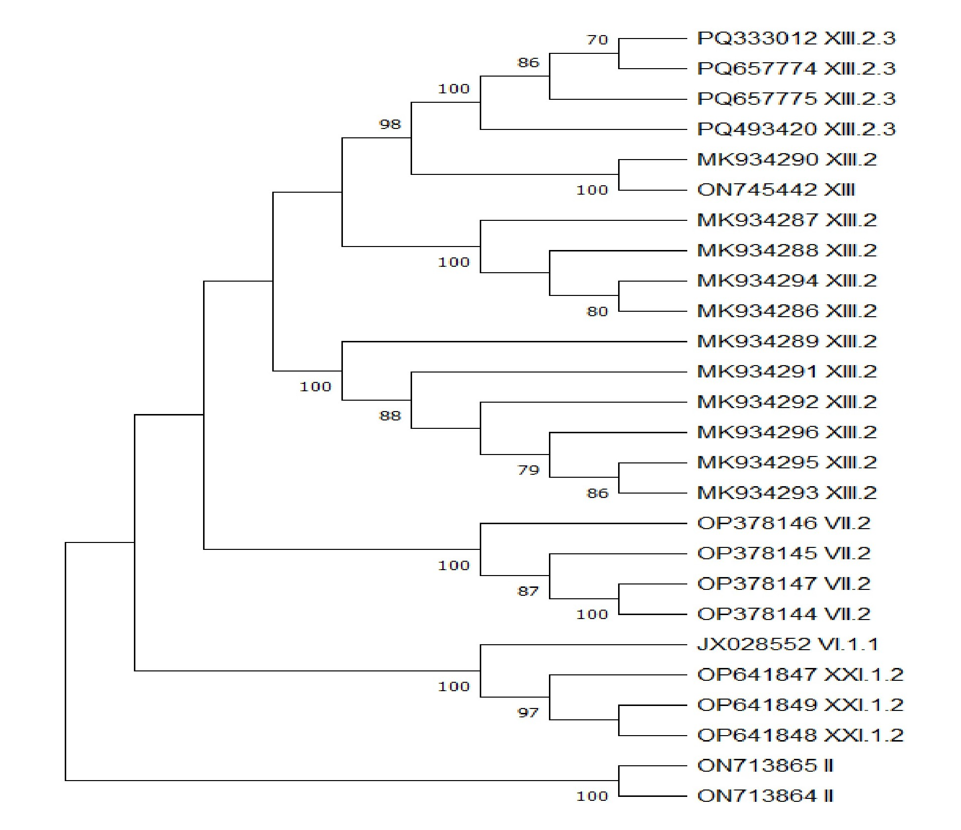
Phylogenetic tree of Bangladeshi NDV isolates, in which the evolutionary history was inferred by using the Maximum Likelihood method and Poisson correction model with 1000 bootstrap replicates. The tree with the highest log likelihood (-3585.91) is shown. The percentage of trees in which the associated taxa clustered together is shown next to the branches. Initial tree(s) for the heuristic search were obtained automatically by applying Neighbour-Joining and BioNJ algorithms to a matrix of pairwise distances estimated using the Poisson model, and then selecting the topology with superior log likelihood value. A discrete Gamma distribution was used to model
evolutionary rate differences among sites (2 categories (+G, parameter = 0.0500)). This analysis involved 26 amino acid sequences. There were total of 553 positions in the final dataset. Evolutionary analyses were conducted in MEGAI1. Only bootstrap values above 70 are shown.
All isolates were predicted to be virulent based on the presence of multibasic amino acid motifs such as “RRQKRF,” “RRKKRF,” or “RTKKRF” at the F protein cleavage site, except for genotype II isolates, which contained the monobasic motif “GRQGRL” characteristic of lentogenic strains. Notably, no complete F gene sequences of Bangladeshi NDV isolates were available in the NCBI GenBank database for the years 2011, 2013, 2014, 2015, 2019, and 2022 (Table 1).
Table 3. Amino acid variability in complete F gene sequences of circulating Bangladeshi NDVs within each genotype/subgenotype
| Isolates under Genotype / Subgenotype | Cleavage site | Variable amino acid position | Variables in neutralizing epitope of F protein |
|---|---|---|---|
| Genotype II | GRQGRL | 17, 25, 264, 429, 441, 486, 520, 549, 550 | Conserved |
| Subgenotype VII.2 | RRKKRF | 19, 24, 25, 31, 78, 97, 113, 170, 186, 190, 193, 235, 402, 442 | Epitope 1 (at 78th ) and epitope 2 (at 170th ) |
| Subgenotype XIII.2 | RRQKRF | 4, 5, 6, 8, 10-14, 18, 19, 24-26, 28, 29, 43, 49, 52, 66, 79, 97, 104, 107, 108, 110, 163, 170, 200, 208, 255, 270, 279, 286, 312, 321, 325, 328, 330, 338, 342, 364, 385, 395, 402, 442, 465, 479, 485, 486, 489, 492, 494, 496, 514, 516, 517, 520, 522, 530, 545 | Epitope 1 (at 79th ) and epitope 2 (at 163th and 170th ) |
| Subgenotype XIII.2.3 | RRQKRF | 4, 13, 63 | Conserved |
| Subgenotype XXI.1.2 | RRQKRF | 16, 17, 19, 21, 150, 235, 294, 425, 470, 479, 487, 530, 537 | Conserved |
Phylogenetic analysis of Bangladeshi NDV isolates
Phylogenetic analysis revealed close evolutionary relationships among isolates within genotype II, subgenotype VII.2, XIII.2.3, and XXI.1.2. By contrast, subgenotype XIII.2 isolates were distributed across separate subclades, indicating greater divergence within this group. Distinct clustering patterns were observed between genotypes and subgenotypes (Figure 1). BLAST analysis further showed that Bangladeshi isolates shared high sequence similarity (93.98%–99.96%) with isolates from India, Pakistan, Japan, Indonesia, and Jordan (Table 2).
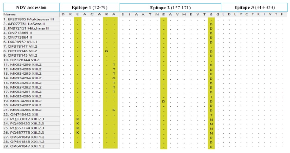
Amino acid variability at antigenic epitopes in F gene sequences of Bangladeshi NDVs compared to NDV vaccine strains used in Bangladesh in which antigenic variation in epitope 1 and epitope 2 were distinguished but epitope 3 remain conserved.
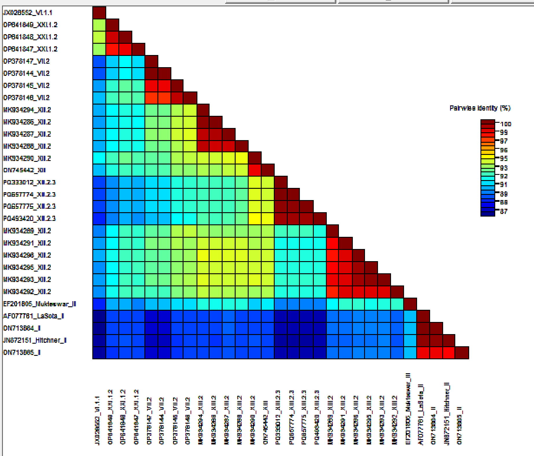
The pairwise identities-based plot of F protein sequences aligned by MAFFT and displayed by Sequence Demarcation Tool (SDT) software.
Evolutionary divergence of NDVs in Bangladesh
Analysis of 26 amino acid sequences of Bangladeshi NDV isolates, with a total alignment length of 553 positions, showed the following pairwise mean evolutionary distances: genotype II (0.02), subgenotype VII.2 (0.00–0.03), subgenotype XIII.2 (0.00–0.07), subgenotype XIII.2.3 (0.00–0.01), and subgenotype XXI.1.2 (0.00–0.02) (Supplementary Tables 1.a–1.e). The overall mean evolutionary distances within each group were 0.02 (II), 0.02 (VII.2), 0.04 (XIII.2), 0.00 (XIII.2.3), and 0.02 (XXI.1.2).
Amino acid variability in F gene sequences of Bangladeshi NDV isolates
Amino acid variability was examined across complete F gene sequences within each genotype and subgenotype. Genotype II isolates exhibited nine substitutions while retaining the conserved lentogenic cleavage site “GRQGRL.” Subgenotype VII.2 isolates, featuring “RRKKRF” and “RTKKRF” motifs, showed 14 variations. Subgenotype XIII.2 isolates carrying the virulent “RRQKRF” motif displayed 61 variable sites, whereas XIII.2.3 isolates showed only three substitutions. Subgenotype XXI.1.2 isolates, which also carried the “RRQKRF” motif, exhibited 13 variations.
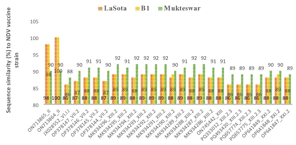
Sequence similarity (%) of NDV vaccine strains used in Bangladesh (LaSota, B1 and Mukteswar) comparing to complete F gene sequences across different genotypes and subgenotypes of circulating Bangladeshi ND V isolates in which orange color indicates LaSota vaccine strain and it's similarity (%) is labelled at inside base; gold color indicates B1 vaccine strain and it's similarity (%) is labelled at centre; and green color indicates Mukteswar vaccine strain and it's similarity (%) is labelled at outside end.
Notably, substitutions were detected in neutralizing epitopes 1 and 2 of subgenotypes VII.2 and XIII.2, whereas epitope 3 remained conserved across all isolates (Table 3). Further comparison with vaccine strains (LaSota, B1, and Mukteswar) confirmed substitutions at epitope 1 in VII.2, XIII.2, and XIII.2.3. Compared with LaSota and B1, substitutions at epitope 2 were observed in VII.2, XIII.2, and XIII.2.3; while compared with Mukteswar, substitutions at epitope 2 were present in all circulating genotypes/subgenotypes in Bangladesh. Epitope 3 remained conserved across all isolates (Figure 2). Additionally, Sequence Demarcation Tool (SDT) based analysis identified similarities across all strains isolated from the country (Figure 3). This highlight the genetic diversity and percussion this might have on the vaccine efficacies in the poultry sector.
Sequence similarity between vaccine strains and Bangladeshi NDV isolates
Comparative sequence similarity analysis revealed a considerable genetic mismatch between circulating NDV isolates and commonly used vaccine strains. LaSota and B1 showed high sequence similarity (98–100%) only with lentogenic strains of genotype II and moderate similarity (86–89%) with virulent strains of genotypes VI.1.1, VII.2, XIII.2, XIII.2.3, and XXI.1.2. By contrast, Mukteswar exhibited 88–92% sequence similarity with all circulating genotypes and subgenotypes in Bangladesh (Figure 4).
Discussion
NDV infects more than 200 avian species, however, chickens are the most susceptible, followed by turkeys [], while other vulnerable species include pheasants, guinea fowl, partridges, and quail. Waterfowl and wild birds often serve as reservoirs [], and pigeons are also frequently affected []. In the present study, NDV was detected only in chickens and pigeons in Bangladesh. Pigeons, as natural reservoirs of lentogenic and mesogenic strains, typically harbor the virus asymptomatically and contribute to genotype diversity through genetic drift, mutations, recombination, and geographic spread. Migratory behavior further facilitates the introduction of new genotypes into naïve regions or the reintroduction of strains into previously controlled areas []. Our findings showed that class II NDV strains circulating in Bangladesh include lentogenic genotype II and virulent genotypes VI (VI.1.1), VII (VII.2), XIII (XIII.2 and XIII.2.3), and XXI (XXI.1.2) [, , ].
The F protein cleavage site remains a critical molecular marker of NDV virulence []. In this study, “RRQKRF” was the most prevalent virulent motif, while alternative motifs “RRKKRF” and “RTKKRF” were identified in subgenotype VII.2. Previous studies have reported that the presence of glutamine (Q) at the third position of the cleavage site enhances viral cell-to-cell spread [, ], suggesting a possible role in viral evolution.
Phylogenetic and BLAST analyses provided insights into viral transmission across species and regions. Phylogenetically, Bangladeshi isolates clustered closely within most genotypes, except for genotype XIII.2 isolates. BLAST searches showed that genotype II isolates from Bangladesh shared strong homology (up to 99.96%) with chicken-derived isolates from Jordan and India. The subgenotype VI.1.1 isolate closely matched (99.4%) a pigeon-derived isolate from Pakistan. Subgenotype VII.2 isolates showed 98% similarity to strains identified in barn owls and eagles from India and Indonesia, respectively. Genotype VII remains of global concern due to its role in multiple panzootics [, ]. Subgenotype XIII.2 isolates displayed 93–97% similarity to isolates from India and Japan, while XIII.2.3 isolates shared 94.3% similarity with a Japanese chicken strain. Subgenotype XXI.1.2 isolates showed 98% similarity to Pakistani pigeon-derived strains. Together with divergence analysis, these findings support intra-genotypic evolution of NDVs in Bangladesh.
Divergence analysis revealed low pairwise and overall mean evolutionary distances (0.00–0.03) among genotypes II, VII.2, XIII.2.3, and XXI.1.2, suggesting limited diversity. By contrast, genotype XIII.2 showed moderate divergence (>0.03), indicating genetic heterogeneity. The overall mean distance of 0.08 across all 26 isolates suggests relatively low similarity among genotypes, highlighting the mutational potential of NDVs.
Several classic experimental studies have investigated single amino acid substitutions in the F protein neutralization epitopes [-]. Amino acid variation within neutralizing epitope 1 (residues 72, 74, 75, 78, 79), epitope 2 (residues 157–171), and epitope 3 (residues 343–353) of the F gene further supports ongoing viral evolution []. Substitutions at these sites can alter antigenic structure by changing side-chain size, polarity, or flexibility, thereby modifying the local 3D conformation of epitopes and their recognition by antibodies. Such mutations may also change surface charge distribution, reducing complementarity with antibody binding sites and lowering binding affinity []. Additionally, substitutions can create N-X-S/T motifs, introducing N-linked glycosylation sites that add glycans capable of shielding epitopes from immune recognition [].
In our study, substitutional analysis across Bangladeshi NDV isolates revealed changes at residue 78 in VII.2 and residue 79 in XIII.2 within epitope 1, and at residue 170 in VII.2 and residues 163 and 170 in XIII.2 within epitope 2. Epitope 3 remained conserved across all isolates. Such variation in the F protein can alter B-cell and T-cell epitope presentation and reduce the efficacy of neutralizing antibodies, which often target conformational epitopes on the F protein. Consequently, antigenic drift resulting from these amino acid substitutions may reduce vaccine effectiveness, contributing to breakthrough infections when mismatches occur between vaccine strains and circulating viruses [, , , -].
Traditional NDV vaccines derived from lentogenic or mesogenic strains of genotypes I, II, III, IV, V, VIII, and XV [-] are still widely used. In Bangladesh, LaSota (GenBank accession no. AF077761) and B1 (GenBank accession no. JN872151), both under genotype II, are administered to chicks, whereas the mesogenic Mukteswar strain (GenBank accession no. EF201805) under genotype III is used in older birds [, ]. In our study, analysis of amino acid variability in complete F gene sequences of Bangladeshi NDV isolates compared with vaccine strains (LaSota, B1, and Mukteswar) revealed substitutions in epitope 1 at residues E74K (XIII.2.3), K78G (VII.2), and A79T/G (XIII.2). For epitope 2, compared with LaSota and B1, substitutions were detected at residues E163D (XIII.2), D170G (VII.2), D170T (XIII.2), and D170N (XIII.2.3). Compared with Mukteswar, substitutions included E163D (XIII.2), G170D (II, VI.1.1, VII.2, XXI.1.2), G170D/T (XIII.2), and G170T/N (XIII.2.3). These variations may compromise immune recognition, which could help explain why NDV remains enzootic and continues to cause significant economic losses despite routine vaccination [, ].
Sequence similarity analysis showed that LaSota and B1 exhibited the highest similarity (100% and 98%) with genotype II field isolates ON713864 and ON713865, respectively. However, similarity decreased markedly against other genotypes: 80–89% with VI.1.1, VII.2, XIII.2, XIII.2.3, and XXI.1.2. Mukteswar showed 88–92% similarity with all circulating genotypes. Such genotypic mismatch likely reduces vaccine-induced immunity due to antigenic variation in surface proteins, particularly the F protein []. In addition, biosecurity weaknesses such as inadequate wild bird control, poor farm hygiene, mixed-species farming and vaccination issues, including incomplete flock coverage, improper administration, irregular booster schedules, and use of genotype II vaccines against genotype VII/XIII strains, create ecological and immunological pressures that allow the virus to evolve, adapt, and persist in poultry populations [].
Recent advances addressing epitope variability in the F protein include the development of genotype-matched antigens, such as genotype VII-matched live/chimeric vaccines [], DNA vaccines with IL-28b adjuvant [], thermostable recombinant GVII live vaccines [], locally matched inactivated GVII.2 vaccines [], non-GVII lentogenic strain–based vaccines [], and viral-vector–based vaccines []. Nevertheless, older genotype II–based vaccines remain vulnerable to epitope drift []. Genotype-matched vaccines have consistently shown superior protection. For example, in China, where ND is endemic, genotype-matched vaccines combined with improved biosecurity have significantly reduced outbreaks [, ]. In India, an inactivated GVII strain vaccine provided 100% protection against GVII challenge in chickens, outperforming the commercial GII LaSota vaccine, which gave 80% protection []. In Bangladesh, A study tested three booster vaccines—live GII LaSota, inactivated GII LaSota, and inactivated GXIII.2 against the local virulent GXIII.2 BD-C161/2010 strain in chickens primed with live LaSota. All boosters prevented death, but the inactivated LaSota outperformed the live version, and the GXIII.2 vaccine gave full clinical protection and significantly reduced virus shedding []. In another experiment, the dominant GVII.2 strain isolated from diseased chickens in Bangladesh was used to develop an inactivated vaccine, which provided nearly 100% protection against the homologous strain. This result contrasted with the only 57% protection against GVII.2 observed in chickens vaccinated with the commercial live GII LaSota strain []. Collectively, these findings emphasize the urgent need for updated, genotype-specific NDV vaccines for Bangladesh.
Future research in Bangladesh should prioritize the development of genotype-matched vaccines using cutting-edge technologies, targeting circulating strains VII and XIII, supported by continuous molecular surveillance. Policymakers should also evaluate the cost-effectiveness of genotype-matched vaccines and enhanced biosecurity strategies to achieve sustainable NDV control in the poultry sector. Full-length F gene sequences were analyzed due to the limited availability of HN or whole-genome data for Bangladeshi NDV strains in the NCBI GenBank database.
Conclusion
The genetic diversity of NDV genotypes and subgenotypes circulating in Bangladesh between 2010 and 2024 reveals the co-circulation of multiple strains, including novel and virulent variants. Subgenotype VII.2, associated with the fifth panzootic, and the recently detected a novel sub-genotype XIII.2.3 pose significant threats to poultry health and production. The ongoing evolution and emergence of new variants complicate control measures based on genotype II and III vaccines, which no longer fully protect against circulating field strains. The reduced effectiveness of NDV vaccines is due to genetic differences between vaccine and circulating strains, which lower cross-protection. To minimize future outbreaks and economic losses, it is essential to develop high-quality, genotype-matched NDV vaccines tailored to the predominant strains (particularly VII and XIII) circulating in Bangladesh.
Development of such genotype-matched vaccines employing advanced technologies, coupled with continuous molecular surveillance, optimized vaccination strategies, and strengthened biosecurity, represents the most effective pathway to sustainable ND control in Bangladesh.
Data Availability Statement
Data sharing does not apply to this article as no new data were generated in this study.
Conflict of Interest
The authors declare that they have no financial or other competing interests with any organization or company, nor among the authors and co-authors.
Acknowledgments
This study was conducted under the project “Development of bacterial outer membrane vesicle (OMV)-based vaccines for Newcastle disease and infectious bursal disease,” funded by BAS-USDA [Grant number 2023/8/BAS]. The authors sincerely thank BAS-USDA, Bangladesh, for their support of this research.
Authors’ Contribution
Farah Zehreen: Methodology, data curation, software, formal analysis, and writing-original draft; Md. Abdur Rahman: Methodology, data curation, software, formal analysis, and writing-original draft; Md. Golzar Hossain: Writing-reviewing & editing, funding acquisition; Masaru Shimada: Writing-reviewing & editing; Sukumar Saha: Conceptualization, supervision, project administration, writing-reviewing & editing, funding acquisition.
Figures and Figure Legends:
References
- Rima B, Balkema-Buschmann A, Dundon WG, Duprex P, Easton A, Fouchier R, et al. (2019) ICTV virus taxonomy profile: paramyxoviridae. J Gen Virol., 100:1593. doi: 10.1099/jgv.0.001328[CrossRef]
- Mansour, S.M.G.; ElBakrey, R.M.; Mohamed, F.F.; Hamouda, E.E.; Abdallah, M.S.; Elbestawy, A.R.; Ismail, M.M.; Abdien, H.M.F.; Eid, A.A.M. (2021) Avian Paramyxovirus Type 1 in Egypt: Epidemiology, Evolutionary Perspective, and Vaccine Approach. Front. Vet. Sci., 8, 647462.
- D. J. Alexander (2001) Newcastle disease. British Poultry Science, vol. 42, no. 1, pp. 5–22.
- D. J. Alexander (2009) Ecology and Epidemiology of Newcastle Disease. Avian Influenza and Newcastle Disease: A Field and Laboratory Manual, pp. 19–26.
- A. Ballagi-Pord´ any, E. Wehmann, J. Herczeg, S. Bel´ak, and B. Lomniczi (1996) Identification and grouping of Newcastle disease virus strains by restriction site analysis of a region from the F gene. Archives of Virology, vol. 141, pp. 243–261.
- D. J. Alexander, E. W. Aldous, and C. M. Fuller (2012) The long view: a selective review of 40 years of Newcastle disease research. Avian Pathology, vol. 41, no. 4, pp. 329–335.
- D. J. Alexander, G. Parsons, and R. Marshall (1986) Avian paramyxovirus type 1 infections of racing pigeons: 4 laboratory assessment of vaccination. Veterinary Record, vol. 118, no. 10, pp. 262–266.
- J. T. Lumeij and J. W. E. Stam (1985) Paramyxovirus disease in racing pigeons. Veterinary Quarterly, vol. 7, no. 1, Article ID 9693954, pp. 60–65.
- G. Cross (1995) Paramyxovirus-1 infection (Newcastle disease) of pigeons. Seminars in Avian and Exotic Pet Medicine, vol. 4, no. 2, pp. 92–95.
- J. Herczeg, E. Wehmann, R. R. Bragg et al., (1999) Two novel genetic groups (VIIb and VIII) responsible for recent Newcastle disease outbreaks in Southern Africa, one (VIIb) of which reached Southern Europe. Archives of Virology, vol. 144, no. 11, pp. 2087–2099.
- E. K. Lee, W. J. Jeon, J. H. Kwon, C. B. Yang, and K. S. Choi (2009) Molecular epidemiological investigation of Newcastle disease virus from domestic ducks in Korea. Veterinary Microbiology, vol. 134, no. 3, pp. 241–248.
- H. Liu, Z. Wang, Y. Wang, C. Sun, D. Zheng, and Y. Wu (2008) Characterization of Newcastle disease virus isolated from waterfowl in China. Avian Diseases, vol. 52, no. 1, pp. 150–155.
- A. Samuel, B. Nayak, A. Paldurai et al., (2013) Phylogenetic and pathotypic characterization of newcastle disease viruses circulating in west africa and efficacy of a current vaccine. Journal of Clinical Microbiology, vol. 51, no. 3, pp. 771–781.
- Azizah AN, Anwar Ch, and Rahardjo AP (2021). Isolation and identification of Newcastle disease virus from ducks sold at traditional livestock market center in Indonesia. Journal of World’sPoultry Research, 11(1): 96-100. DOI: https://www.doi.org/10.36380/jwpr.2021.12[CrossRef]
- Chowdhury, T.I.M.F.R.; Sarker, A.J.; Amin, M.M.; Hossain, W.I.M.A. (1981) Studies of Newcastle disease in Bangladesh. Bangladesh Vet. J., 15, 1–9.
- Rehman, Z.U.; Ren, S.; Butt, S.L.; Manzoor, Z.; Iqbal, J.; Anwar, M.N.; Sun, Y.; Qiu, X.; Tan, L.; Liao, Y.; et al. (2021) Newcastle Disease Virus Induced Pathologies Severely Affect the Exocrine and Endocrine Functions of the Pancreas in Chickens. Genes, 12, 495.
- Walker, P.J., Siddell, S.G., Lefkowitz, E.G., Mushegian,A.R., Adriaenssens, E.M., Alfenas-Zerbini, P., Dempsey, D.M., Dutilh, B.E., García, M.L., Hendrickson, R.C., Junglen, S., Krupovic, M., Kuhn, J.H., Lambert, A.J., Łobocka, M., Oksanen, H.M., Orton, R.J., Robertson, D.L., Rubino, L., Sabanadzovic, S., Simmonds, P., Smith, D.P., Suzuki, N., Van Doorslaer, K., Vandamme, A.M., Varsani, A. and Zerbini, F.M. (2022) Recent changes to virus taxonomy ratified by the International Committee on Taxonomy of Viruses. Arch. Virol., 167(11): 2429–2440.
- Xiao S, Nayak B, Samuel A, et al. (2012) Generation by reverse genetics of an effective, stable, live-attenuated Newcastle disease virus vaccine based on a currently circulating, highly virulent Indonesian strain. PLoS One., 7(12):e52751.
- Jin J, Zhao J, Ren Y, Zhong Q, Zhang G. (2016) Contribution of HN protein length diversity to Newcastle disease virus virulence, replication and biological activities. Sci Rep., 6(1):36890.
- Nagai, Y.; Hamaguchi, M.; Toyoda, T. (1989) Molecular Biology of Newcastle Disease Virus. Prog. Vet. Microbiol. Immunol., 5, 16–64.
- Steward, M.; Vipond, I.B.; Millar, N.S.; Emmerson, P.T. (1993) RNA Editing in Newcastle Disease Virus. J. Gen. Virol., 74 Pt 12, 2539–2547.
- Chang, A.; Dutch, R.E. (2012) Paramyxovirus Fusion and Entry: Multiple Paths to a Common End. Viruses, 4, 613–636.
- Ganar, K.; Das, M.; Sinha, S.; Kumar, S. (2014) Newcastle Disease Virus: Current Status and Our Understanding. Virus Res., 184, 71–81.
- Snoeck, C.J.; Owoade, A.A.; Couacy-Hymann, E.; Alkali, B.R.; Okwen, M.P.; Adeyanju, A.T.; Komoyo, G.F.; Nakouné, E.; Le Faou, A.; Muller, C.P. (2013) High Genetic Diversity of Newcastle Disease Virus in Poultry in West and Central Africa: Cocirculation of Genotype XIV and Newly Defined Genotypes XVII and XVIII. J. Clin. Microbiol., 51, 2250–2260.
- WOAH (2024) Newcastle Disease (Infection with Newcastle Disease Virus), in Manual of Diagnostic Tests and Vaccines for Terrestrial Animals: (Mammals, Birds and Bees), vol. 1, pp. 555–574.
- Ali AAH, Abdallah FM, Farag GK, and Fatehi S (2022). An overview on virological pathognesis, clinical manifestation, and molecular studies on newcastle disease virus. European Journal of Molecular & Clinical Medicine, 9(4): 2807-2824. Available at: https://ejmcm.com/pdf_19147_252376f6f087e9fbf06385528c6f4108.html
- W. Fan, Y. Wang, S. Wang et al., (2017) Virulence in Newcastle disease virus: A genotyping and molecular evolution spectrum perspective. Research in Veterinary Science, vol. 111, pp. 49–54.
- V. M. B. D. Moura, L. Susta, S. Cardenas-Garcia et al., (2016) Neuropathogenic capacity of lentogenic, mesogenic, and velogenic newcastle disease virus strains in day-old chickens. Veterinary Pathology, vol. 53, no. 1, pp. 53–64.
- H. Hamid, R. S. F. Campbell, and C. Lamichhane (1990) “The Pathology of infection of chickens with the lentogenic V4 strain of newcastle disease virus,” Avian Pathology, vol. 19, no. 4, pp. 687–696.
- Czeglédi, A.; Ujvári, D.; Somogyi, E.; Wehmann, E.; Werner, O.; Lomniczi, B. (2006) Third genome size category of avian paramyxovirus serotype 1 (Newcastle disease virus) and evolutionary implications. Virus Res., 120, 36–48.
- Kim, L.M.; King, D.J.; Suarez, D.L.; Wong, C.W.; Afonso, C.L. (2007) Characterization of class I Newcastle disease virus isolates from Hong Kong live bird markets and detection using real-time reverse transcription-PCR. J. Clin. Microbiol., 45, 1310–1314.
- Kim, L.M.; King, D.J.; Curry, P.E.; Suarez, D.L.; Swayne, D.E.; Stallknecht, D.E.; Slemons, R.D.; Pedersen, J.C.; Senne, D.A.; Winker, K.; et al. (2007) Phylogenetic diversity among low-virulence newcastle disease viruses from waterfowl and shorebirds and comparison of genotype distributions to those of poultry-origin isolates. J. Virol., 81, 12641–12653.
- Welch, C.N.; Shittu, I.; Abolnik, C.; Solomon, P.; Dimitrov, K.M.; Taylor, T.L.; Williams-Coplin, D.; Goraichuk, I.V.; Meseko, C.A.; Ibu, J.O.; et al. (2019) Genomic comparison of Newcastle disease viruses isolated in Nigeria between 2002 and 2015 reveals circulation of highly diverse genotypes and spillover into wild birds. Arch. Virol., 164, 2031–2047.
- Suarez, D.L.; Miller, P.J.; Koch, G.; Mundt, E.; Rautenschlein, S. (2020) Newcastle Disease, Other Avian Paramyxoviruses, and Avian Metapneumovirus Infections. In Diseases of Poultry, 14th ed.; Swayne, D.E., Ed.; Wiley-Blackwell: Hoboken, NJ, USA, pp. 111–166.
- Dimitrov, K.M.; Abolnik, C.; Afonso, C.L.; Albina, E.; Bahl, J.; Berg, M.; Briand, F.X.; Brown, I.H.; Choi, K.S.; Chvala, I.; et al. (2019) Updated unified phylogenetic classification system and revised nomenclature for Newcastle disease virus. Infect. Genet. Evol., 74, 103917.
- Barnali Nath, Sachin Kumar (2017) Emerging variant of genotype XIII Newcastle disease virus from Northeast India. Acta Trop. 2017 Aug:172: 64-69. doi: 10.1016/j.actatropica.2017.04.018.[CrossRef]
- A. Panda, Z. Huang, S. Elankumaran, D. D. Rockemann, and S. K. Samal (2004) “Role of fusion protein cleavage site in the virulence of Newcastle disease virus,” Microbial Pathogenesis, vol. 36, no.1, pp. 1–10.
- Puro K and Sen A (2022). Newcastle disease in backyard poultry rearing in the Northeastern States of India: Challenges and control strategies. Frontiers in Veterinary Science, 9: 799813. Available at: https://www.frontiersin.org/articles/10.3389/fvets.2022.799813/full
- Zeb, M.T.; Dumont, E.; Khan, M.T.; Shehzadi, A.; Ahmad, I. (2024) Multi-Epitopic Peptide Vaccine Against Newcastle Disease Virus: Molecular Dynamics Simulation and Experimental Validation. Vaccines, 12, 1250. https://doi.org/10.3390/vaccines12111250[CrossRef]
- Tamura K., Stecher G., and Kumar S. (2021). MEGA 11: Molecular Evolutionary Genetics Analysis Version 11. Molecular Biology and Evolution https://doi.org/10.1093/molbev/msab120[CrossRef]
- Zuckerkandl E. and Pauling L. (1965). Evolutionary divergence and convergence in proteins. Edited in Evolving Genes and Proteins by V. Bryson and H.J. Vogel, pp. 97-166. Academic Press, New York.
- Felsenstein J. (1985). Confidence limits on phylogenies: An approach using the bootstrap. Evolution 39:783-791.
- Getabalew M, Alemneh T, Akeberegn D, Getahun D, and Zewdie D (2019). Epidemiology, diagnosis & prevention of Newcastle disease in poultry. American Journal of Biomedical Science and Research, 3(1): 50-59. DOI: https://www.doi.org/10.34297/AJBSR.2019.03.000632[CrossRef]
- C. J. Snoeck, A. T. Adeyanju, A. a. Owoade et al., (2013) “Genetic diversity of newcastle disease virus in wild birds and pigeons in West Africa,” Applied and Environmental Microbiology, vol. 79, no. 24, pp. 7867–7874.
- Hany F. Ellakany, Ahmed R. Elbestawy, Hatem S. Abd El-Hamid, Rasha E. Zedan, Ahmed R. Gado, Ayman E. Taha, Mohamed A. Soliman, Mohamed E. Abd El-Hack, Ayman A. Swelum, Islam M. Saadeldin, Hani Ba-Awadh and Elsayed O.S. Hussein. (2019) Role of Pigeons in the Transmission of Avian Avulavirus (Newcastle Disease-Genotype VIId) to Chickens. Animals, 9, 338; doi:10.3390/ani9060338[CrossRef]
- Kochiganti, S., Rajkhowa, T.K., Kiran, J., Zodinpui, D. and Choudhury, F.A. (2024). Molecular Characterization of Class II Newcastle Disease Virus from Field Outbreaks in Mizoram, India. Indian Journal of Animal Research. doi: 10.18805/IJAR.B-5241[CrossRef]
- F. Zereen, M.A. Rahman, M.G. Hossain, et al., (2024) First report of the emergence of novel sub-genotype XIII.2.3 of Newcastle disease virus in chickens from selected regions of Bangladesh, Infection, Genetics and Evolution, https://doi.org/ 10.1016/j.meegid.2025.105742
- Anat Wisemana,b, Mali Shabbata , Aliza Cohena , Ido Froma , Yael Almog Saminaa , Einat Sade Levya , Shahar Szera , Nadav Zeltcera , Eyal Klementb and Yaniv Pimaa (2022) Point mutation revealed the resurgence of sub-genotype VII.2 Newcastle disease virus in Israel. Avian Pathology 2022, VOL. 51, NO. 3, 236–243. https://doi.org/10.1080/03079457.2022.2040730[CrossRef]
- Samal S, Khattar SK, Paldurai A, Palaniyandi S, Zhu X, Collins PL, and Samal SK (2013). Mutations in the cytoplasmic domain of the Newcastle disease virus fusion protein confer hyperfusogenic phenotypes modulating viral replication and pathogenicity. Journal of Virology, 87(18):10083-10093. DOI: https://www.doi.org/10.1128/jvi.01446-13[CrossRef]
- Y. Wang, W. Yu, N. Huo et al., (2017) “Comprehensive analysis of amino acid sequence diversity at the F protein cleavage site of Newcastle disease virus in fusogenic activity,” PLoS ONE, vol.12, no. 9, Article ID e0183923.
- F. Sabouri, M. Vasfi Marandi, and M. Bashashati (2017) “Characterization of a novel VIIl sub-genotype of Newcastle disease virus circulating in Iran,” Avian Pathology, vol. 47, no. 1.
- P. J. Miller, R. Haddas, L. Simanov et al., (2015) “Identification of new sub-genotypes of virulent Newcastle disease virus with potential panzootic features,” Infection, Genetics and Evolution, vol. 29, pp. 216–229.
- Tetsuya Toyoda, Bin Gotoh, Takemasa Sakaguchi, Hiroshi Kida, And Yoshiyuki Nagai (1998) Identification of Amino Acids Relevant to Three Antigenic Determinants on the Fusion Protein of Newcastle Disease Virus That Are Involved in Fusion Inhibition and Neutralization. Journal of Virology, Vol. 62, No. 11 p. 4427-4430.
- [54]. Yusoff K, Nesbit M, McCartney H, Meulemans G, Alexander DJ. (1989) Location of neutralizing epitopes on the fusion protein of Newcastle disease virus strain Beaudette C. J Genl Virol; 70:3105–3109.
- C. Neyt, J. Geliebter, M. Slaoui, D. Morales, G. Meulemans, A. Burny (1989) Mutations Located on Both Fl and F2 Subunits of the Newcastle Disease Virus Fusion Protein Confer Resistance to Neutralization with Monoclonal Antibodies. Journal of Virology, Vol. 63, No. 2, p. 952-954
- Omony JB, Wanyana A, Mugimba KK, Kirunda H, Nakavuma LJ,Otim-Onapa M and Byarugaba KD (2021) Epitope Peptide-Based Predication and Other Functional Regions of Antigenic F and HN Proteins of Waterfowl and Poultry Avian Avulavirus Serotype-1 Isolates From Uganda. Front. Vet. Sci. 8:610375. doi: 10.3389/fvets.2021.610375[CrossRef]
- Shalit Y, Tuvi-Arad I (2020) Side chain flexibility and the symmetry of protein homodimers. PLoS ONE 15(7): e0235863. https://doi.org/10.1371
- Karim M Selim, Abdullah Selim, Abdelsatar Arafa, Hussein A Hussein, Ahmed A Elsanousi (2018) Molecular characterization of full fusion protein (F) of Newcastle disease virus genotype VIId isolated from Egypt during 2012-2016. Vet World;11(7):930–938. doi: 10.14202/vetworld.2018.930-938[CrossRef]
- Mohammed Nooruzzaman, Tanjin Tamanna Mumu, Congriev Kumar Kabiraj, Azmary Hasnat, Md Mijanur Rahman, Emdadul Haque Chowdhury, Kiril M. Dimitrov and Mohammad Rafiqul Islam (2021) Genetic and biological characterization of Newcastle disease viruses circulating in Bangladesh during 2010–2017: further genetic diversification of class II genotype XIII in Southcentral Asia. Journal of General Virology; 102:001554. DOI 10.1099/jgv.0.001554
- Gong Y, Cui Z (2011) Epitope variation in the Newcastle disease virus HN gene under antibody immune selective pressure in cell culture. Sci China Life Sci 54:474–479
- Seyedeh Somayeh Hosseini, Khosrow Aghaiypour Kolyani, Robab Rafiei Tabatabaei, Hossein Goudarzi, Abbas Akhavan Sepahi, Maryam Salemi (2021) In silico prediction of B and T cell epitopes based on NDV fusion protein for vaccine development against Newcastle disease virus. Veterinary Research Forum; 12
- 157 – 165. doi: 10.30466/vrf.2019.98625.2351[CrossRef]
- Bello MB, Yusoff K, Ideris A, Hair-Bejo M, Peeters BPH, Omar AR (2018) Diagnostic and vaccination approaches for Newcastle disease virus in poultry: the current and emerging perspectives. Biomed Res Int 2018:7278459
- Habibi H, Firuzi S, Nili H, Asasi K, Mosleh N (2020) Efficacy of thermostable Newcastle disease virus strain I-2 in broiler chickens challenged with highly virulent Newcastle virus. Arch Razi Inst 75:31–37
- Hu Z, He X, Deng J, Hu J, and Liu X (2022). Current situation and future direction of Newcastle disease vaccines. Veterinary Research, 53: 99.
- https://www.doi.org/10.1186/s13567-022-01118-w[CrossRef]
- L. R. Barman, M. N. Islam, M. F. Flensburg, A. Permin, S. L. Petersen and M. R. Islam (2010) Newcastle disease vaccination regimen comprising both Lentogenic and Mesogenic strains is more effective than Lentogenic strain only. The Bangladesh Veterinarian; 27(1): 1 – 7
- Hossain, I.; Subarna, J.F.; Kabiraj, C.K.; Begum, J.A.; Parvin, R.; Martins, M.; Diel, D.G.; Chowdhury, E.H.; Islam, M.R.; Nooruzzaman, M. (2023) A Booster with a Genotype-Matched Inactivated Newcastle Disease Virus (NDV) Vaccine Candidate Provides Better Protection against a Virulent Genotype XIII.2 Virus. Vaccines; 11:1005. https://doi.org/10.3390/vaccines11051005[CrossRef]
- Saha S. MI, Rahman MM, Alam KMT. (1998) Efficacy of an inactivated Newcastle disease virus vaccine prepared from a local isolate. Bangladesh Veterinary Journal. 32:57-62
- Arooba Fatima, Maaria Raheel Spaul , Hira Khan, Muhammad Muneeb, Mian Muhammad Awais, Zahid Kamran, Zeeshan Ahmad Bhutta (2025) Unveiling the Hidden Link of Biosecurity in Preventing Vaccine Failure in Livestock: An Updated Review. Bio Commun.;1(1):18-29. https://doi.org/10.71320/bcs.0003[CrossRef]
- Wang, X.; Yao, Y.; Yang, W.; Lu, X.; Gao, R.; Liu, K.; Chen, Y.; Gu, M.; Hu, J.; Hu, S.; et al. (2024) Development and Evaluation of a Novel Chimeric Genotype VII Newcastle Disease Vaccine: Overcoming Maternal Antibody Interference and Spray Administration. Vet. Sci., 11, 532. https://doi.org/10.3390/ vetsci11110532
- Charlie F. Amoia, Augustino A. Chengula, Jean N. Hakizimana, Philemon N. Wambura, Muhammad Munir, · Gerald Misinzo, James Weger-Lucarell (2024) Development of a genotype-matched Newcastle disease DNA vaccine candidate adjuvanted with IL-28b for the control of targeted velogenic strains of Newcastle disease virus in Africa. Veterinary Research Communications, 49:33. https://doi.org/10.1007/s11259-024-10590-y[CrossRef]
- Amal Abdallah Mouhamed, Jiho Lee, Deok-Hwan Kim, Chang-Seon Song (2024) Comparative protective efficacy of a newly generated live recombinant thermostable highly attenuated vaccine rK148/GVII-F using a single regimen against lethal NDV GVII.1.1. Avian Pathology, Volume 53, Issue 1. https://doi.org/10.1080/03079457.2023.2263395[CrossRef]
- Haque MA, Haque ME, Parvin MK, Kamal MM, Islam TR, Sadekuzzaman M, Islam MA, Khatun MM, Hossain MT, Uddin MA, Nahar SS, Khasruzzaman AKM and Islam MA (2024) Determination of immunogenicity of an inactivated ND-vaccine developed experimentally with Newcastle disease virus (Genotype VII.2) local isolates of Bangladesh. Front. Immunol. 15:1482314. doi: 10.3389/fimmu.2024.1482314[CrossRef]
- Boravleva, E.; Treshchalina, A.; Gordeeva, D.; Gambaryan, A.; Belyakova, A.; Gafarova, I.; Prilipov, A.; Sadykova, G.; Adams, S.; Timofeeva, T.; et al. (2025) Genotype I Newcastle Disease Virus, Isolated from Wild Duck, Can Protect Chickens Against Newcastle Disease Caused by Genotype VII. Pathogens, 14, 380. https://doi.org/10.3390/ pathogens14040380
- Chengyao Hou, Ruiqi Ni, Lijun Zhao, Wenjun Yan, Kailu Wang, Qinyuan Chu, Xinggui Chen, Hongning Wang and Xin Yang (2025) A novel chimpanzee adenovirus vector vaccine for protection against infectious bronchitis and Newcastle disease in chickens. Veterinary Research, 56:100. doi.org/10.1186/s13567-025-01528-6[CrossRef]
- Mohammad Hassanzadeh , Mehran Abedi, Mohsen Bashashati, Ali Reza Yousefi, Mohammad Abdoshah, Sara Mirzai (2024) Evaluation of the Newcastle disease virus genotype VII–mismatched vaccines in SPF chickens: A challenge efficacy study. Veterinary and Animal Science, 24:100348. doi.org/10.1016/j.vas.2024.100348[CrossRef]
- Li Y, Rehman ZU, Li M, Manzoor Z, Liu W, Qiu X, Sun Y, Liao Y, Tan L, Song C, Yu S, Ding C, Meng C (2021) Comparison of the protective antigen variabilities of prevalent Newcastle disease viruses in response to homologous/heterologous genotype vaccines. Poult Sci 100:101267
- Ananda Kumar, B.S.; Panickan, S.; Bindu, S.; Kumar, V.; Ramakrishnan, S.; Saxena, S.; Shrivastava, S.; Dandapat, S. (2023) Immunogenicity and Protective Efficacy of an Inactivated Newcastle Disease Virus Vaccine Encapsulated in Poly-(Lactic-Co-Glycolic Acid) Nanoparticles. Poult. Sci., 102, 102679.
- Supplementary Table 1.a Evolutionary Divergence between genotype II isolates
- Supplementary table 1.b Evolutionary Divergence among sub-genotype VII.2 isolates
- Supplementary table 1.c Evolutionary Divergence among sub-genotype XIII.2 isolates
- Supplementary table 1.d Evolutionary Divergence among sub-genotype XIII.2.3 isolates
- Supplementary table 1.e Evolutionary Divergence among sub-genotype XXI.1.2 isolates
Services
Article usage statistics
Loading…