Despite significant research, many aspects of EHV-1, including the specific host and environmental factors contributing to myeloencephalopathy, remain poorly understood.
While in vitro studies have shed light on how EHV-1 modulates host immune responses, more in vivo research is needed to understand these effects in susceptible hosts.
Further research should focus on identifying potential therapeutic targets for EHV-1.
Advancements in EHV-1 therapeutics and regulations hinge on integrating comprehensive epidemiological data with a deep understanding of the virus's complex biology and pathogenesis.
Selected from the article's own text.
Abstract
Equid herpesviruses (EHVs) are a group of highly impactful viral pathogens that affect horses, presenting a substantial risk to the global equine industry. Among these, equid herpesvirus-1 (EHV-1) primarily causes respiratory infections. However, its ability to spread to distant organs can lead to severe consequences such as abortion and neurological diseases. These viruses can enter a dormant phase, with minimal activity, and later reactivate to trigger active infections at any time. Recently, there has been a notable rise in the prevalence of a particularly devastating strains of EHV-1 known as equid herpesviral myeloencephalopathy (EHM). In the light of dynamic nature of EHV-1, this review provides a thorough overview of EHV-1 and explores how advances in viral biology affect the pathophysiology of viral infection. The information presented here is crucial for understanding the dynamics of EHV-1 infections and creating practical plans to stop the virus's global spread among equid populations.
Keywords: Equid herpesviruses; Abortion; Myeloencephalopathy; Viral biology; Global spread
Introduction
Equid herpesvirus-1 (EHV-1) is one of the most important and widespread viral diseases of horses, posing a significant threat to the global equine industry . The EHV-1 has garnered increased attention over the last decade as a result of frequent outbreaks accompanied by neurologic symptoms and abortion causing severe economic losses in the horse industry worldwide. Moreover, it is associated with respiratory signs, and neonatal death . EHV-1 belongs to Herpesviridae family, sub-family Alphaherpesvirinae and genus Varicellovirus . The virus has a double-stranded DNA genome consisting of two distinct regions: unique long (UL) and unique short (US). Both regions are bordered by an inverted internal (IR) and terminal (TR) repeat sequences. The genome consists of 150,223 bp comprising at least 80 open reading frames (ORFs) that encode 76 distinct genes where four ORFs (ORF64, 65, 66, and 67) in the TRS are duplicated . Nucleocapsid and tegument of the virus are enclosed by an envelope presenting twelve viral glycoproteins on the surface (gB-gp14, gC-gp13, gD-gp18, gE, gG, gH, gI, gK, gL, gM, gN and gp2) .
Herpesviruses have undergone significant diversification concerning their virion morphology, biological characteristics, and antigenic properties. They are classified into three subfamilies within the Herpesviridae family: Alphaherpesvirinae, Betaherpesvirinae, and Gammaherpesvirinae, based on their biological properties and morphology . All nine equid herpesviruses identified so far belong to either the Alphaherpesvirinae or Gammaherpesvirinae subfamilies. Alphaherpesviruses include EHV-1, EHV-3, EHV-4, EHV-6, EHV-8, and EHV-9. Gammaherpesviruses include EHV-2, EHV-5, and EHV-7. Among these, only EHV-1, 2, 3, 4, and 5 can cause diseases in horses. EHV-6 to 8 cause diseases in donkeys and are referred to as asinine herpesviruses (AHV: AHV-1 to 3), while EHV-9 or gazelle herpesvirus (GHV) infects Thomson's gazelles (Hassanien et al., 2020)(Table 1).
Alphaherpesviruses are found in various host species and are known for their rapid replication process, often establishing latency in sensory neurons or lymphocytes of their infected hosts . They efficiently spread through intercellular communication but tend to evade cellular containment and replicate within host cells. This replication process leads to cytopathic effects and the formation of intranuclear eosinophilic inclusion bodies . Alphaherpesviruses show a preference for specific host species despite their ability to infect various hosts. These viruses can enter latency periods where their pathogenicity remains dormant. They are thought to disseminate most efficiently through the nervous system, primarily via intra-axonal transmission. The Alphaherpesvirinae subfamily includes four genera including Simplexvirus, Varicellovirus, Mardivirus, and Iltovirus, with EHV-1 belonging to the Varicellovirus .
Betaherpesviruses have a limited host range and a longer replication cycle unlike the Alphaherpesviruses . They only replicate in cells derived from their specific host and have a slow replication cycle, lasting several days . Infected cells in tissue culture do not lyse but become larger, and they contain intranuclear inclusion bodies (Roizmann and Knipe, 2001; . Latent infections are predominantly established in monocytes or macrophages, as well as in renal tubular epithelial cells and salivary gland ducts. Betaherpesviruses do not exhibit preferential neural expansion. This subfamily comprises four genera including Roseolovirus, Proboscivirus, Cytomegalovirus, and Muromegalovirus .
Gammaherpesviruses are characterized by slow replication kinetics, lymphotropism, and a narrow host tropism. They tend to establish latency in T or B cells during the initial infection stages, with limited expression of lytic proteins in specific cell subsets . Gammaherpesviruses exhibit a higher degree of conservation of homologous genes compared to the other subfamilies and feature a distinct set of genes at the genome termini that play a significant role in viral pathogenesis . This subfamily consists of four genera which include Percavirus, Macavirus, Rhadinovirus, and Lymphocryptovirus .
Significance of EHV-1 to the National Equid Industry: An Economic Perspective
EHV-1 has notable consequences of on the equine industry. The occurrence of abortions in pregnant mares during the later stages of gestation represents a significant setback for the equine industry, leading to substantial reductions in productivity. According to data from the UK and the USA, the incidence of abortions induced by EHV-1 ranges from 4.5% to 8.9% . However, this percentage could be inflated to create outbreaks that lead to abortion storms with rates as high as 75% ; . In addition to the loss of replacement stock and the spread of viruses in the environment, the average cost of losing a single foal that dies at birth is around €3,000 .
Additionally, neurological outbreaks of equid herpesviral myeloencephalopathy (EHM) are a severe manifestation of the disease, resulting in horse fatalities, disruption of breeding or training schedules, event cancellations, and imposing significant restrictions on horse mobility ; Taghi et al., 2015). This, in turn, exacerbates management challenges at racetracks, training facilities, and other equestrian events. Although equine recovery from EHV-1 is possible, it is common for the productivity of affected horses to be severely compromised. The associated expenses for the care and management of infected horses can amount to a substantial sum, often reaching several thousand dollars, depending on the scale of the equine operation . One horse infected with the EHV-1’s neurological form can result in expenses of up to €22,000 (Taghi et al., 2015). Meanwhile, one prominent viral cause of acute respiratory disease, particularly in young horses, is rhinopeumonitis resulting from EHV infection . Exercise regimens may be interrupted, weight loss, missed training days, and medical expenses related to treating these respiratory infections may occur. In severe situations, a horse's life may be lost during treatment with antibiotics, anti-inflammatory drugs, and antiviral medications, which costs about €200 per week .
Horses account for 40% of the equine population of around 1.5 million in Egypt . While accurate figures are not available, horses play an important role in Egypt's economy. The horse industry's economic impact includes both direct and indirect contributions. The industry directly encompasses breeding, exporting, and other operations that create revenue and job possibilities in addition to educational initiatives pertaining to the management and care of horses. Indirect economic consequences of the horse business can include the planning of social events and equestrian activities. The region's overall economic vibrancy can be enhanced by these events, which can also increase local company growth, tourism, and cultural participation .
Epidemiology and Spread of EHV-1: Understanding Transmission Dynamics
Horses commonly encounter either EHV-1 or its closely related variant, EHV-4, at a young age. Research findings indicate that a significant portion of the equine population, ranging from 80 to 90 %, contracts one of these two pathogens by the time they reach two years of age. The high degree of antigenic similarity between EHV-1 and EHV-4 often poses challenges for sero-epidemiological investigations. This is due to the absence of specific antibodies for each type and the presence of significant antigenic cross-reactivity in natural infections .
In the early 1990s, it was observed that the envelope glycoprotein, gG, of EHV-4 elicits a type-specific antibody response. This discovery has played a crucial role in identifying antibodies in polyclonal sera from infection cases, even when a mixture of both EHV-1 and EHV-4 is present. Researchers have utilized antigenic determinants located in the carboxyl terminal region of the gG glycoproteins of EHV-1 and EHV-4 as an effective method to distinguish between these two viral species. This differentiation is based on the distinct humoral responses they trigger in their respective hosts. The accurate annual incidence of EHV-1 remains uncertain due to concurrent infections with EHV-4 and the ability of both viruses to enter a latent state. Exploiting latency, a critical survival strategy employed by Alphaherpesviruses, allows for persistent and widespread dissemination within host populations. The reactivation of a virus in a host previously infected and experiencing a latency period can lead to the manifestation of clinical symptoms and subsequent viral shedding at any time .
EHV-1 is highly contagious, and infection can be easily contracted through direct or indirect contact with contaminated materials, including fomites, feces and aerosols . Usually, direct contact with the nasal secretions of infected animals, which have the potential to turn into aerosols carrying infectious particles is how EHV is spread between hosts. EHVs may be transmitted by airborne transmission (particles < 5 µM) or droplet transmission (particles > 5 µM) . Additional means of viral transmission to susceptible equines include contact with a horse that is severely infected or that is shedding a reactivated virus. Contact with a placenta or foetus that has been aborted that has a lot of infectious virus particles can also result in transmission. Because the virus sticks to the cellular layer surrounding the embryo, transferring equines to another person increases the risk of EHV-1 transmission and .
The transmission cycle of EHV-1 between mare and foal has been extensively studied, revealing that populations of mares and foals play a significant role as reservoirs for the virus. This allows for viral transmission both before and after the weaning period. Foals have been found to become infected within the first 30 days of life, and even after a comprehensive vaccination program in mares, viral shedding may persist in foals as young as 22 days old. Based on empirical evidence, it can be inferred that mares carrying the virus, especially those with latent EHV-1, serve as a continuous means of viral propagation to foals through horizontal transmission facilitated by contact with the nursing dam (Foote et al., 2004 and .
Latent virus may be reactivated due to the physiological stress factors associated with pregnancy, parturition, weaning, strenuous exercise, long-distance transport and administration of corticosteroids and . Consequently, there is a potential risk of young foals being exposed to EHV-1 infections originating from the active shedding of the virus by affected mares, there by posing a potential hazard. The available data illustrates a cyclic but largely unremarkable epidemiological pattern concerning EHV-1 infection. This pattern emerges when an infected mare serves as a continuous reservoir for virulent viral particles transmitted to its offspring during non-breeding periods.
Mapping the Global Distribution of EHV: A Regional Emphasis on Africa and the Middle East
EHVs are endemic in nearly all countries where large horse populations are maintained, often as part of cultural traditions or agricultural economics (OIE, 2018). EHV-1 and EHV-4 are established as significant pathogens causing acute respiratory diseases in equids . Although surveillance studies for these viruses in healthy horse populations are limited, reports of EHV-1 detection linked to respiratory issues are relatively infrequent in recent literature, especially compared to instances associated with neurological disease or abortion .
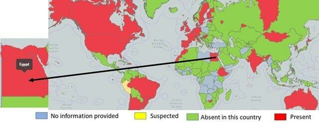
Globally, outbreaks of EHVs are becoming more frequent (Figure 1). The most recent outbreak of equid herpesvirus in Europe occurred in 2021, originating from a show-jumping event in Spain with related outbreaks in other European countries such as France, Belgium, and Germany. The rapid spread of the neurological form of the virus (EHV-1) prompted the cancellation of several international events in Europe by the governing body of Equestrian Sports (FEI). Additionally, a significant outbreak of EHV-4 took place at a Standardbred horse-breeding farm in northern Germany in 2017 .
In Africa, where about 60% of the world's horse population and over 95% of all donkeys and mules are concentrated , Ethiopia stands out with approximately half of the continent's equine population. FAO statistics indicate that Ethiopia is home to over 7 million donkeys, mules, and horses . A recent study focusing on equids exhibiting respiratory symptoms in Ethiopia revealed the presence of EHV-1, 2, and 5. Notably, EHV-1 was identified as the predominant type, accounting for 62% of all equid species .
However, despite the economic importance of horses in Egypt, there is currently a lack of accurate and official documentation regarding the health status of horses and vaccination programs. This information gap can pose challenges in managing and preventing equine diseases, including those caused by viruses like EHV-1. Establishing comprehensive health monitoring and vaccination programs can help protect the health of horses and the sustainability of the equine industry in Egypt . Given the diverse sources of income generated by the horse industry at the national level, the outbreak of any disease affecting the equine population has the potential to significantly impact the economic well-being of the industry.
EHVs, which are endemic viruses in Egypt, pose a continuous and ongoing threat to the horse industry in the country. EHV-1, EHV-2 and EHV-4 were detected in clinical samples collected from horses in 2005 and 2006 (Amer et al., 2011). Furthermore, isolation of EHV-1 from aborted fetuses in Egypt was documented (Abd El-Hafeiz et al., 2010), and isolation of EHV-4 was documented also from aborted fetuses in Egypt during 2017 . These viruses can have detrimental effects on the health and productivity of horses, potentially leading to economic losses for breeders, competitors, and exporters alike. It is essential for the Egyptian equine industry to continue implementing effective disease management and biosecurity measures to mitigate the risks associated with EHV outbreaks and safeguard the industry's economic stability .
Genome Organization and Biological Functions of Viral Proteins
The complete genome sequence of EHV-1 has been published, providing valuable information about the virus's genomic organization. EHV-1 possesses a linear double-stranded DNA genome that is approximately 150.2 kilobasepairs (kbp) in size and has a base composition with around 56.7% G+C content. The genome structure of EHV-1 comprises of a long unique region (UL) spanning 112,870 base pairs (bp) that flanked by a small, inverted repeat sequence (TRL/IRL) with a length of 32 bp, a short unique region (US) covering 11,861 bp, which is flanked by a large inverted repeat (TRS/IRS) spanning 12,714 bp. Within the EHV-1 genome, there are 80 open reading frames (ORFs), which encode 76 different genes. Notably, four ORFs are duplicated and present in the terminal repeat sequence (TRS). These duplicated ORFs are named ORF 64, 65, 66, and 67 and are found in the sequences flanking the short unique region , 1998).
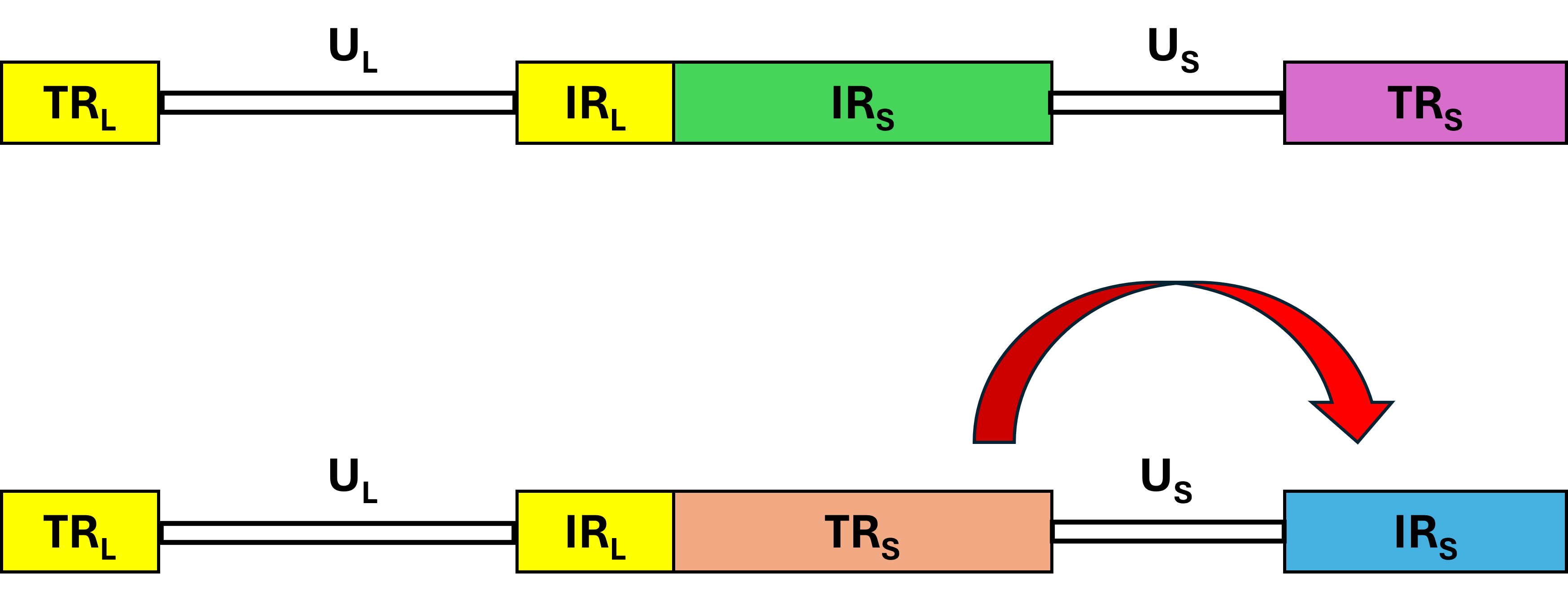
The presence of inverted repeats allows for the generation of two isomeric DNA molecules from the short components, contributing to the existence of virion populations with two orientations (Figure 2). The gene arrangement in EHV-1 is characterized by tightly arranged ORFs with minimal intervening sequences, limited ORF overlap, and relatively few instances of exon splicing. This gene layout in EHV-1 is generally similar to other sequenced herpesviruses, with the primary distinction being that EHV-1 encodes five genes (ORF1, 2, 67, 71, and 75) that lack structural homologs in comparison to all other herpesviruses sequenced thus far. While the functions of some of these genes remain unknown, they are predicted to play a significant role in the unique biology of EHV-1, enabling the virus to adapt to the horse as its natural host . The genomic details of EHV-1 ORFs including the functions of individual genes were illustrated in Table 2.
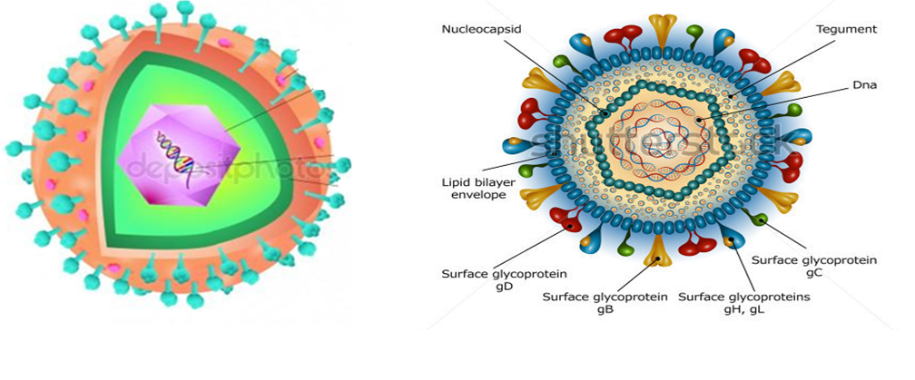
The structural composition of a typical EHV-1 particle encompasses around 30 distinct polypeptides. The virion's core consists of a linear double-stranded DNA genome, meticulously packaged within an icosahedral capsid that displays a T = 16 symmetry. This capsid has an estimated diameter ranging from 100- 110 nm . The nucleocapsid, responsible for housing the viral genome, is composed of six distinct proteins encoded by open reading frames (ORFs) 22, 25, 35, 42, 43, and 56. All herpesvirus capsids share a common structure, characterized by a uniform composition of 162 capsomers, consisting of 12 pentons and 150 hexons. The nucleocapsid also features a ring-shaped assembly of 12 portal proteins that facilitate the entry of viral DNA into the capsid. This conservation in capsid protein structure and organization persists across various herpesvirus lineages, despite differences in nomenclature . The structural architecture of atypical herpesvirus is illustrated in Figure 3.
The region situated between the nucleocapsid and the envelope, known as the amorphous tegument, comprises approximately twelve discrete proteins. These proteins are expressed by ORFs such as ORF 11, 12, 13, 14, 15, 23, 24, 40, 46, 49, 51, and 76. Notably, UL36, a significant tegument protein, has a propensity to bind with the capsid's pentons (VP5), contributing to the establishment of icosahedral symmetry within the innermost segment of the tegument. The viral envelope, which encases the nucleocapsid and tegument, originates from modified host-derived cell membrane regions . EHV-1's envelope contains eleven glycoproteins, which serve as homologs to those found in HSV-1. The study highlights the conservation of these eleven glycoproteins, designated as gB-gp14, gC-gp13, gD-gp18, gE, gG, gH, gI, gK, gL, gM, and gN, across all members of the Alphaherpesvirus genus. These glycoproteins have been named consistently with HSV-1 nomenclature due to their widespread presence. Like other herpesviruses, EHV-1's envelope glycoproteins play a critical role in determining viral entry into host cells, the range of host susceptibility, intercellular viral spread, pathogenic potential, and the subsequent immune responses elicited during infection . Notably, EHV-1 possesses an additional gp2 protein, sharing sequence similarity exclusively with AHV-3 and EHV-4 viruses. When both the tegument and viral envelope are present, the virion size significantly increases to approximately 300 nm, surpassing its nominal size of 120 nm ; Paillot et al., 2008).
Cellular Invasion and Virus Replication: Exploring the Intricacies
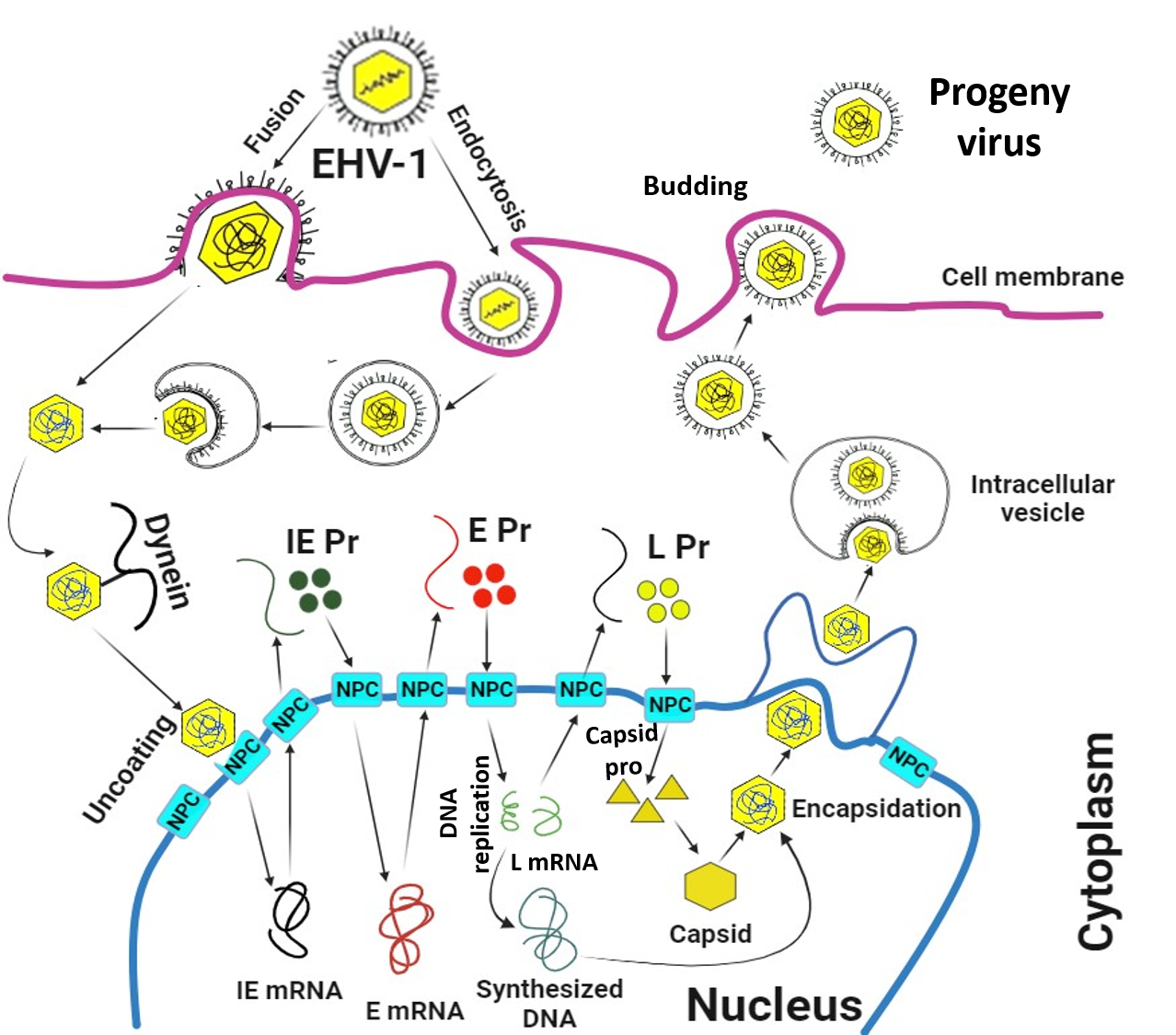
The lytic replication cycle of EHV-1 involves several key steps (Figures 4 and 5) that include entry into a permissive host cell, virus uncoating, virus replication, virion assembly and virion particle egress. The EHV-1 is known to infect various cell types in horses, including endothelial cells in inner organs, epithelial cells in the respiratory tract, mononuclear cells in lymphoid organs and peripheral blood. Similar to other Alphaherpesviruses, EHV-1 initiates infection by attaching to heparan sulfate molecules on the cell surface. This initial attachment is mediated by glycoproteins C and B (gC and gB). Following this attachment, gD on the virus binds to specific receptors on the cell surface. However, EHV-1 has a unique feature in that it utilizes Major Histocompatibility Complex I (MHC-I) molecules on certain equine cells as entry receptors. These MHC-I molecules directly interact with gD on the viral envelope, facilitating the entry of EHV-1 into those cells. This process of viral entry is essential for the initiation of infection and the subsequent replication of the virus within host cells ; ; ; ; Osterrieder and Van de Walle, 2010; .
EHV-1 has the capability to invade permissive cells through two distinct mechanisms, either through direct fusion with the host cell membrane or via cell-mediated endocytosis resulting in a productive infection . The direct fusing of the viral envelope with cellular membranes has been proposed as a function of glycoprotein H (gH) . It has also been known for a long time that gL is necessary for proper gH folding, trafficking, and operation. Activating gH/gL to prime gB for fusion is the first step in the sequential process of fusion, according to other research, which begins with gD attaching to its corresponding receptors .
These entry routes facilitate the release of viral nucleocapsid and tegument proteins into the host cell upon successful entry. As is common with Alphaherpesviruses, once the virus has entered the host cell, the tegument proteins separate from the nucleocapsid. Subsequently, the capsid embarks on a journey along microtubules, guided by dynein, a motor protein that moves in the direction of the microtubules minus end. This mode of capsid transport is particularly significant in infections involving cell types like neurons, where the virus may need to traverse a considerable distance from the infection site to reach the cell nucleus . Upon reaching the cell nucleus, the capsid directly interacts with the nuclear pore complex (NPC) and proceeds to release its contents into the nucleus, leaving the capsid itself in the cytoplasm. In the context of HSV-1, this process is facilitated by the inner tegument protein UL36 (ICP1/2), which carries a nuclear localization signal. Additionally, nucleoporins Nup358 and Nup214 play crucial roles by binding either directly or indirectly to the capsid during this process . All these associations and interactions are essential for the successful nuclear import of the viral DNA, a process that relies on importin β . Once inside the nucleus, the virus embarks on crucial processes such as transcription and replication of its genome, which are pivotal steps in the assembly of virus progeny . These events lead to substantial reorganization of the nucleus, resulting in an enlarged nucleus, disruption of the nucleolus, perturbation of nuclear domain-10 (ND-10) structures, chromatin condensation, and eventual chromatin degradation, along with the destruction of the nuclear lamina, especially during late infection .
The coordinated transcription of EHV-1 genes is regulated by a group of proteins, including one immediate early (IE) protein (IEP), four early proteins (EICP0, EICP22, EICP27, and IR2), and the late EHV-1 α-gene trans-inducing factor (ETIF or VP16) . This cascade of regulatory events is initiated by a protein in EHV-1 analogous to the tegument VP16 protein, which serves as a transactivating agent for IE (α) gene expression. During viral entry, the tegument protein VP16 is introduced into the host cell and plays a pivotal role in facilitating the efficient activation of the virus's lytic replicative cycle. The Inducible Expression Promoter (IEP) of EHV-1, consisting of 1487 amino acids, is encoded within both of the inverted repeats, and this discovery holds paramount importance in understanding the virus replication process. This passage delves into various aspects, including the induction of heterologous viral promoters during the early stages of infection, the intrinsic regulatory mechanisms governing gene expression, and the cooperative activation of both early (E or β) and late (L or γ) viral genes through synergistic interactions involving EICP22 and EICP27.
Regarding EHV-1, research has documented the trans-activating abilities of the IE and EICP0 proteins. Interestingly, their interaction is characterized by an antagonistic dynamic rather than a synergistic one. Within the genome of EHV-1, genes designated as E have been found to be significantly involved in facilitating the virus's replication activities. Conversely, genes labeled as L are responsible for encoding the structural proteins that constitute the viral structure. Drawing from the paradigm of HSV-1 replication, current research suggests that once the E proteins are synthesized, viral DNA replication initiation occurs. This process involves the intricate interplay of at least seven early origin gene products: UL5, UL8, UL9, UL29, UL30, UL42, and UL52 ; .
Like other herpesviruses, EHV-1 can exist for the entirety of a susceptible host's life after first infection in their cells. Following the initial phases of EHV-1 infection in the epithelial upper respiratory tract, infected horses go through a latency period in which they do not exhibit any symptoms of disease, viral shedding, or cell-associated viremia. Although a well-coordinated, productive EHV-1 infection results in active viral gene expression, latency is distinguished by viral gene expression limitation, which prevents the synthesis of viral components and eliminates infectious virus particles ; . It has been determined that the primary sites of EHV-1 latency in horses are in lymphocytes, both circulating and draining lymph nodes, and sensory nerve cell bodies inside the trigeminal ganglia. The latency-associated transcript (LAT) antisense to the immediate-early viral gene (ORF 64) or a regulatory early gene (ORF 63) is the only instance of the repressed transcription and translation of the virus during latency ; .
Unraveling the Pathobiology of EHV-1 infection
The pathogenesis associated with EHV-1 infection has been elucidated using an experimental infection model utilizing the AB4 strain of EHV-1. EHV-1 is known for its high virulence and extreme transmissibility, primarily affecting equine species. The primary mode of transmission typically involves direct contact with contaminated materials, such as nasal secretions and tissues from aborted fetuses, or indirect transmission through inanimate objects known as fomites. EHV-1 primarily replicates in nasal and mucosal epithelial cells, particularly in horses with a deficient mucosal immune response. Following viral replication, these epithelial cells in the upper respiratory tract (URT) undergo rapid necrosis, leading to robust inflammatory responses and the development of erosions. Consequently, infectious virus shedding occurs through the nasal route . EHV-1 is known for efficiently spreading within the upper respiratory tract (URT) by exploiting infected mucosal monocytes, which serve as a means to penetrate deeper connective tissues. This phenomenon has been well-documented in previous research (Gryspeerdt et al., in 2010; .
Within the first 24 hours of infection, infected mononuclear leukocytes are observed in lymphoid organs associated with the respiratory tract, both in the parenchyma and sinuses . EHV-1 then undergoes a subsequent replication phase, resulting in increased viral particle production. This process leads to the release of infected leukocytes through efferent lymphatic vessels, allowing entry into the systemic circulation and resulting in viremia, where virus particles are associated with cells. Viremia is a critical factor in the progression of EHV-1 pathogenesis, leading to further replication at secondary sites, such as the endothelial tissue of the gravid uterus or the central nervous system.
The development of viremia is associated with two significant clinical outcomes often seen in EHV-1 infections: the cessation of pregnancy and the emergence of a neurological syndrome (equid herpesviral myeloencephalopathy [EHM]), both of which are closely related to respiratory pathologies caused by EHV-1.Thrombo-ischemic necrosis in the spinal cord and vascular endothelial infection are associated with EHM, a severe neurological disease caused by EHV-1 infection. A single nucleotide polymorphism (A2254 → G2254) in ORF30, which enables the viral DNA polymerase to replace aspartic acid for asparagine (N752 → D752), has been connected to EHV-1 strains that are associated with EHM. It is debatable, nevertheless, how important this mutation was in the development of EHM in infected horses. Additional host and viral factors could potentially exacerbate the diseases ; ; .
EHV-1 interactions with the host immune system
Understanding the interaction between EHV-1 and the host's immune response is crucial for developing effective immunotherapy. First, the mucosa of the upper respiratory tract acts as a barrier to prevent the infection of EHV-1. Therefore, in order to recognise and neutralise the virus at the site of entry and to later induce adaptive immunity, a timely and effective mucosal innate immune response is crucial . When equine subjects were experimentally infected with a virulent strain of EHV-1, viral components were detected in the regional lymph nodes (LNs) of the respiratory system within 12 hours post-infection (hpi). This early presence of viral components suggests that EHV-1 engages with the host's immune system, triggering an immediate host response characterized by the release of inflammatory cytokines.
EHV-1 infected horses typically display virus-neutralizing (VN) and complement-fixing (CF) antibodies within two weeks of infection. While VN antibodies provide longer protection (up to a year) and are type-specific, CF antibodies have cross-reactivity between EHV-1 and EHV-4 but only last for about three months. The host's humoral immunity primarily targets epitopes on the surface of envelope glycoproteins of EHV-1 (. Various antibody isotypes, including IgGa, IgGb, IgGc, IgGd, IgM, and IgG (T), have been detected in EHV-1-infected horses. While circulating antibodies can mitigate the initial respiratory infection caused by EHV-1, they are less effective against the more severe outcomes of abortion and neurological pathologies, which result from cell-associated viremia (Rusli et al., 2014; . After initial and repeated experimental infections with a virulent EHV 1, nasal wash samples from weanling horses were collected for analysis of EHV-1 specific antibody responses using the ELISA assay to monitor IgGa, IgGb, IgG (T), IgA, and IgM. In the nasal mucosa over the duration of the 13-week sample period, IgA was the most common antibody isotype detected. Induced mucosal IgA with VN activity was the experimental initial EHV-1 infection. After experimental primary infection with EHV-1, mucosal IgA with VN activity was induced. The duration of the IgA response lasts for at least 26 weeks with repeated infections ,
Numerous investigations have examined the presence of cytokines' nucleic acid or protein, including interferon gamma (IFNγ), interferon alpha (IFNα), interleukin-4 (IL-4), IL-10, and tumour necrosis factor (TNF-α), in order to characterise cytokine responses to EHV-1 infection. In the first ten days following an experimental EHV-1 infection in ponies, interferons were found in serum and nasal secretions . Prior studies have reported an increase in the expression of pro-inflammatory cytokines in an in vitro model of equine endothelial cells (EECs) at 10 hpi with neuropathogenic or non- neuropathogenic strains of EHV-1. This early up-regulation of inflammatory cytokines is essential for stimulating and activating the adaptive component of the host immune response, aiding in the elimination of the viral antigen (Johnstone et al., 2016). However, there is concern in the academic community that this pro-inflammatory response may also induce pathological effects that could counteract its antiviral benefits. Furthermore, the host's humoral immunity to EHV-1 infection is reported to be temporary, making horses susceptible to reinfection even after vaccination (van der Meulen et al., 2006).
After cell-associated viremia has been established, eliminating EHV-1 effectively requires an active cytotoxic T lymphocyte (CTL) response. After experimental EHV-1 infection, there is an increased CTL response and interferon-gamma (IFN-γ) that are essential for triggering antigen-presenting cells and boosting the antiviral activity of circulating cytotoxic CD8 T cells. The frequency and magnitude of circulating CTLs are correlated with protection against EHV-1-associated diseases .
In the context of EHV-1 exposure, adult ponies with an increased production of EHV-1-specific circulating CTLs exhibit fewer clinical symptoms compared to young ponies with low EHV-1 activity in their circulating CTLs. This highlights the importance of CTL precursor or memory cells in EHV-1 re-infection, offering a reasonable approach to evaluate the efficacy of EHV-1 vaccines in equine populations. Epitopes of the gene product of EHV-1 IEP, encoded by ORF 64, have been specifically recognized by CTLs derived from horses expressing the MHC class 1 A3/B2 serological haplotype . To identify host-specific factors leading to EHM, elderly mares were used as an experimental equine EHV-1 model in a study by Giessler et al. (2024). After an EHV-1 Ab4 neuropathogenic strain infection was used in an investigation, non-EHM horses showed early elevations of IFN-α (nasal secretions), IRF7/IRF9, IL-1β, CXCL10, and TBET (blood), along with an increase in IFN-γ throughout viremia. In contrast, EHM horses' nasal secretions had low amounts of IFN-α, but their blood showed a correlation between viremia and peak levels of IRF7, IRF9, CXCL10, and TGF-β. In addition, compared to non-EHM horses, EHM horses had significantly higher serum IgG3/5 antibody titres and levels of IL-10 in nasal secretions, peripheral blood mononuclear cells, and CSF. These results suggest that the upregulation of cytokines and chemokines linked to cellular immunity, as well as the prompt activation of type 1 IFN, are necessary for protection against EHM. The activation of TH-2 or regulatory immunity, on the other hand, seemed to be associated with an increased risk of EHM .
Previous research has identified several EHV-1 modulatory genes that could account for the absence of long-term immunity (Van ; ; . Crucially, the products of two genes have been found to interfere with innate immunity and MHC-I expression, both of which are necessary for the production of CTL responses. UL49.5 protein is one of these genes; it has been shown to control MHC-1 expression by blocking the transporter linked to antigen processing (TAP) . The second is the EHV-1 ORF1 gene, which codes for a protein like the UL56 protein (pUL56) of the herpes simplex virus 1. In vitro, MHC-1 cell surface expression was reduced by EHV-1 pUL56 . Deletions in the ORF1/2 regions impact TH-1- and IL-8-specific T box transcription factor (T-bet) responses, nasal viral shedding, and clinical illness in vivo in experimentally infected ponies . EHV-1 pUL56 has been shown in another investigation to modulate immunity in equine respiratory epithelial cells (EREC) but not in peripheral blood mononuclear cells (PBMCs). According to the study, EHV-1 pUL56 changed the amounts of IFN-alpha and IL-10 mRNA expression while downregulating MHC-I and MHC-II expression in ERECs. Furthermore, ERECs with deletion of EHV-1 pUL56 had higher levels of chemokines and neutrophil and monocyte chemotaxis .
Exploring Therapeutic Approaches, Vaccines, and Control Strategies for EHV-1
Currently, there is no known pharmaceutical agent that can comprehensively mitigate the adverse effects of EHV-1-related ailments. However, the implementation of rigorous hygiene and management protocols, in conjunction with the appropriate administration of therapies aimed at alleviating specific symptoms in infected equids, has the potential to effectively limit the transmission of the viral pathogen. The current guidelines for managing recumbent equines involve providing supportive care, ensuring proper nutritional support, and maintaining adequate hydration. Additionally, it is advisable to perform regular bladder and rectal evacuations to prevent colic. Furthermore, efforts should be made to reduce inflammation in the central nervous system . Symptomatic treatment with non- steroidal anti-inflammatory agents as an adjunct therapy may be helpful ; .
Pharmacological interventions, including the use of corticosteroids and immunomodulatory agents, may be considered for symptomatic management of initial signs in cases of EHM. However, there is no empirical study to definitively establish the efficacy of either drug class. Therefore, caution should be exercised to avoid triggering the reactivation of virus shedding in latently infected horses. Corticosteroids have been suggested to have a protective effect on the cellular response to central nervous system (CNS) infection, which may help mitigate the development of hemorrhagic, edematous, vasculitic, and thrombotic manifestations commonly seen in the early stages of EHM. As a result, the administration of corticosteroids is typically recommended only for severe cases of EHM .
Similarly, the use of immunostimulants administered to horses before exposure to stressors might help prevent viral reactivation and replication, but their specific value in treating EHV-1 infection remains uncertain and requires further research . Antiviral drugs, especially virustatic agents like acyclovir derivatives, are theoretically beneficial for EHV-1 infection. Beside acyclovir, prophylactic administration of valacyclovir hydrochloride has been tried in experimentally infected horses with demonstrable benefits . Ganciclovir has been demonstrated to be the most potent inhibitor of EHV-1 infection in an in vitro study that investigated the efficacy of many antivirals against EHV-1 . The antiviral efficacy of many porphyrin compounds against EHV-1 was assessed. Specifically, Cu (III) and Fe (II) tetrasulfonated phenylporphyrin demonstrated potent antiviral activity against EHV-1. Porphyrin-containing substances inhibited cell-to-cell fusion, gB-mediated viral entry, and free virus . More recently, a study found that giving ponies experimentally infected with the EHV-1 C(2254) variation valganciclovir orally lowers clinical symptoms, virus shedding, and cell-associated viremia (Thieulent et al., 2022).
EHV-1 infection, like other herpesviruses, is a complex viral infection characterized by the establishment of persistent latent infection, which allows the virus to naturally persist in horse populations throughout the year. EHV-1 has evolved various strategies to evade components of the host's innate and adaptive immune responses (van . As a result, the development of an effective EHV-1 vaccine requires the ability to trigger strong and persistent humoral (antibody-based) and cell-mediated (involving immune cells) immune responses against the virus. Furthermore, the induction of cell-associated viremia is a crucial step in the development of abortion and EHM. Therefore, a promising vaccine candidate must be capable of eliciting specific immune responses that can effectively inhibit the progression of cell-associated viremia and prevent the occurrence of these pathological conditions.
Despite the limited effectiveness of subunit, live, inactivated, and DNA vaccines, vaccination has proven to be the most effective preventive measure against EHV-1 infection in horses during the past few decades. However, as of right now, there is no vaccine that can protect against the neurological type of EHV-1 . Currently, available vaccines for EHV-1 include inactivated whole virus vaccines and modified live vaccines (MLV). In a recent research study, three groups of horses were either given a saline placebo or immunized with two different vaccines. The Rhinomune (Boehringer Ingelheim), which is formulated from the Rac-H strain, and Pneumabort K-1B (Zoetis), containing EHV-1 1P and 1B strains. After administering these vaccines, their effectiveness was evaluated through an EHV-1 challenge experiment using the Findlay OH03 strain. The study found that the saline control group displayed clinical symptoms of EHV-1 disease, including fever, lethargy, reduced appetite, coughing, nasal discharge, and labored breathing. Both Rhinomune (Boehringer Ingelheim), a modified live vaccine (MLV), and Pneumabort K-1B (Zoetis), an inactivated vaccine, demonstrated efficacy in reducing the clinical incidence of the disease. However, the MLV vaccine showed better protection against EHV-1 disease . While both Rhinomune (Boehringer Ingelheim), a modified live vaccine (MLV), and Pneumabort K-1B (Zoetis), an inactivated vaccine, showed efficacy in reducing the clinical incidence of EHV-1 disease, their effectiveness in preventing EHV-1-induced abortion or EHM is still not definitively proven.
EHV-1 antigens are also included in some multivalent vaccines available globally, typically in their inactivated forms. Additionally, there have been attempts to develop recombinant vaccine models that express EHV-1 glycoproteins (gB, gC, and gD). While these recombinant vaccines have been shown to reduce initial nasal viral shedding in vaccinated horses, they have demonstrated less efficacy in protecting against cell-associated viremia and clinical signs of the disease. While there have been advancements in EHV-1 vaccine development, there is still a need for further research to develop vaccines that provide comprehensive protection against EHV-1-induced abortion, EHM, and other clinical manifestations of the disease . Intriguingly, a recombinant vaccine expressing an EHV-1 IE gene, encoded by ORF 64, significantly reduced cell-associated viremia in vaccinated ponies, however, its effect on EHV-1 induced abortion and EHM remain inconclusive . There is currently no available vaccine that completely prevents EHV-1 infection, or EHV-1-induced cell-associated viremia or latency, and EHV-1 myeloencephalopathy has been reported in vaccinated horses . Vaccinating horses at risk of exposure to equid herpesvirus-1 (EHV-1) is recommended as a preventive measure to reduce the severity of EHV-1-related clinical manifestations. An exhaustive breakdown of all vaccines currently accessible in the field are mentioned in Table 3. The American Association of Equine Practitioners (AAEP) has updated its guidelines for vaccinating adult horses, providing detailed recommendations for EHV-1 vaccination. These guidelines serve as a valuable resource for horse owners and veterinarians to ensure the appropriate and effective vaccination of horses to protect them against EHV-1.
Control measures for managing equid herpesvirus type 1 (EHV-1) infection are designed to achieve two main objectives: mitigating the spread of the virus among susceptible horses and preventing virus reactivation in horses with latent infections. By effectively implementing these control measures, the spread of EHV-1 can be minimized, protecting both susceptible equine individuals and the overall equine population from the virus. These additional control measures are crucial for effectively managing and preventing the spread of EHV-1 within equine populations and farms. They aim to minimize the risk of transmission and maintain the health of the equine population .
Conclusions
EHV-1 remains a persistent threat to the equine industry, necessitating proactive prevention measures. Despite significant research, many aspects of EHV-1, including the specific host and environmental factors contributing to myeloencephalopathy, remain poorly understood. More extensive epidemiological investigations are essential to uncover the determinants of this disease. While in vitro studies have shed light on how EHV-1 modulates host immune responses, more in vivo research is needed to understand these effects in susceptible hosts. Further research should focus on identifying potential therapeutic targets for EHV-1. Advancements in EHV-1 therapeutics and regulations hinge on integrating comprehensive epidemiological data with a deep understanding of the virus's complex biology and pathogenesis.
Compliance with Ethical Standards: NA
Funding source:
The authors did not receive any funds for this study; it was financially supported by the Authors' efforts.
CRediT Authorship contribution statement:
Conceptualization, AFA, MAR; Writing original draft, AFA, RTH, RFE, MAR, and MM; Writing-Review and Editing, AFA, and MAR, Visualization, AFA, RTH, RFE, MAR, and MM; Supervision, and Funding Acquisition, MM
Declaration of competing interest:
The authors declare that they have no known competing financial interests or personal relationships that could have appeared to influence the work reported in this paper.
Data availability:
We have provided all data in the supplemented material.
Figures
Figure 1.
Mapping the Worldwide Distribution of EHV (Ata et al., 2022)
Figure 2.
Inversion in the orientation of the TRS and the IRS of EHV-1
Figure 3.
Schematic illustration of EHV-1 structure.
Figure 4.
EHV-1's lytic life cycle. The virus gains entry into susceptible cells, either through membrane fusion or through the non-classical endocytosis pathway. Subsequently, the nucleocapsid is released into the cytoplasm of the infected cell, then transported to the nucleus by dynein, attaches to the nuclear pore complex (NPC) and introduces the viral DNA directly into the nucleus. This initiate the viral gene expression via the transcription of the Immediate Early (IE) (a) gene. Then, immediate early proteins are produced in the cytoplasm and migrate to the nucleus, where they oversee the transcription of Early (E) (b) genes. Early proteins, synthesized in the cytoplasm, move to the nucleus to kick start virus DNA replication and the expression of Late (L) (g) genes. Afterwards, some of the L proteins produced in the cytoplasm migrate to the nucleus to assemble the capsid before encapsulating the new virus DNA. The newly constructed virion subsequently traverses the nuclear and cytoplasmic membranes before its eventual release from the cell.
Figure 5.
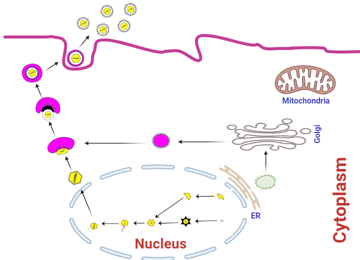
Herpesvirus egress pathway. Once the virus genome is encapsulated within the nucleus, the herpesvirus nucleocapsid will emerge through the inner nuclear membrane, leading to the positioning of an enveloped primary virion near the nucleus. This initial envelope is de-envelopment as the virus moves into the cytosol, where the nucleocapsid obtains tegument proteins. Subsequently, a final envelopment happens within the cytoplasm, originating from the trans-Golgi network. The enveloped virion is then enclosed in a vesicle and conveyed to the plasma membrane for eventual release.
References
Ackermann, M., 2006. Pathogenesis of gammaherpesvirus infections. Vet. Microbiol. 113, 211–222.
Afify, A.F., Ahmed, B.M., Salem, S.A., El-Sanousi, A.A., Shalaby, M.A., 2017. First Isolation and identification of EHV-4 during abortion outbreak among Arabian horses in Egypt reflects an alteration in pathogenic potentiality of EHV-4. J. Virol. Sci. 2, 92-101.
Ahdy, A.M., Ahmed, B.M., Elgamal, M.A., Shaalan, M., Farag, I.M., Mahfouz, E.R., El-Sanousi, A. A., 2022. Detection of Equid Alphaherpesvirus 1 from Arabian Horses with different clinical presentations between 2016-2019 in Egypt. J. Equine Vet. Sci. 114, 103960.
Ahmed, B.M., Bayoumi, M.M., Farrag, M.A., Elgamal, M.A., Daly, J.M., Amer, H.M., 2022. Emergence of equine influenza virus H3Nx Florida clade 2 in Arabian racehorses in Egypt. Virol J. 12, 19(1), 185.
Allen, G. P., Kydd, J. H., Slater, J. D., Smith, K. C., 2004. Equid herpesvirus 1 and equid herpesvirus 4 infections. Infectious disease of livestock. R. C. T. J. A. W. Coetzer. Oxford, New York, OUP Southern Africa, 2nd Edition, 2: 829-859
Al-Shammari, Z., Ahmed, B., Haroun, M., Afify, A.F., Elsanousi, A.A., and Shalaby, M., 2016. A first molecular phylogeny of an Egyptian equine herpesvirus-4 strain derived from a fetal Arabian horse. J. Vet. Sci. Med. Diagn. 5(10.4172), 2325-9590.
Amer, H.M., Shaltout, A.K., El-Sabagh, I.M., El-Sanousi, A.A., Shalaby, M.A., 2011. Prevalence of equine herpes viruses 1, 2 and 4 in Arabian horse population in Egypt. Afr. J. Microbiol. Res. 5(27), 4805-4811.
Asteraye GB, Pinchbeck G, Knight-Jones T, Saville K, Temesgen W, Hailemariam A, Rushton J. Population, distribution, biomass, and economic value of Equids in Ethiopia. PLoS One. 2024 Mar 22;19(3):e0295388.
Ata, E. B., Shaapan, R. M., Ghazy, A. A., Kandil, O. M., Abou-Zeina, H. A., 2022. Epidemiological aspects of some equine viral diseases. Iraqi j. vet. sci. 37. 121-127.
Azab, W., Bedair, S., Abdelgawad, A., Eschke, K., Farag, G.K., Abdel‐Raheim, A., Ali, A.A., 2019. Detection of equid herpes viruses among different Arabian horse populations in Egypt. Vet. Med. Sci. 5(3), 361-371.
Azab, W., Osterrieder, N., 2012. Glycoprotein B of equine herpesvirus type 1 (EHV-1) is required for entry and cell-to-cell spread of the virus. Vet. Microbiol. 156(1-2), 55-63.
Azab, W., Tsujimura, K., Maeda, K., Kobayashi, K., Mohamed, Y. M., Kato, K., et al., 2010. Glycoprotein C of equine herpesvirus 4 plays a role in viral binding to cell surface heparan sulfate. Virus Res. 151, 1–9.
Black, J. B., Frampton, A. R. 2023. Anti-inflammatory compounds reduce equine herpesvirus type 1 replication and cell-to-cell spread. Front. Vet. Sci. 10, 1165917.
Breathnach, C., Yeargan, M., Timoney, J., Allen, G., 2006. Detection of equine herpesvirus-specific effector and memory cytotoxic immunity in the equine upper respiratory tract. Vet. Immunol. Immunopathol. 111, 117–125.
Breathnach, C.C., Yeargan, M.R., Sheoran, A.S., Allen, G,P., 2001. The mucosal humoral immune response of the horse to infective challenge and vaccination with equine herpesvirus-1 antigens. Equine Vet. J. 33, 651–7.
Callé, A., Ugrinova, I., Epstein, A.L., Bouvet, P., Diaz, J.-J., Greco, A., 2008. Nucleolin is required for an efficient herpes simplex virus type 1 infection. J. Virol. 82, 4762–4773.
Carlson, J.K., Traub-Dargatz, J.L., Lunn, D.P., Morley, P.S., Kohler, A., Kasper, K., Lunn, K.F., 2013. Equine viral respiratory pathogen surveillance at horse shows and sales. J. Equine Vet. Sci. 33(4), 229-237.
Copeland, A.M., Newcomb, W.W., Brown, J.C., 2009. Herpes simplex virus replication: roles of viral proteins and nucleoporins in capsid-nucleus attachment. J. Virol. 83, 1660–1668.
Crabb B. S., Studdert M. J., 1993. Epitopes of glycoprotein G of equine herpesviruses 4 and 1 located near the C termini elicit type-specific antibody responses in the natural host. J. Virol. 67 6332–6338.
Davison, A. J., 2010. Herpesvirus systematics. Vet. Microbiol. 143, 52–69.
Dayaram, A., Seeber, P. A., Greenwood, A. D., 2021. Environmental Detection and Potential Transmission of Equine Herpesviruses. Pathogens (Basel, Switzerland), 10(4), 423
Derbal, S., 2021. Epidemiology of EHV-1 and EHV-4 infections: A review. Int. J. Vet. Sci. 7(2), 108-112.
Derbigny, W.A., Kim, S.K., Jang, H.K., O’Callaghan, D.J., 2002. EHV- 1 EICP22 protein sequences that mediate its physical interaction with the immediate-early protein are not sufficient to enhance the trans-activation activity of the IE protein. Virus Res. 84, 1–15.
Edington, N., Welch, H. M., Griffiths, L., 1994. The prevalence of latent equid herpesviruses in the tissues of 40 abattoir horses. Equine Vet. J. 26, 140–142.
Frampton, A.R., Stolz, D.B., Uchida, H., Goins, W.F., Cohen, J.B., Glorioso, J.C., 2007. Equine herpesvirus 1 enters cells by two different pathways, and infection requires the activation of the cellular kinase ROCK1. J. Virol. 81, 10879–10889.
Garré, B., Van Der Meulen, K., Nugent, J., Neyts, J., Croubels, S., De Backer, P., 2007. In vitro susceptibility of six isolates of equine herpesvirus 1 to acyclovir, ganciclovir, cidofovir, adefovir, PMEDAP and foscarnet. Vet. Microbiol. 122, 43–51.
Giessler, K.S., Goehring, L S., Jacob, S.I., Davis, A., Esser, M.M., et al. 2024. Impact of the host immune response on the development of equine herpesvirus myeloencephalopathy in horses. J Gen Virol. 105(5).
Gilkerson, J. R., Bailey, K. E., Diaz-Méndez, A. and Hartley, C. A., 2015. Update on Viral Diseases of the Equine Respiratory Tract.Veterinary Clinic North America Equine Practice, 31:91–104.)
Goehring, L.S., Lunn, D.P., 2008. Equine herpesvirus myeloencephalopathy,” in Current Therapy in Equine Medicine, 6th Edn, eds Robinson, N.E, Sprayberry, K. (St Louis, MO: Saunders), 77–181.
Goehring, L.S., Wagner, B., Bigbie, R., Hussey, S.B., Rao, S., Morley, P.S., 2010. Control of EHV-1 viremia and nasal shedding by commercial vaccines. Vaccine. 28, 5203–11.
Goodman, L.B., Wagner, B., Flaminio, M.J., Sussman, K.H., Metzger, S.M., Holland, R. et al. 2006. Comparison of the efficacy of inactivated combination and modified-live virus vaccines against challenge infection with neuropathogenic equine herpesvirus type 1 (EHV-1). Vaccine. 24, 3636–45.
Gryspeerdt, A.C., Vandekerckhove, A.P., Garré, B., et al., 2010. Differences in replication kinetics and cell tropism between neurovirulent and non-neurovirulent EHV1 strains during the acute phase of equine herpesvirus myeloencephalopathy. Vet. Microbiol. 141(3-4), 207-217.
Hannant, D., Jessett, D.M., O'Neill, T., Dolby, C.A., Cook, R.F., Mumford, J.A., 1993. Responses of ponies to equid herpesvirus-1 ISCOM vaccination and challenge with virus of the homologous strain Res Vet Sci. 54, 299–305.
Hassanien, R. T., Thieulent, C. J., Carossino, M., Li, G., & Balasuriya, U. B. R., 2024. Modulation of Equid Herpesvirus-1 Replication Dynamics In Vitro Using CRISPR/Cas9-Assisted Genome Editing. Viruses. 16(3), 409.
Hassanien, R.T., El-Nahas, E.M., Mahmoud, N.A., El-Bagoury, G.F. 2000. Molecular Characterization of Equine Herpesvirus-1 and Asinine Herpesvirus-5 Isolated from Aborted Fetuses of Arabian Horses. Thai J. Vet. Med. 50, 397–403.
Heldens, J.G., Hannant, D., Cullinane, A.A., Prendergast, M.J., Mumford, J.A., Nelly, M., 2001. Clinical and virological evaluation of the efficacy of an inactivated EHV1 and EHV4 whole virus vaccine (Duvaxyn EHV1,4). Vaccination/challenge experiments in foals and pregnant mares. Vaccine.19, 4307–17.
Henninger, R. W., Reed, S.M., Saville, W.J., Allen, G.P., Hass, G.F., Kohn, C.W., et al., 2007. Outbreak of neurologic disease caused by equine herpesvirus-1 at a university equestrian center. J. Vet. Intern. Med. 21, 157–165.
Huffman, J.B., Newcomb, W.W., Brown, J.C., Homa, F.L., 2008. Amino acids 143 to 150 of the herpes simplex virus type 1 scaffold protein are required for the formation of portal-containing capsids. J. Virol. 82, 6778–6781.
Hussey, G.S., Giessler, K.S., 2022, Contribution of the immune response to the pathogenesis of equine herpesvirus-1 (EHV-1): Are there immune correlates that predict increased risk or protection from EHV-1 myeloencephalopathy? Vet J. 282, 105827.
Johnstone, S., Barsova, J., Campos, I., Frampton, A.R., 2016. Equine herpesvirus type 1 modulates inflammatory host immune response genes in equine endothelial cells. Vet. Microbiol. 192, 52–59.
Kamel, M., Nauwynck, H., Delva, J., 2020. Equine Herpes viruses Type 1 (EHV-1) and Type 4 (EHV-4)—Masters of Co-evolution and Immune Evasion. Viruses. 12(8), 825.
Kang, H.W., Lee, E.Y., Lee, K.K., Ko, M.K., Park, J.Y., Kim, Y.H., Jeoung, H.Y., 2021. Evaluation of the Variability of the ORF34, ORF68, and MLST Genes in EHV-1 from South Korea. Pathogens. 10(4), 425.
Koppers-Lalic D., Reits, E.A., Ressing, M.E., Lipinska, A.D., Abele, R., et al. 2005. Varicelloviruses avoid T cell recognition by UL49.5-mediated inactivation of the transporter associated with antigen processing. Proc. Natl. Acad. Sci. USA.102, 5144–5149
Kukhanova, M., Korovina, A., Kochetkov, S., 2014. Human herpes simplex virus: life cycle and development of inhibitors. Biochemistry. 79, 1635–1652.
Kurtz, B.M., Singletary, L.B., Kelly, S.D., Frampton, A.R., Jr., 2010. Equine herpesvirus 1 entry via endocytosis is facilitated by αV integrins and an RSD motif in glycoprotein D. J. Virol. 84(1), 1075-1086.
Kydd, J.H., Davis-Poynter, N., Birch, J., Hannant, D., Minke, J., Audonnet, J.-C., et al., 2006. A molecular approach to the identification of cytotoxic T-lymphocyte epitopes within equine herpesvirus 1. J. Gen. Virol. 87, 2507–2515.
Khusro, A., Aarti, C., Rivas-Caceres, R.R., Barbabosa-Pliego, A., 2020. Equine Herpesvirus-I Infection in Horses: Recent Updates on its Pathogenicity, Vaccination, and Preventive Management Strategies. J Equine Vet Sci. 87, 102923.
Laval, K., Poelaert, K.C., Van Cleemput, J., Zhao, J., Vandekerckhove, A.P., Gryspeerdt, A.C., Nauwynck, H.J., 2021. The pathogenesis and immune evasive mechanisms of equine herpesvirus type 1. Fontiers in Microbiology, 12, 435.
Léon, A. G., Fortier, C. F., Tapprest, J., Leclercq, R., Pronost, S., 2008. Detection of equine herpesviruses in aborted foetuses by consensus PCR. Vet Microbiol. 126(1-3), 20-9.
Liu, S. A. (2015). Development and Characterization of a Live Attenuated Vaccine to Combat Equine Herpesvirus Type 1 Infection (PhD thesis, Faculty of the Louisiana State University and Agricultural and Mechanical College, Department of Pathobiology).
Lunn, D., Davis-Poynter, N., Flaminio, M., Horohov, D., Osterrieder, K., Pusterla, N., et al., 2009. Equine herpesvirus-1 consensus statement. J. Vet. Intern. Med. 23, 450–461.
Ma, G., Feineis, S., Osterrieder, N., Van de Walle, G. R., 2012. Identification and characterization of equine herpesvirus type 1 pUL56 and its role in virus-induced downregulation of major histocompatibility complex class I. J. Virol. 86, 3554–3563.
Marzo, A. D. (2013). Is Latent Equine Herpesvirus Type 1 (EHV-1) Reactivated by Triggering Activation of the Unfolded Protein Response in Equine Peripheral Blood Leukocytes? (Master's thesis, College of Graduate Studies and Research, Department of Large Animal Clinical Science, University of Saskatchewan, Saskatoon).
Maxwell, L.K., Bentz, B.G., Gilliam, L.L., Ritchey, J.W., Pusterla, N., Eberle, R., et al., 2017. Efficacy of the early administration of valacyclovir hydrochloride for the treatment of neuropathogenic equine herpesvirus type-1 infection in horses. Am. J. Vet. Res. 78, 1126–1139.
Meselhy, N.K., El-khair, M.A.A., Ahmed, B.M., El-Soally, S.A., Fares, A.M., Nayel, M.A., Hussein, H.A., 2019. Sequence analysis of Seven Equine Herpes Type 1 Viruses circulating in non-vaccinated Arabian and Foreign horses in Egypt. J. Virol. Sci. 5, 11-21.
Minke, J.M., Fischer, L., Baudu, P., Guigal, P.M., Sindle, T., Mumford, J.A., 2006.Use of DNA and recombinant canarypox viral (ALVAC) vectors for equine herpes virus vaccination Vet. Immunol Immunopathol.111, 47–57.
Muylaert, I., Tang, K.-W., and Elias, P., 2011. Replication and recombination of herpes simplex virus DNA. J. Biol. Chem. 286, 15619–15624.
Oladunni, F.S., Horohov, D.W., Chambers, T.M., 2019. EHV-1: A constant threat to the horse industry. Front. Microbiol. 10, 2668
Osterrieder, N., Van de Walle, G.R., 2010. Pathogenic potential of equine Alphaherpes viruses: the importance of the mononuclear cell compartment in disease outcome. Vet. Microbiol. 143, 21–28.
Paillot, R., Case, R., Ross, J., Newton, R., Nugent J. 2008. Equine Herpes Virus-1: Virus, Immunity and Vaccines. Open Vet. Sci. J. 2, 68-91.
Patel, J., Heldens, J., 2005. Equine herpes viruses 1 (EHV-1) and 4 (EHV-4)–epidemiology, disease and immunoprophylaxis: a brief review. Vet. J. 170, 14–23.
Pavulraj, S., Eschke, K., Theisen, J., Westhoff, S., Reimers, G., Andreotti, S., Azab, W., 2021. Equine herpesvirus type 4 (EHV-4) outbreak in Germany: Virological, serological, and molecular investigations. Pathogens. 10(7), 810.
Pellet, P., 2007. The family Herpesviridae: a brief introduction. Fields Virol. 5, 3137–3166.
Perkins, G., Babasyan, S., Stout, A.E., Freer, H., Rollins, A., Wimer, C.L., 2019. Intranasal IgG4/7 antibody responses protect horses against equid herpesvirus-1 (EHV-1) infection including nasal virus shedding and cell-associated viremia. Virology. 531, 219–32.
Pritchard, J.,A. Lindberg, D. Main, Whay, H., 2005. Assessment of the welfare of working horses, mules and donkeys, using health and behaviour parameters. Prev. Vet. Med. 69, 265–283.
Pusterla N., Mapes S., 2008. Evaluation of an Air Tester for the Sampling of Aerosolised Equine Herpesvirus Type 1. Vet. Rec. 163, 306–308.
Pusterla, N., Hussey, G. S., 2014. Equine Herpesvirus 1 Myeloencephalopathy. Veterinary Clinics of North America Equine Practice, 30, 489-506.
Rajcani, J., Durmanova, V., 2001. Mechanisms of replication of alpha-and betaherpes viruses and their pathogenesis. Bratisl. Lek. Listy 102, 505–514.
Reed, S.M., Toribio, R.E., 2004. Equine herpesvirus 1 and 4. Vet. Clin. North Am. Equine Pract. 20, 631–642.
Riaz, A., Murtaz-ul-Hasan, K., Akhtar, N., 2017. Recent understanding of the classification and life cycle of herpes viruses: a review. Sci. Lett. 5, 195–207.
Roizman, B., 1982. The family herpesviridae: general description, taxonomy, and classification, in The Herpes viruses, ed. B. Roizman, (Boston, MA: Springer), 1–23.
Roizman, B., Knipe, D.M., 2001. Herpes simplex viruses and their replication, in Fields Virology, eds. Knipe, D.M., Howley, P.M. (Philladelphia, PA: Lippincott Williams & Wilkins), 2399–2460.
Roizmann, B., Desrosiers, R., Fleckenstein, B., Lopez, C., Minson, A., Studdert, M., 1992. The familyHerpesviridae: an update. Arch. Virol. 123, 425–449.
Rusli, N.D., Mat, K.B., Harun, H.C., 2014. A review: interactions of equine herpesvirus-1 with immune system and equine lymphocyte. Open J. Vet. Med. 4, 294–307.
Rybachuk, G. V. 2009. Antiviral chemotherapeutic agents against equine herpesvirus type 1: the mechanism of antiviral effects of porphyrin derivatives. LSU Doctoral Dissertations. 2545
Sasaki, M., Hasebe, R., Makino, Y., Suzuki, T., Fukushi, H., Okamoto, M., et al., 2011. Equine major histocompatibility complex class I molecules act as entry receptors that bind to equine herpesvirus-1 glycoprotein D. Genes Cells. 16, 343–357.
Schnabel, C. L., Babasyan, S., Rollins, A., Freer, H., Wimer, C. L., et al., 2019. An Equine Herpesvirus Type 1 (EHV-1) Ab4 Open Reading Frame 2 Deletion Mutant Provides Immunity and Protection from EHV-1 Infection and Disease. J. Virol. 93(22), e01011-19.
Slater, J. D., 2014. Equine herpesviruses. In: Sellon, D., Long, M. (Eds.), Equine Infectious Diseases. Saunders Elsevier, St. Louis, MO, USA, pp., 151-168.
Slater, J. D., Borchers, K., Thackray, A. M., Field, H. J., 1994. The trigeminal ganglion is a location for equine herpesvirus 1 latency and reactivation in the horse. J. Gen. Virol. 75, 2007-2016.
Soboll Hussey, G., Ashton, L.V., Quintana, A.M., Van de Walle, G.R., et al., 2014. Equine herpesvirus type 1 pUL56 modulates innate responses of airway epithelial cells. Virology. 464-465, 76-86.
Soboll Hussey, G., Hussey, S.B., Wagner, B., Horohov, D.W., Van De Walle, et al. 2011. Evaluation of Immune Responses Following Infection of Ponies with an EHV-1 ORF1/2 Deletion Mutant. Vet. Res. 42, 23.
Soboll, G., Breathnach, C., Kydd, J., Hussey, S., Mealey, R., Lunn, D., 2010. Vaccination of ponies with the IE gene of EHV-1 in a recombinant modified live vaccinia vector protects against clinical and virological disease. Vet. Immunol. Immunopathol. 135, 108–117.
Spear, P.G., 2004. Herpes simplex virus: receptors and ligands for cell entry. Cell Microbiol. 6, 401–410.
Sutton, G., Garvey, M., Cullinane, A., Jourdan, M., Fortier, C., Moreau, P., et al. 2019. Molecular Surveillance of EHV-1 Strains Circulating in France during and after the Major 2009 Outbreak in Normandy Involving Respiratory Infection, Neurological Disorder, and Abortion. Viruses. 11, 916.
Taktaz Hafshejani, T., Nekoei, S., Vazirian, B., Doosti, A., Khamesipour, F., Anyanwu, M. U. 2015. Molecular Detection of Equine Herpesvirus Types 1 and 4 Infection in Healthy Horses in Isfahan Central and Shahrekord Southwest Regions, Iran. Biomed Res Int. 2015, 917854.
Telford, E. A. R., Watson, M. S., McBride, K., Davison, A. J., 1992. The DNA sequence of equine herpesvirus-1. Virology. 89, 304-316.
Telford, E. A., Watson, M. S., Perry, J., Cullinane, A. A., Davison, A. J., 1998. The DNA sequence of equine herpesvirus-4. J. Gen. Virol. 79 (5), 1197- 1203.
Temesgen, T., Getachew, Y., Negussie, H., 2021. Molecular identification of equine herpesvirus 1, 2, and 5 in equids with signs of respiratory disease in central Ethiopia. Vet. Med. Res. 12, 337-345.
Thieulent, C.J., Sutton, G., Toquet, M.P., Fremaux, S., Hue, E., Fortier, C., et al. 2022. Oral administration of valganciclovir reduces clinical signs, virus shedding and cell-associated viremia in ponies experimentally infected with the equid herpesvirus-1 C(2254) variant. Pathogens. 11(5), 539.
Van De Walle, G.R., Goupil, R., Wishon, C., Damiani, A., Perkins, G.A., Osterrieder, N., 2009. A Single-Nucleo- tide Polymorphism in a Herpesvirus DNA Polymerase Is Sufficient to Cause Lethal Neurological Disease. J. Infect. Dis. 200, 20-25.
van der Meulen, K.M., Favoreel, H.W., Pensaert, M.B., and Nauwynck, H.J., 2006. Immune escape of equine herpesvirus 1 and other herpes viruses of veterinary importance. Vet. Immunol. Immunopathol. 111, 31–40.
Vandekerckhove, A.P., Glorieux, S., Gryspeerdt, A., Steukers, L., Van Doorsselaere, J., Osterrieder, N., et al., 2011. Equine Alphaherpesviruses (EHV-1 and EHV-4) differ in their efficiency to infect mononuclear cells during early steps of infection in nasal mucosal explants. Vet. Microbiol. 152, 21–28.
Vissani, M.A., Perglione, C.O., Zabal, O., Alvarez, G., Thiry, E., Barrandeguy, M., Parreño, V., 2020.Topical Ganciclovir Reduces Viral Excretion in Mares With Equine Coital Exanthema. J. Equine Vet. Sci., 94, 103199.
Vollebregt, T., 2014. Economic analysis of Equine Herpes Virus within the Dutch commercial horse sector. Thesis Business Economics BEC-80424, Wageningen University.
Wagner, B., Goodman, L. B., Babasyan, S., Freer, H., Torsteinsdottir, S., Svansson, V., et al., 2015. Antibody and cellular immune responses of naive mares to repeated vaccination with an inactivated equine herpesvirus vaccine. Vaccine. 33, 5588–5597.