Special Issue: Emerging and Re-Emerging Animal Health Challenges in Low and Middle-Income Countries · Volume 13, Issue 1, 7 August 2025 · Open Access
The Effect of Nano Saffron Extract on Opened Wounds in Rats
·
Affiliations & Notes ⌄
1Department of Surgery and Obstetrics, College of Veterinary Medicine, University of Al-Qadisiyah, Al-Qadisiyah, Iraq. *Correspondence | Muslim F. Diwan, Department of Surgery and Obstetrics, College of Veterinary Medicine, University of Al-Qadisiyah, Al-Qadisiyah, Iraq
The finding of this study supports the efficacy of Nano saffron extract in promoting wound healing compared with the control and vaseline-treated group, the Vaseline treated group exhibit slightly improve wound healing compared to control group with thick epidermis and a thin keratinized layer.
The 5% saffron extract nano particles ointment for treating skin wounds in rats significantly accelerate the healing process by enhancing cellular proliferation, wound contraction, maturation, and remodeling phase.
Further experiments are suggested to assess the effect of Nano saffron on the healing process of chronic open wounds.
Selected from the article's own text.
Abstract
Wound healing is a complex procedure, especially in animals where self-care is naturally limited and exposure to environmental aggravators is high. The present study aims to demonstrate the role of Nano-saffron in natural therapy and alternative medicine. To illustrate this, thirty male Wistar rats, housed in plastic cages, were randomly divided into three groups with each group containing 10 rats. Group 1 (G1) served as control, while group 2 (G2) was treated with Vaseline, and group 3 (3) was treated with 5% Nano-saffron. Rats were anesthetized with IM injection of ketamine (50mg / K.G BW), xylazine (10mg/K.G BW) mixture. A surgical incision was performed at the dorsal region about 10 mm of their back, and treatments were topically applied once a day for seven consecutive days. Thereafter, biopsies were taken on the 7th, 14th, and 21st days and examined for histopathological evaluation of the healing process. Histopathologic evaluation revealed the superiority of healing in group 3, treated with Nano saffron, highlighting its wound healing potential. Histological changes following wound injury in Nano saffron treated skin indicated complete healing of all skin layers, including the epidermis, reticular dermis, papillary dermis, hypodermis, and sebaceous glands. Collectively, Nano saffron demonstrated impressive wound healing capabilities and may offer an economical and safer option for post-wound care agents.
Keywords | Open wound, Nano saffron, Healing, Rat model
Introduction
Wound is a breakdown of the skin with or without loss of underlying connective tissue caused by disease, like an ulcer, or injury, like surgery, chemicals, cold, heat, or pressure ; . Wounds can be classified according to various factors including cause, wither acute or chronic, location, depth, and contamination . Knowing the class of wound is essential for diagnosis, selecting therapy, and determining treatment regimens (Muhamed, 2016).
Wound healing is a natural physiological and biological process in response to tissue injury resulting in the restoration of tissue integrity . It is highly coordinated, multilayered process that begins as an inflammatory response , followed by proliferative phase, and finally a remodeling phase . The participation of numerous cell types, cytokines, mediators and the vascular system . Various conventional approaches are being used to promote wound healing. Recent developments in wound care technology include Nano-therapies, stem cell therapy, and bio-engineered skin grafts .
Nanoparticles are materials with dimensions less than 100 nm with various shapes . It can result in stimulating numerous molecular and cellular processes that aid in creating a wound microenvironment with antibacterial, angiogenic, and anti-inflammatory effects (Anamik et al., 2022). The use of nanoparticles in products for topical applications improves the penetration of these materials compounds deep into skin layers .
The saffron plant belongs to the Iridaceae family. This herbaceous perennial plant can reach heights of 10 cm to 25 cm, developing from its bulbs . Crocus sativus flowers begin to appear in autumn, and they contain a thin style that ends with a single stigma composed of three filaments with an intense red color, which are the plant parts that interest humans . The main bioactive components in saffron are crocetin, crocin, saffranal, and picrocrocin . Saffron has a long history in medicinal use; recent studies validate its potential to lower the risk of many diseases . It exhibits anti-inflammatory activity and antioxidant activity . In wound healing, saffron has various mechanisms, including the enhancement of fibroblasts, epithelialization, and wound closure .
Materials and Methods
The work was conducted to evaluate the effect of Nano-saffron in treatment of open wounds in rats, thirty adult male rate were randomly divided into three groups and each group contain 10 rats (G1 not treated, G2 treated with Vaseline and G3 treated with Nano-saffron), all are anesthetized (by IM injection mix of ketamine 50mg/kg BW and xylazine 10mg/kg BW) to make surgical incision at the dorsal region about 10 mm of their back. The treatment began on the first day of the surgical procedure and continued for 7 consecutive days. The specimens were collected and preserved in a 10% buffered formalin solution then sent to the histological laboratory for evaluation at 7, 14 and 21 days.
A 90-gram sample of saffron was used to obtain 25 grams of saffron extract by using soxhlet device. Then, the saffron extract powder was converted Nano particles by the ultrasonic method (Jinllto and Kenneth, 2010). 10 g of saffron extract was dissolved in 100 ml of ethanol, and then the solution was filtered three times (two times with filter paper and the third time with a syringe filter (22 mm). The filter solution was treated with ultrasonic waves by mean of ultrasonic cell disruptor device (ultrasonic cell disruptor: UCD-150; Ultrasonic power 150 W, Probe 6mm) (Faithful Intelligent Ultrasonic processor) using wave energy 50 watts for 30 min where the saffron particles reached to 62 nm in size (Qais et al., 202), the treated solution was poured in a petri dishes and placed in oven at 50 oC to dry. The dried extract was scraped and collected from the petri dish, ground, and then made into a 5% ointment of Nano saffron 5 gm. of Nano saffron was crushed with 95 gm. of Vaseline in a glass mortar and pestle. Several tests were made to assess the Nano saffron structures, including, Scanning Electron Microscopy, UV vis. absorption spectroscopy, and FTIR.
Results
UV-spectrophotometer
UV-Vis Spectroscopy can be described as an analytical technique used for identifying the absorbance of colloidal solutions and the optical properties of prepared compounds over a range of wave lengths (200 nm to 800nm). Approximately 2 mL of the prepared nanoparticles has been added to a 1 cm path-length quartz cuvette to record the UV–vis spectrum. showed UV–vis spectrum of synthesized nanomaterial with maximum absorption at 440 nm.
FTIR analysis
The Fourier Transform Infrared spectrometer (FTIR) spectrum of synthesized nanoparticles is presented. A vast vibration peak at 3391.68 cm−1 corresponds to the O-H stretch of the alcohol group. The absorption peaks at 2353.12 and 2319.29 cm−1 correspond to the strong O=C=O stretching of carbon dioxide. The strong band at 1712.59 cm−1 represents carboxylic acid (C=O stretch) present in the nanomaterial. The medium band at 1663.17 cm−1 relates to the imine/oxime (C=N stretching). The small vibrational peak at 1374.24 cm−1 corresponds to the medium C–H bending mode attributed to the alkane group (methyl group). Other bonds present in the sample were observed at 1230.16 cm−1 and 1160.52 cm−1, corresponding to strong C-O stretching in alkyl aryl and ester groups, respectively. The prominent vibration peak at 1074.56 cm−1 represents a strong C-O stretching of alcohol.
SEM image analysis
FE-SEM can be described as an analysis that provides information about the morphology of the surface and particle size of prepared samples. It displays FE-SEM analysis of prepared NPs. The formed nanomaterial is uniform in shape with a smooth surface morphology. The diameter of the NPs is about 49.18±3.24 nm for 100 particles.
Clinical evaluation
All animals throughout the experiment time (21 days) post-wounding (PW). Clinically have been seen energetic, healthy, active, and with good healing processes at the site of injury. No infections developed in any of the experimental animals, wounds, and no deaths were recorded. Primarily, all of the wound areas increased in size within 4 hours after wound creation and continued to swell and increase in size for the following 24 hours- PW with inflammatory signs’ exaggeration. The entire wound was swollen, and the wound edges were elevated and red. From the second day, PW showed a thick scab on the wound that persisted for longer than 7 days. The wound size decreased gradually until the 21st day, when it began to form scar tissue.
Surface area
The initial wound surface area (mm2) on the first day of the experiment was (93.1). Skin wounds were measured on days 0, 7, 14, and 21 post wound injury. The average area of wound on the 7th day in G1, G2, and G3 was 60.38, 62.40, and 49.99, respectively (Table 1; Figure 2). There were significant differences between G2 and G3, while they recorded a significant difference in comparison to G1 and G2.
Table 1: Surface area (mm2).
P. G.
0 day
7 days
14 days
21 days
G1
93.1 ± 0 Aa
60.38 ± 0.878 Ab
43.007 ± 0.735 Ac
29.68 ± 0.785Ad
G2
93.1 ±0 Aa
62.40 ± 0.686 Bb
40.19 ± 0853 Bc
26.62± 0.673 Bb
G3
93.1 ±0 Aa
49.99 ± 0.978Db
10.9 ± 0.652 Dc
5.06 ± 0.362 Dd
2.70 < LSD (P 0.05)
Mean± standard error. Means with different capital letters in the same column and small letters in the same row denote a significant difference at P<0.05.
Histopathologic assessment of wound healing
The histopathologic assessment reveals the following changes in every group for 3 weeks.
On the 7th day post-wound injury
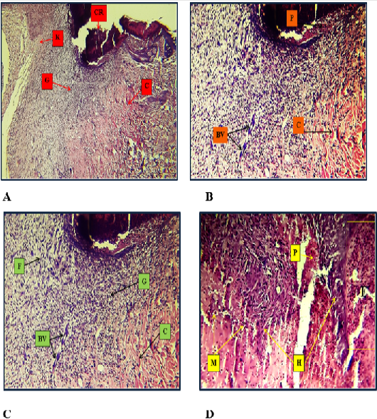
G1: Histopathological changes after wound injury after one week showed that there is a Thick crust attached closely to the edges of the wound, profuse purulent exudate, and wide scar tissue with profuse granulation tissue in the dermis. Profuse purulent exudate which formed from dead neutrophils and tissue debris and thick crust above the edges of wound Thin and branched network of collagen fibers in the dermis (Figure 1A).
G2: Histopathological changes following wound injury, one week after, showed a Thick crust with purulent exudate and complete sloughing of the epidermal layers. They are High granulation tissue with profuse fibrosis, a thin and branched network of collagen, and wide scar tissue (Figure 1B). Showed was thick crust above the edges of the wound. There were profused granulation tissue with few blood vessels, newly formed, and fibrosis. Additionally, a branched network of collagen is present (Figure 1C).
G3: Histopathological changes observed one week after wound injury showed marked downward hyperplasia of the stratum basal in the epidermis. There were Presence of purulent exudate above the site of wound characterized by dead neutrophils and tissue debris. That was hyperemia and inflammatory cells in the dermis. There is hyperemia, as well as the presence of inflammatory cell, primarily macrophages, in the dermis (Figure 1D).
On the 14th day post-wound injury
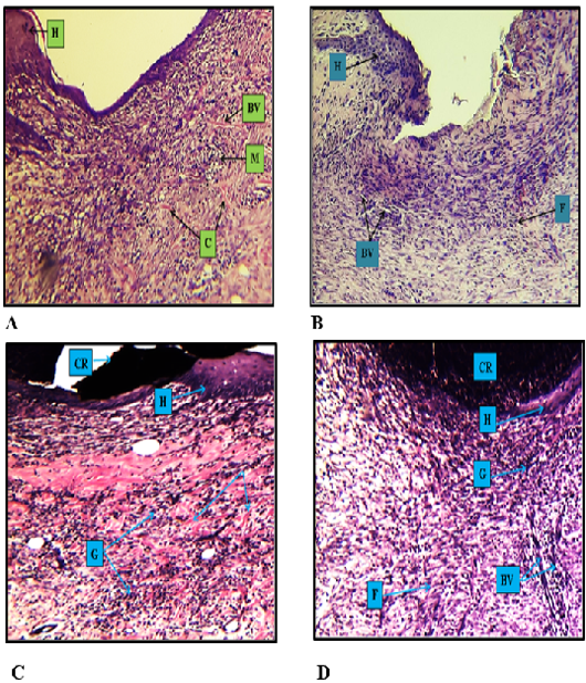
G1: Histopathological changes that follow wound injury after the second week showed Complete sloughing of epidermal layers with slight hyperplasia on both edges of the wound. They are profuse granulation tissue with wide scar tissue. Few hyperplastic areas are present on the edge of the wound, which contains profuse granulation tissue with high fibrosis and newly formed small blood vessels (Figure 2A).
G2: Histopathological changes following wound injury after the second week showed wide scar tissue with complete sloughing of crust and a few purulent exudates above the epidermis. In the dermis, there is profuse fibrosis and few, newly-formed blood vessels with a small amount of collagen fibers. Thin and little hyperplasia of the stratum basal above the edges of the wound. They are Infiltration of inflammatory cells, mainly macrophages, new formation of small blood vessels with fibrosis, and a thin network of collagen fibers in the dermis. With higher magnification, there is hyperplasia of basal cells in the stratum basal, accompanied by infiltration of macrophage and the formation of new small blood vessels (Figure 2B).
G3: Histopathological changes observed in the second week after wound injury revealed the presence of a thick crust closely adhered to the epidermal layers, as well as hyperplasia and proliferation of basal cells in the epidermis. In the dermis, there is high granulation tissue with a fine, irregular network of collagen fibers (Figure 2C). A thick crust is present above the epidermis, and there is hyperplasia of the stratum basale. They are profound granulation tissue with small new blood vessel formation above the site of the wound and profuse fibrosis (Figure 2D).
On the 21st day post-wound injury
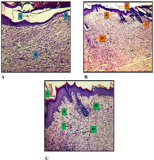
G1: Histopathological changes that follow wound injury after the 3rd week showed thin and proliferating epidermal layers above the wound edges. Wide scar tissue in the dermis with profuse fibrosis horizontally on the wound edges, and small, newly formed blood vessels were found vertically on it. New and few hyperplasia of the stratum basal with a thin keratinized layer were observed. In the dermis, there is scattered infiltration of macrophages, accompanied by profuse fibrosis and a limited number of blood vessel formations (Figure 3A).
G2: Histopathological changes observed after the third week following wound injury revealed a thin keratinized layer with thick epidermal layers, wide scar tissue with severe and profuse fibrosis, and newly formed small blood vessels. Slightly downward hyperplasia of epidermal layers with a thin keratinized layer above it was observed. In the dermis, there is profuse fibrosis and newly formed blood vessels that are vertically oriented on the edges of the wound (Figure 3B).
G3: Histopathological changes following wound injury, observed after the third week revealed normal epidermal layers with a thin, keratinized layer above the epidermis, narrow scar tissue with profuse fibrosis, and newly formed hair follicles in the dermis. Normal epidermal layers with slight hyperplasia of the stratum basal were seen. A thin keratinized layer is closely attached to the epidermis, with newly formed blood vessels and profuse fibrosis, along with small hair follicles in the dermis (Figure 3C).
Discussion
In this study, medicinal plants were used to enhance wound healing processes and reduce the bacterial burden . After preparation of Nano saffron extract, the samples were analyzed for Nano tests to improve and analyze Nano saffron. UV-vis. The spectrum shows a maximum absorption 245 nm. As shown. This is accompanied by . All molecules give similar UV absorption spectra.
FT-IR analyze of Nano saffron sample show O=C=O stretching bond, strong stretch bond of C=O of carboxylic acid, C-N bond of imine/oxime, C-H peak of alkane, the largest vibration peak of C-O starching of alcohol, these features of bonds improve successful preparation of Nano saffron is agreed with . SEM analysis showed the surface morphology and particles size, saffron Nano particles were uniform in shape with smooth surface morphology, the diameter of 100 particles were 49.18±3.24nm indicating Nano size particles, Nano size increase the potency of these particles for penetration of body cells and enhanced the pharmaceutical efficacy, these result agreed with .
During the treatment period, the animals exhibited energetic, healthy, and active behavior, with a good healing process. No infections developed in any wounds, and no deaths were recorded. After 4 hours of wound creation, the wounds increase in size and swelling within 24 hours post wound, which is a sign of inflammation as reported by .
After 7 days post wounding, the wound size decreased gradually till the 21th day becoming a small scar tissue as circular or liner in shape, the superiority of healing was obvious in G3 than other groups, indicate powerful healing characters of Nano saffron this is agreed with . This result may be due to smooth transition from the exudative inflammation phase to the proliferative phase, characterized by reduced leukocyte infiltration, especially at the wound edges.
The finding of this study supports the efficacy of Nano saffron extract in promoting wound healing compared with the control and vaseline-treated group, the Vaseline treated group exhibit slightly improve wound healing compared to control group with thick epidermis and a thin keratinized layer. However, sever and extensive fibrosis in the dermis with only a few newly formed small blood vessels, suggest that Vaseline alone doesn’t significantly promote angiogenesis or collagen deposition as confirmed by , their study highlighted the limitation of conventional topical treatments, which lack bioactive compounds that actively stimulate the wound healing process.
The histopathologic evaluation shows that wound healing in G3 is superior to that in other treated groups at the end of the study. G3 showed normal epidermal layers with slightly hyperplastic stratum basal, a thin keratinized layer closely attached to the epidermis, newly. They were Formed blood vessels profuse fibrosis with small hair follicles in the dermis. This result is satisfactory . Our results show that the Nano saffron was better at wound healing compared to the other groups. The enhance wound healing effects of Nano saffron can be attributed to multiple biological mechanisms, first saffron contain powerful antioxidants which reduce oxidative stress and promote fibroblast proliferation , secondly, anti-inflammatory properties help regulate the secretion of inflammatory mediators such as interleukins and tumor necrosis factors, thereby minimizing prolong inflammation and facilitating tissue repair, thirdly, saffron stimulate collagen synthesis which play a crucial role in skin remodeling and scar tissue formation (Comino-Sanz et al., 2021).
Conclusions and Recommendations
The 5% saffron extract nano particles ointment for treating skin wounds in rats significantly accelerate the healing process by enhancing cellular proliferation, wound contraction, maturation, and remodeling phase. Further experiments are suggested to assess the effect of Nano saffron on the healing process of chronic open wounds.
Acknowledgments
The author is grateful to the University of Al-Qadisiyha, College of Veterinary Medicine for all the facilities to achieve this study.
Novelty Statement
This study introduces a novel approach by using the Nano saffron extract in treating wounds in rat. The findings highlight the positive effect of the Nano saffron extract on wound healing process, suggest their potential utility in wound healing.
Author’s Contribution
Muslim F. Diwan conceptualized and design the experiment. Mohammed A. Mohammed carried out the experimental work. All authors analyzed the data and drafted the manuscript. All authors reviewed and approved the final version of the manuscript.
Conflict of interest
The authors have declared no conflict of interest.
Figures
Figure 1:.
(A) Control group (G1) 7 days post wound shows profuse purulent exudate (P) which formed from dead neutrophils and tissue debris and thick crust above the edges of wound. Thin and branched network of collagen fibers (C) and newly formed blood vessels (BV) in the dermis. 10X H & E. (B) Positive control group (G2) 7 days post wound Shows thick crust (CR) with purulent exudate and complete sloughing at epidermal layers. High granulation tissue (G) with profuse fibrosis, thin and branched network of collagen (C) and wide scar tissue (K). 4X H & E. (C) Positive control group (G2) 7 days post wound shows thick crust above the edges of wound. Profuse granulation tissue (G) with few blood vessels newly formed (BV) and fibrosis (F). Also there is branched network of collagen (C). 10X H & E. (D) Treatment group (G3) 7 days post wound shows there is marked downward hyperplasia (H) of stratum basale in the epidermis. Presence of purulent exudate (E) beneath the epidermis characterized by dead neutrophils and tissue debris. Hyperemia (P) and inflammatory cells (M) in the dermis. 10X H & E.
Figure 2:.
(A) Control group (G1) 14 days post wound shows few hyperplasia (H) on the edge of the wound with profuse granulation tissue which contain high fibrosis (F) and newly formed, small blood vessels (BV). 10X H & E. (B) Positive control (G2) 14 days post wound shows thin and few hyperplasia (H) of stratum basale above the edges of wound. Infiltration of inflammatory cells mainly macrophages (M), new formation of small blood vessels (BV) with fibrosis and thin network of collagen fibers (C) in the dermis. 10X H & E. (C) Treatment group (G3) 14 days post wound shows presence of thick crust (CR) attached closely with the epidermal layers. Hyperplasia (H) and proliferation of basal cells in the epidermis. In the dermis, there is high granulation tissue (G) with fine irregular network of collagen fibers (C). 10X H & E. (D) Treatment group (G3) 14 days post wound shows thick crust (CR) above the epidermis and hyperplasia (H) of stratum basale. Profuse granulation tissue (G) with small new blood vessels formation (BV) above the site of wound and profuse fibrosis (F). 10X H & E.
Figure 3:.
(A) Control group (G1) 21 days post wound shows new and few hyperplasia of stratum basale (H) with thin keratinized layer above it (K). In the dermis, there is scattered infiltration of macrophages (M) with profuse fibrosis and few blood vessels formation. 10X H & E. (B) Positive control (G2) 21 days post wound shows thin keratinized layer (K) with thick epidermal layers (H). Wide scar tissue with severe and profuse fibrosis (F) and newly-formed small blood vessels (BV). 4X H & E. (C) Treatment group (G3) 21 days post wound shows normal epidermal layers with slightly hyperplasia (H) of stratum basale. Thin keratinized layer (K) closely attached with the epidermis. Newly-formed blood vessels (BV) and profuse fibrosis (F) with small hair follicles (HF) in the dermis. 10X H & E.
References
Alistair Y, Clare-Ellen M (2011). The physiology of wound healing. Surgery (Oxford), 29(10) 475-479. https://doi.org/10.1016/j.mpsur.2011.06.011[CrossRef]
Altammar, K.A., 2023. A review on Nano particles: Characteristics, synthesis, applications and challenges. Front. Microbiol., 14: 1155622. https://doi.org/10.3389/fmicb.2023.1155622[CrossRef]
Anamika K, Lalit G, Beom SK (2022). Nanomaterial based therapy for wound healing. Nanomaterials, 12(4): 618. https://doi.org/10.3390/nano12040618[CrossRef]
Angela C, Andrea C, Caterina C, Fernada S (2024). Role of the skin immune system in wound healing. Cell, 13(7): 624. https://doi.org/10.3390/cells13070624[CrossRef]
Bharati KP, Kadamb HP, Ryan YH, Chuen NL, Shabbir MM (2022). The gut-skin microbiota axis and its role in diabetic wound healing. A review based on current literature. Int. J. Mol. Sci., 23(4): 2375. https://doi.org/10.3390/ijms23042375[CrossRef]
Christodoulou E, Nikolaos P, Kadolglou NK, Georgia V (2015). Saffron: A natural product with potential pharmaceutical applications. J. Pharma. Pharmacol., 67(12): 1634-1649. https://doi.org/10.1111/jphp.12456[CrossRef]
Comino SIM, Lopez FMD, Castro B, Pancorbo-Hidalgo PL (2021). The role of antioxidant on wound healing: A review of the current evidence. J. Clin. Med., 10(16): 35-58. https://doi.org/10.3390/jcm10163558[CrossRef]
Dowling A, Cliff R, Grobert N, Hutton D, Oliver R, Oneill O, Pethica J, Pidgeon N, Porritt J, Ryan J (2004). Nanoscience and Nanotechnologies: Opportunities and uncertainties. R. Soc. R. Acad. Eng., 44(1): 7-10.
Edraki M, Akbarzadeh A, Hosseinzaheh M, Tanidh N, Salehi A, Abadi O (2014). Healing effect of sea buckthorn, olive and their mixture on full- thickness burn wounds. Adv. Skin Wound Care, 27(7): 317-323. https://doi.org/10.1097/01.ASW.0000451061.85540.f9[CrossRef]
Ghasemali K, Seyed JH, Peyman Z, Maryam G, Amirhossein A (2008).The effect of saffron (Crocus sativus) extract for healing of second-degree burn wounds in rats. Keio J. Med., 57(4): 190-195. https://doi.org/10.2302/kjm.57.190[CrossRef]
Ibtissam M, Mohamed A, Abdelbasset B (2019). Traditional and modern uses of saffron (Crocus sativus) Cosmetics, 6(4): 1-11. https://doi.org/10.3390/cosmetics6040063[CrossRef]
Jin HB, Kenneth SS (2010). Application ultrasound to the synthesis of nano structured materials. Adv. Mater., 22(10): 1039-1059. https://doi.org/10.1002/adma.200904093[CrossRef]
Lee K, Lee B, Lee M-H, Kim B, Chinannai KS, Ham I, Choi HY (2015). Effect of ampelopsis radix on wound healing in scalded rats. BMC Complement. Altern. Med., 15: 1-9. https://doi.org/10.1186/s12906-015-0751-z[CrossRef]
Loriana C, Donato C, Michele P, Laura S, Nunzia C, Vincezo C (2020). The influence of soil physical and chemical properties on saffron (Crocus sativus L.) growth, yield and quality. Aronomy, 10(8): 1-22. https://doi.org/10.3390/agronomy10081154[CrossRef]
Magdalena R, Jolanta F (2022). Nanoparticales for topical application in the treatment of skin dysfunction- An overview of dermo- cosmetic and dermatological products. Int. Mol. Sci., 23(24): 15980. https://doi.org/10.3390/ijms232415980[CrossRef]
Marc B, Adrian W, Robert PC, Brendan B, Jon L, Charles E, Lynn A, William AM, Mubinah B, Jasmin H, Claire B, Raguel V, Florencia G, Margarida M (2024). Topically applied therapies for the treatment of skin disease: Past present and future. Pharmacol. Rev. J., 76(5): 689-790. https://doi.org/10.1124/pharmrev.123.000549[CrossRef]
Mojtaba H, Nafise D, Malihezaman M, Mohsen SP, Mohadeseh O (2021). Wound healing potential of crocin and safranal, main saffron (Crocus sativus L.) the active constituents in exscioin wound model in rat. Galen Med. J., 10(19). https://doi.org/10.31661/gmj.v10i.1900[CrossRef]
Montserrat F, Maria LH, Stefano B (2023). Cellular and molecular processes in wound healing. Biomedicines, 11(9): 2526. https://doi.org/10.3390/biomedicines11092526[CrossRef]
Morteza A, Azadeh G, Hamid R, Safa MB, Yaser M (2022). A systemic review on classification, identification, and healing process of burn wound healing. Int. J. Lower Extrem. Wounds, 21(1): 18-30. https://doi.org/10.1177/1534734620924857[CrossRef]
Muhamed IM (2016). Classification of wounds: Know before research research and clinical practice. Cell Ther. Regenerat. Med. J., 1(2): 79. https://doi.org/10.15562/ctrm.21[CrossRef]
Oztuk F, Aylin TE (2011). Wound healing: Anew approach to the topical wound care. Cutaneous Ocular Toxicol., 30(2): 92-99. https://doi.org/10.3109/15569527.2010.539586[CrossRef]
Parida C, Dash SK (2015). Analysis of UV-vis. absorption spectra of luffa cylindrical- resorcinol composties. Mater. Sci. Indian J., 12(9): 311-316. https://www.tsijournals.com/abstract/analysis-of-uvvis-absorption-spectra-of-luffa-cylindrica-resorcinol-composites-4896.html
Poma A, Fontecchio G, Carlucci G, Chichiricco G (2012). Anti-inflammatory properties of drug from saffron crocus. Anti-inflamm. Anti-allergy Agents Med. Chem., 11(1): 37-51. https://doi.org/10.2174/187152312803476282[CrossRef]
Praveen K, Sagar N, Dinesh N, Ahmed AAY, Narendar D (2022). Innovative treatment strategies to accelerate wound healing: Trajectory and recent advancements. Cells, 11(15): 2349. https://doi.org/10.3390/cells11152439[CrossRef]
Qais I. M. Al Dulemy, Muhammad M. A., M. Abdul-Razak. 2021. Effect of nano seaweed extract on tillaring pattrin, growth and yield of barley varieties. Int. J. Aquatic Sci., 12(2): 5385-5400. https://www.journal-aquaticscience.com/article_145191.html
Shirin P, Akbar D, Mina KH, Stephan W, Siamak K, Shahrbanoo M, Fernando GA, Maria AA (2018). Description and genetic variation of a distinct species of potyvirus infection saffron (Crocus sativus L.) Plants in major production regions in Iran. Annala Appl. Biol., 173(3): 233-242. https://doi.org/10.1111/aab.12456[CrossRef]
Soheilifar MH, Dastan D, Masoudi-Khoram N, Neghab HK, Nobari S, Tabaie SM, Amini R (2024). In vitro and in vivo evaluation of the diabetic wound healing properties of saffron (Crocus sativus L.) petals. Sci. Rep., 14(1): 1-11. https://doi.org/10.1038/s41598-024-70010-8[CrossRef]
Tawanda JZ, Kathleen EF, James FS, Abhishek R, Michael AH, Christopher MS (2018). Quantifying carboxylic acid concentration in model polyamide desalination membranes via fourier transform infrared spectroscopy. Macromolecules, 51(17): 6623-6629. https://doi.org/10.1021/acs.macromol.8b01194[CrossRef]
Tibor W (2022). History of saffron. Longhua Chinese Med., 5(15): 21-67. https://doi.org/10.21037/lcm-21-67[CrossRef]
Wilkins RG, Martin U (2013). Wound cleaning and wound healing: A concise Review. Adv. Skin Wound Care, 26(4): 160-163. DOI: 10.1097/01.ASW.0000428861.26671.41[CrossRef]