Research Article · Volume 13, Issue 4, 27 February 2025 · Open Access
Effects of Nano-Cerium Oxide and Alpha Lipoic Acid on Sciatic Nerve Regeneration in Rats
· ·
Affiliations & Notes ⌄
1Department of Surgery and Obstetrics, College of Veterinary Medicine, University of Basrah, Basrah, Iraq. *Correspondence | Dhuha Adel Yasser, College of Veterinary Medicine, Basrah university, Iraq
The histopathological changes reported for the ALA group after 4 weeks of treatment showed decreased perineural fibrosis, large vacuolation 2(50%), degenerated axon 3(75%), and prominent perineural and Schwann cells as in (Table 1) and (Figure 3A).
The histopathological changes of the mix group- treated group after 4 weeks revealed the presence of almost normal myelinated nerve fibers1(25%), slightly disarranged, normal Schwann cells 1(25%), axons1(25%), congestion1(25%), increased blood vessels1(25%), and mild edema1(25%), (Figure 4A).
The Mix-Treated Group appears to be the best, showing the most normal histological features, minimal degeneration, and early signs of nerve regeneration across all time points.
The control groups exhibit significant and progressive nerve damage, making them the least favor.
Selected from the article's own text.
Abstract
Peripheral nerve injuries are diverse and are primarily the result of trauma. Blunt trauma comprises contusion, laceration, stretching, traction, penetrating, and perforating injuries Cerium oxide nanoparticles possess antioxidant qualities, and utilizing this nanoparticle can safeguard neuronal cells from cell death caused by Alzheimer’s disease. Objective, the objective of this research was to assess how Alpha lipoic acid and Nano cerium Oxide impact the regrowth of sciatic nerve rats. Methodology, thirty-six adult animals using, the animals were separated into three groups (n=12 of each one). The first group (Control)(CG) was left without treated. The second group was treated orally with alpha lipoic acid. a third or (mix group) treated with alpha lipoic acid and nano-cerium oxide orally. Samples from the nerve were collected at 4th, 8th, and 12th weeks after the surgery. Results, the histopathological findings showed that the (Mix group) had the best response compared to the control group and (alpha group) and showed notable nerve damage and some signs of regeneration as time passed. Although there are signs of significant high regeneration specifically at 12 weeks of Mix group showed only 9(25%) of still nerve damage, while the ALA group showed 15(41.6%) at 12 weeks, in addition, the control group showed 24(66%) of still nerve damage which appeared as delayed nerve regeneration at the end of the experiment. This variability indicates that the control and alpha lipoic acid group may have some advantages, but it is not as reliable and successful as the (mix group), which demonstrates more consistent and strong indications of nerve regeneration. Conclusion, the use of nano-cerium oxide and alpha lipoic acid enhanced the nerve regeneration due to Therapeutic potential of nano-cerium oxide and alpha lipoic acid making them a possible new treatment for nervous system regeneration.
Peripheral nerves can be damaged by various factors such as stretching, squeezing, reduced blood flow, severing, and fractures in long bones, leading to axonotmesis or, more severe, neurotmesis Sciatic nerve neuropathy is considered a peripheral nerve condition. The second most frequent nerve damage in the lower limbs is injury A crush injury causes physical trauma and deprivation of oxygen to the tissues. It results in generating reactive oxygen species and thus neuropraxia and axonotmesis without damaging the connective tissue unharmed Injury to the peripheral nerve causes additional loss of muscle mass, leading to varying levels of disability. When the sciatic nerve is injured, degeneration occurs both beyond and before the injury site via Wallerian and retrograde degeneration, impacting the neurons involved . Neuropathy on length begins with harm to the nerve’s most sensitive part and progresses toward the center. Although motor problems are common, sensory issues are even more prevalent . Although the peripheral nervous system can regenerate after serious injuries, the regeneration process is usually insufficient and the recovery of function is not complete ; . Schwann cells play a crucial role in stimulating the healing and renewal process following damage to a peripheral nerve According to researchers ancient Chinese traditional Chinese medicine (TCM) could potentially impact the function of peripheral nerves. stimulating Schwann cell growth to promote regeneration increasing the output of several cells The main method of treatment for severe or complete nerve damage is surgery. Innovative techniques such as allograft, autograft, and new materials science and engineering methods are being developed to repair damaged nerves (Gu X et al., 2011) Nevertheless, alternative treatment options have been developed to assist in nerve regrowth, either as the primary solution for axonotmesis or as an additional choice after undergoing surgery. Therapies utilizing nanoparticles such as Nano cerium oxide have been recommended for their positive effects on nerve regeneration and functional enhancement. It was also mentioned that growth factors and transferring neural stem cells have important neuroprotective impacts ; Nanoparticles have been incorporated into medical applications, offering a non-intrusive treatment method used in various fields. It demonstrates effectiveness in easing pain and assisting in the recovery of conditions such as tendinopathies, osteoarthritis, rheumatoid arthritis, wound healing, and nerve damage Nanoparticles are utilized for various purposes like catalysts, drug delivery, antibacterial functions. Nanoparticles of cerium oxide (CeONPs) are nanostructured materials that are not harmful and can function as a catalyst to eliminate reactive oxygen species ; Due to their antioxidant properties it is anticipated that CeONPs will support the generation of nerves. Recent studies proved that nanoparticles of cerium oxide are able to provide protection for a variety of purposes varieties of mammalian cells (like neural, hepatic, and epidermal cells) caused by inflammation and oxidative stresses On the other side, Alpha-lipoic acid (ALA) shows a potent ability to protect the nervous system. It is utilized for managing conditions resulting from oxidative stress, like diabetic neuropathy In the beginning of 2008, Melli and colleagues found that ALA has a protective effect on sensory neurons and stops the programmed cell death of DRG cells Alpha-lipoic acid (ALA) shows potent neuroprotective properties. protective benefit. It is utilized for the treatment of illnesses resulting from oxidation. OS, such as diabetic neuropathy, is a type of stress that is harmful. treating multiple sclerosis and reducing inflammation response ; ;
Aims of the Study
This study’s goal had been to evaluate the effect of Nano cerium oxide and alpha lipoic acid on nerve regeneration on (neurotmesis) on sciatic nerve experimental model on rats utilizing histopathological Evaluation of the nerves.
MATERIALS AND METHODS
Ethics Statement
This study involving animals was conducted in accordance with the ethical guidelines set by the local Animal Care and Use Committee at the College of Veterinary Medicine, University of Basrah. The study was reviewed and approved by the Research Ethics Committee (Protocol No. 16/37/2024) with confirmation that all procedures conducted in this study were in accordance with ethical standards.
Experimental Groups
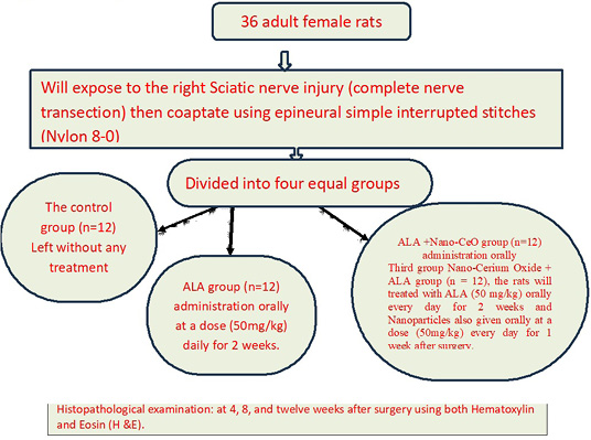
thirty-six adult female healthy Wistar rats were included in this study. having bodyweight average (230 ± 20g). They were housed in suitable cages with favorable conditions at the animal facility of the College of Veterinary Medicine at the University of Basrah. They were fed commercial pellets and given water while being kept in their designated cages for 15 days before the examination. The right Sciatic nerve was completely transected in all animals, and then instant coaptation was done. Then it was divided equally and randomly into three groups (n=12).
The first group, The control group (CG) (n=12) was left without any extra interferences after coaptating the nerve.
The second group (the Alpha Lipoic Acid group) (ALA group) (n = 12) after surgery administration orally of ALA at a dose (50 mg/kg) every day for 2 weeks. The dose of ALA (50 mg/kg) was selected according to the previous rat studies.
Third group Nano-Cerium Oxide + ALA group (n = 12), the rats will treated with ALA (50 mg/kg) orally every day for 2 weeks and Nanoparticles also given orally at a dose (50mg/kg) every day for 1 week after surgery.
Experimental Design
Preparation of cerium oxide nanoparticles
Cerium oxide nanoparticles were individually created and then aseptically thinned as listed:
We weighed about 300mg of Nano Cerium Oxide powder using a sensitive balance.
Then the 300mg of Ceo2 nanoparticles powder was dissolved in 3ml of distilled water.
We then dosage all rats with 0.5ml of diluted cerium oxide solution.
The process was repeated once daily for seven days.
Preparation of Alpha Lipoic Acid
Each capsule contains 300mg of ALA was diluted in 6ml of DW.
Each rat was given 1 ml of the diluted solution orally.
This method was repeated for 14 days for each the ALA group and mix group.
Surgical Procedure
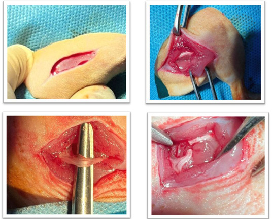
The animals will be anesthetized based on a protocol by injection of 10 mg/kg Xylazine HCL (2%) and 75 mg/kg ketamine HCL (10%). ; . All animals in this study will have their right limb selected. The region between the middle of the abdomen and the back of the hind limbs was clipped and shaved then washed thoroughly with distal water; followed by a 2-3 minutes surgical scrub of the area using diluted liquid soap and then antisepsis (ethanol alcohol 70%) was applied on the whole clipping area and finally, the site of the incision was applied with 2.5% tincture iodine A sterile towel covered the distal end of the right limb (the target limb) and secured with towel clips; the surgical area was then draped for the surgery. By locating the sciatic nerve through feeling the greater trochanter of the femur. and stifles as landmarks, at the posterior-lateral thigh about 1 cm caudo-lateral to the greater trochanter, about one-third of the femur, at the distal level. A three cm incision length was made on the proximal half of the distance between the stifle joint and the trochanter major. The biceps femoris and The semitendinosus muscles were blindly dissected with the dull tips of Mayo scissors to reveal the nerve , and a scalpel was used to make a complete cut. (using blade 15 sizes) was proximally made at about 1-1.5 cm to the site of the bifurcation of the nerve (Figure 1). The coaptate of the two extremities will be created utilizing a basic interrupted suture technique (3 stitches) using 8-0 Nylon suture material, the suturing involves only the epineurium in the area between the distal and proximal segment. A regular closure was performed on the muscles and skin .
The animals were then given Meloxicam (0.25 mg/kg BW) as analgesic and (2 mg/kg BW) Ceftiofur HCL once daily for 3 days and 5 days post-surgery for Meloxicam and Ceftiofur HCL respectively .
At the end of the experiment, we euthanized all groups at periods of 4, 8, and 12 weeks by giving a overdose of anesthesia to the rats.
Neuro Histopathological Examination
Three centimeters of Sciatic nerve (1.5 cm above and 1.5 cm below the nerve transected) were taken at 4, 8, and 12 post-surgery for neurohistopathological examination secondly The specimen was immersed in 10% buffered formalin for 24 hours, then trimmed to size, placed in a special cassette, washed with tap water, sliced using a microtome, and treated with ethanol and xylene to eliminate formalin. The astronauts then put the tissue in paraffin wax for preservation. than the paraffin templates sectioning by the microtome and fixation of the small spaceman to the glass slid and staining the section by using Hematoxylin and Eosin stain (H and E) .
Table 1: Comparing the groups over time and emphasizing pathological abnormalities in the sciatic nerve using the comprehensive scoring system.
Parameter
Time period
4 Weeks
8 Weeks
12 Weeks
Positive Control
Vacuolation
2
3
3
Degeneration of nerve fibers
2
2
3
Edema
2
3
3
Inflammatory Cells
2
2
3
Cell Nuclei abnormality
1
1
2
Congestion of Blood Vessels
2
2
2
Status of Schwann Cells
2
2
2
Condition of Axon Sheath
2
3
3
Presence of Macrophages/Debris
2
3
3
Total
17
21
24
ALA-treated
Vacuolation
2
3
2
Degeneration of nerve fibers
3
3
3
Edema
3
3
2
Inflammatory Cells
3
3
2
Cell Nuclei abnormality
1
2
1
Congestion of Blood Vessels
2
2
1
Status of Schwann Cells
1
2
1
Condition of Axon Sheath
1
3
2
Presence of Macrophages/Debris
1
3
1
Total
17
24
15
mix-treated
Vacuolation
1
2
1
Degeneration of nerve fibers
1
2
1
Edema
1
2
1
Inflammatory Cells
1
2
1
Cell Nuclei abnormality
1
1
1
Congestion of Blood Vessels
1
2
1
Status of Schwann Cells
1
2
1
Condition of Axon Sheath
1
2
1
Presence of Macrophages/Debris
1
2
1
Total
9
17
9
Analysis of Histopathological Examination Using Different Scoring System
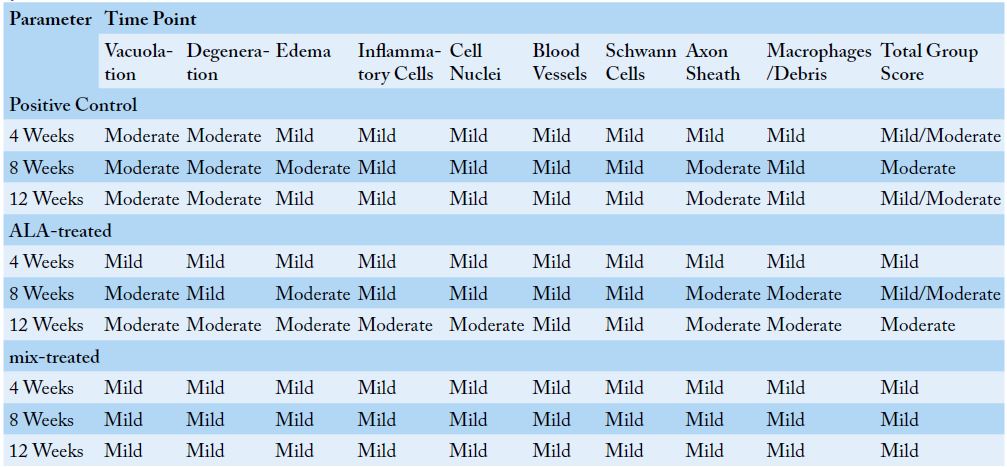
Using Gibson-Corley et al.’s principles and based on the model of we designed various scoring systems to evaluate histopathological changes and compare them between groups. In the current study, we discovered a link between the designed scoring system and the obvious histopathological changes in different groups over time, including vaccination, degeneration, edema, inflammatory cells, cell nuclei morphology, blood vessel congestion, Schwann cells, axis sheath, macrophages, and debris at 4 weeks, 8 weeks, and 12 weeks (Table 1). We used a descriptive scoring method to confirm research findings, facilitate direct comparison of histopathological alterations between control and experimental groups (Schafer., 2018) as well as between treatment groups, and ease the interpretation of our histological data (Meyerholz and Beck., 2018) Histopathological characteristics such as vacuolation, degeneration, and edema were examined for each group at particular time points (4 weeks, 8 weeks, and 12 weeks). Each parameter score in this scoring system is classified as mild, moderate, or severe according to the grouping criteria. The dominant category of individual criteria determines the total severity for each group at any given time point (Table 2).
RESULTS AND DISCUSSION
The Results of Histopathological Changes
The histological analysis of sciatic nerve sections was collected from different experimental groups at various time intervals (4, 8, and 12 weeks). These categories consist of the positive control, ALA (Alpha-Lipoic Acid), and mix group Hematoxylin and Eosin (H and E) staining allowing for examination at different magnifications within each group, with detailed analysis based on the given descriptions.
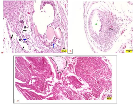
The control group’s histopathological examination at 4 weeks revealed the existence of small vacuolated areas with bigger multilocular vacuoles containing less intense eosinophilic material and edema 2(50%) (Figure 2A). Moreover, the distribution of fibers, degraded nerve fibers2(50%), swollen or absent axons 2(50%), vacuolation 2(50%) and differing levels of edema 2(50%) are also observed (Figure 2A). During the 8 weeks, histopathological changes included edema 3(75%), inflammatory cell infiltration 2(50%), vacuolation 3(75%), distorted cell nuclei 1(25%), and extravasated RBCs (Figure 2B). Additionally, Fiber disorganization, nerve fiber atrophy, blockage, and swelling (Figure 2B). However, the examination of tissue changes in this group after 12 weeks showed disorganized fibers, damaged cells 2(50%), swollen axon sheaths containing debris 3(75%), healthy Schwann cells2(50%) and axons, intense blood vessel engorgement, extensive degeneration, nerve fiber necrosis 3(75%), inflammatory cells 3(75%), and blood vessel congestion 2(50%) (Figure 2C).
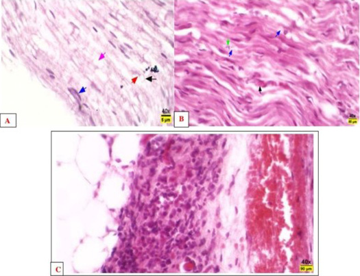
The histopathological changes reported for the ALA group after 4 weeks of treatment showed decreased perineural fibrosis, large vacuolation 2(50%), degenerated axon 3(75%), and prominent perineural and Schwann cells as in (Table 1) and (Figure 3A). While, after 8 weeks of treatment, the alteration was severe disarrangement and degeneration of fibers3(75%), notable degeneration of Schwann cells2(50%), edema3(75%), mild degeneration, dilated axon sheaths3(75%) with debris, macrophages3(75%), infiltration of inflammatory cells 3(75%), and congestion 2(50%) (Figure 3B). However, after 12 weeks of treatment with ALA, the microscopical lesions were thickening of the perineurium layer, infiltration of inflammatory cells2(50%) (, severe degeneration, and necrosis of nerve fibers3(75%) (Figure 3C).
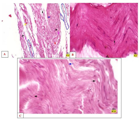
The histopathological changes of the mix group- treated group after 4 weeks revealed the presence of almost normal myelinated nerve fibers1(25%), slightly disarranged, normal Schwann cells 1(25%), axons1(25%), congestion1(25%), increased blood vessels1(25%), and mild edema1(25%), (Figure 4A). However, after 8 weeks the microscopical features include the detection of normal arrangements of nerve fiber, normal Schwann cells2(50%), slight degeneration2(50%), expanded axon sheaths harboring debris, macrophages2(50%), mild degeneration, normal axon sheaths2(50%), and congested blood vessels2(50%). (Figure 4B) After 12 week the results of the current study recorded that almost Schwann cells were normal1(25%), the blood vessels were congested 1(25%), and the nerve fibers were normally arranged (Figures 4C).
Scoring Index
The positive control group experienced a mild rise in vacuolation from 4 to 8 weeks, followed by a severe increase at 12 weeks. Degeneration occurs progressively over time, showing progressive nerve tissue deterioration. Edema, inflammatory cells, blood vessels, Schwann cell state, axon sheath condition, and the presence of macrophages and debris all had reasonably constant scores throughout time, with modest changes that tend to rise. While the ALAL-treated group exhibits varying trends across parameters, some improve over time (e.g., vacuolation decreases at 4 weeks and then increases), while others worsen (e.g., degeneration, edema, and inflammatory cells increase over time) after 8 weeks and then slightly decrease in the final period (12 weeks). While the mix-treated group exhibits minor variations in scores across criteria, with a little increase in the total score at 8 weeks and a noteworthy decrease in the severity of scores in the final. The results of this scoring system were consistent with the comprehensive scoring system. The positive control group shows moderate alterations with a higher overall severity than the other groups. While the ALA group demonstrates a trend from mild to moderate severity, particularly at 8 weeks, In contrast, period (12 weeks), indicating a potentially favorable effect or encouraging response to treatment. In contrast, the mix-treated group consistently exhibits significant improvement compared to alterations in ALA and control groups, indicating improved histopathology outcomes.
In this study, histopathological changes in the sciatic nerve across control, ALA-treated, and Mix-treated groups were evaluated at intervals of 4, 8, and 12 weeks. The results provide insights into the effectiveness of each treatment in mitigating nerve damage and promoting regeneration. The observations from Hematoxylin and Eosin (H and E) staining revealed distinct patterns of degeneration, vacuolation, and inflammation, highlighting the comparative benefits and limitations of each therapeutic approach. Over time, the control group showed progressive, severe damage to the sciatic nerve, beginning with small vacuolated foci and larger multilocular vacuoles containing eosinophilic material at 4 weeks. By 8 weeks, degeneration worsened, with widespread edema, inflammatory cell infiltration, and disrupted cell nuclei. By 12 weeks, extensive fiber disarrangement, nerve fiber necrosis, and vascular congestion were observed, suggesting persistent and severe nerve damage due to the lack of therapeutic intervention. Such progressive degeneration aligns with previously reported findings on nerve damage in untreated models of neuropathy ; supported these findings, where in the study of , the progressive degeneration seen in untreated neuropathy models is well-documented, and they report similar patterns of cellular and structural deterioration over time, including vacuolation, edema, and necrosis. They emphasize that, without intervention, nerve tissues typically undergo increasing disorganization and damage due to chronic inflammation and oxidative stress, which aligns with the observations in this study’s control group. Thus, their research supports the idea that untreated neuropathic conditions lead to severe, cumulative damage, which is reflected in the histological findings. On the other hand, raised concerns primarily about the methods used to assess the severity of vacuolation and necrosis. They argue that relying on (H and E) staining alone can limit the accuracy of detecting and quantifying these changes, as (H and E) staining may not distinctly capture subtle variances in vacuole formation or cell death stages . recommend utilizing supplementary, more precise indicators like specific immunohistochemical stains or electron microscopy for a clearer and more comprehensive evaluation. Their concerns suggest that although the observed outcomes may indicate real degeneration, more intricate details might need different methods to fully understand the extent of the damage, especially about vacuolation and necrosis. The ALAtreated group showed early therapeutic benefits, exhibiting decreased perineural fibrosis and less severe degeneration at 4 and 8 weeks. At 4 weeks, the group exhibited a reduced number of degenerated axons and notable Schwann cells, potentially indicating partial nerve preservation by ALA, recognized for its antioxidant characteristics ; Nevertheless, by 12 weeks, indications of significant degeneration and necrosis remained, implying that although ALA provided some initial neuroprotective benefits, these were inadequate over an extended duration. Such outcomes are consistent with studies that have documented moderate efficacy of ALA in mitigating nerve damage who acknowledged the possible immediate advantages of ALA but contended that these effects were inadequate for preventing long-term deterioration. They concluded that while ALA might delay nerve degeneration initially, it does not completely prevent the long-term progression of neuropathy, which aligns with the observed severe degeneration and necrosis at 12 weeks in the current study. Their agreement stems from the similarity in outcomes showing that ALA’s moderate neuroprotective effect can provide partial relief but is insufficient for sustained nerve protection in chronic or prolonged injury settings.
The Mix-treated group exhibited the most favorable outcomes, demonstrating negligible deterioration at all time intervals. At 4 weeks, the nerve fibers appeared largely normal, with only slight disarrangement and mild edema.
At 8 weeks, nerve fibers remained well-arranged, with normal Schwann cells and only mild degeneration. By 12 weeks, the sciatic nerve tissue was nearly normal, with minimal degeneration and well-preserved histology. These results indicate that the Mix treatment may promote nerve regeneration and maintain histological integrity over time. This group’s performance aligns with studies suggesting that Mix-based therapies can enhance cellular repair and reduce inflammation in neural tissues . They completely endorsed these results because their investigation showed that Mix based therapies possess robust regenerative capabilities and can reliably preserve tissue integrity in neural injury models. They noticed that Mix compounds aided in decreasing inflammation, oxidative stress, and cellular degeneration, leading to ongoing nerve protection and structural maintenance over time. credited these results to Mix’s capacity to improve cellular repair mechanisms and adjust immune responses, which renders it remarkably efficient for neural regeneration. Their findings demonstrated comparable stability in preserving nerve structure, particularly over long durations, prompting them to support the outcomes of the present study. concurred on the efficacy of Mixbased treatments in enhancing nerve regeneration and minimizing inflammation while emphasizing the necessity for additional validation across various forms of neural injuries. They discovered that although Mix demonstrated beneficial outcomes in certain models, like peripheral nerve injuries, there was insufficient evidence regarding its efficacy across a wider variety of neural damage types (e.g., central nervous system injuries or severe chronic neuropathies). Farinas et al. suggested that further research in various neural injury scenarios is needed to verify Mix’s therapeutic consistency and applicability. This caution is evident in their limited support, acknowledging the encouraging outcomes in the current study while simultaneously suggesting wider testing for complete validation. The results of the study emphasize a distinct difference in the neuroprotective impacts of various treatments, particularly when contrasting the untreated control group’s significant and advancing damage with the more varied outcomes in the treated groups. The results from the control group highlight the swift deterioration that takes place in neuropathic conditions without treatment, evidenced by disordered nerve fibers, necrosis, and ongoing inflammation, which can worsen nerve injury through cycles of cellular stress and immune response .In the ALAtreated group, the noted initial neuroprotective effects are probably due to ALA’s antioxidant properties, which might temporarily alleviate oxidative stress and inflammation, both significant factors in nerve degeneration..Nonetheless, the results also indicate the shortcomings of ALA in delivering lasting protection, implying that while it could be effective at first, its advantages wane over time, as evidenced by the resurgence of notable degeneration by 12 weeks. This result corresponds with other studies suggesting that antioxidants by themselves might not completely avert ongoing nerve injury in chronic models The consistently favorable results in the Mix-treated group characterized by slight degeneration, well-maintained histology, and early indication of regeneration at all time intervals emerge as the most encouraging finding. The effectiveness of Mix in maintaining nerve structure indicates strong anti-inflammatory and potentially healing qualities that can promote nerve health for longer durations, in contrast to the more short-lived advantages seen with ALA and nano-treatments. strongly endorsed this conclusion, highlighting Mix’s ability for long-lasting regeneration because of its anti-inflammatory and regenerative properties. Nonetheless, as highlighted by , conducting additional trials across different neural injury models is crucial to confirm the generalizability of Mix and fine-tune dosing and treatment parameters for wider clinical relevance.
CONCLUSIONS AND RECOMMENDATIONS
The Mix-Treated Group appears to be the best, showing the most normal histological features, minimal degeneration, and early signs of nerve regeneration across all time points.
The ALA group shows some beneficial effects but is less effective than the Mix-treated group.
The control groups exhibit significant and progressive nerve damage, making them the least favor.
ACKNOWLEDGEMENTS
We would like to thank the Head of the Department of Surgery and Obstetrics, the Dean of the College of Veterinary Medicine, and the President of the University of Basra for their support in the college laboratory and facilitating the scientific research process and everyone who contributed to the completion of this research
NOVELTY STATEMENTS
New in our work entitled (Study the efficacy of Nano-Cerium Oxide and Alpha Lipoic Acid on Regeneration of sciatic Nerve Injury (Neurotmesis) in Rats) Despite the existence of treatment, it is still a challenge in veterinary medicine and few researchers shed light on the use of dual therapy in neurological treatment.
AUTHOR’S CONTRIBUTIONS
Dhuha Adel: Contributed to preparing the animals, preparing the surgical site, and monitoring after the surgery period. As well as writing and reviewing the manuscript.
Ammar M. Hashim: Contributed to performing the surgery. And taking nerve images.
Rafid Majeed: Analysis and interpretation of data.
Figures
Figure 1. Figure 1:.
showed the surgical site, showed exposure of sciatic nerve, sciatic nerve a complete transect.
Figure 2:.
A-control-4 weeks. Photomicrographs of the sciatic nerve the positive control showed distributed of the arrangement of fibers, degenerated nerve fibers (blue arrows), and they had swollen or missing axons with various degrees of edema (black arrows). H and E, 4x, B-control-8 weeks. Photomicrographs showed edema (ED), and infiltration of Inflammatory cells; H and E, 4X. C-control-12 weeks. Photomicrographs showed severe disarrangement of fibers, notable degeneration of axon sheaths, edema, and infiltration of inflammatory cells (black circle). H and E, 4x.
Table 2:.
Comparison of histopathological changes across different groups and time points using the descriptive scoring system.
Figure 3:.
A-4 week. Photomicrographs of the sciatic nerve in the ALA group showed a significant decrease in the thickness of perineural fibrosis compared to the control group. There is significant large vacuolation (pink arrow), degenerated axon (red arrow), prominent perineural cells (blue arrow), and Schwann cells (black arrows). H and E, 40x. B-8 weeks. Sections of the Sciatic nerve of the ALA group showed mild degeneration, dilated axon sheaths (blue arrows) containing debris (fragmented axons and/or myelin) (green arrow), and macrophages (black arrow) that engulf this debris. H and E, 40x. C-12 weeks. Sections of the Sciatic nerve of the ALA group showed thickening of the perineurium layer, infiltration of the inflammatory cells (black arrow), and congestion (yellow arrows). H and E, 40x.
Figure 4:.
A-4 weeks. Photomicrographs of the sciatic nerve of the mix-treated group showed almost normal myelinated nerve fibers (blue arrow), but slightly disarranged, atrophied, and myelin sheath loss (blue circled area) were also detected, normal Schwann cells (pink arrows), normal axon (black arrows), congestion and increase blood vessels (vellow arrow), and edema also evident. H and E, 40x. B-8 weeks. Photomicrographs of the sciatic nerve of the mix-treated group showed epineurium (black arrows), and normal nerve fiber arrangements (black arrows) in the endoneurium. H and E, 40x. C-12 weeks. Sections from the Sciatic nerves of the mix-treated group revealed almost normally arranged nerve fibers (blue arrow), normal Schwan cells and axons (black arrows). H and E, 10x.
References
Al-Haideri DH, Al-Timmemi HA (2024). Efficacy of chitosan nanoparticles and mesenchymal stem cells in rabbit models for sciatic nerve regeneration. Iraqi J. Vet. Sci., 38(2): 369-377. https://doi.org/10.33899/ijvs.2023.142572.3186[CrossRef]
Ali ZA, Kadhum HJ, Dawood Q, Kadhim L (2023). Clinical and electrophysiological charactrastic of peripheral neuropathy in patients with hematological malignancies receiving chemotherapy Abstract. Med. J. Basrah Univ., 41(2): 165-176. https://doi.org/10.33762/mjbu.2023.145070.1175[CrossRef]
Allodi I, Udina E, Navarro X (2012). Specificity of peripheral nerve regeneration: interactions at the axon level. Prog. Neurobiol., 98: 16–37. https://doi.org/10.1016/j.pneurobio.2012.05.005[CrossRef]
Bannai JAA (2015). Clinical and histopathological study of the effect of the pulsed magnetic field and low level laser therapy on the regeneration of the sciatic nerve in rabbits.
Celardo I, Traversa E, Ghibelli L (2011). Cerium oxide nanoparticles: A promise for applications in therapy. J. Exp. Ther. Oncol.,9:47–51.
Chaudhary P, Marracci G, Galipeau D, Pocius E, Morris B, Bourdette D (2015). Lipoic acid reduces inflammation in a mouse focal cortical experimental autoimmune encephalomyelitis model. J. Neuroimmunol., 289:68–74. https://doi.org/10.1016/j.jneuroim.2015.10.011[CrossRef]
Datta A, Mishra S, Manna K, Saha KD, Mukherjee S, Roy S (2020). Pro-oxidant therapeutic activities of cerium oxide nanoparticles in colorectal carcinoma cells. ACS omega, 5(17): 9714-9723. https://doi.org/10.1021/acsomega.9b04006[CrossRef]
Distad BJ, Weiss MD (2013). Clinical and electrodiagnostic features of sciatic neuropathies. Phys. Med. Rehabil. Clin. N. Am., 24:107–20. https://doi.org/10.1016/j.pmr.2012.08.023[CrossRef]
Farinas AF, Esteve IVM, Pollins AC, Cardwell NL, Kaoutzanis C, Nussenbaum ME, Thayer WP (2020). Diffusion magnetic resonance imaging predicts peripheral nerve recovery in a rat sciatic nerve injury model. Plast. Reconstr. Surg., 145(4): 949-956. https://doi.org/10.1097/PRS.0000000000006638[CrossRef]
Ghayour MB, Abdolmaleki A, Behnam-Rassouli M (2017). The effect of memantine on functional recovery of the sciatic nerve crush injury in rats. Turk. Neurosurg., 27(4). https://doi.org/10.5137/1019-5149.JTN.16792-15.1[CrossRef]
Gibson-Corley KN, Olivier AK, Meyerholz DK (2013). Principles for valid histopathologic scoring in research. Vet. Pathol., 50(6): 1007-1015. https://doi.org/10.1177/0300985813485099[CrossRef]
Gonçalves M, Vale N, Silva P (2024). Neuroprotective Effects of Olive Oil: A Comprehensive Review of Antioxidant Properties. Antioxidants, 13(7): 762. https://doi.org/10.3390/antiox13070762[CrossRef]
Gu X, Ding F, Yang Y, Liu J (2011). Construction of tissue-engineered nerve grafts and their application in peripheral nerve regeneration. Prog. Neurobiol., 93: 204–230. https://doi.org/10.1016/j.pneurobio.2010.11.002[CrossRef]
Helal MM, Hussein A (2022). The Effect of Local Application of Magnesium Oxide Powder on the Blood Parameters During Nerve Regeneration of Injured Sciatic Nerve in Rat. Indian J. Forensic Med. Toxicol., 16:1. https://doi.org/10.37506/ijfmt.v16i1.17946[CrossRef]
Hussein TJ, Waheed Z, Majeed SK (2014). Effects of different dose of cypermthrin on serum acetylcholine concentration, spinal cord and sciatic nerve histopathology in adult rats. Basrah J. Vet. Res., 13(1). https://doi.org/10.33762/bvetr.2014.88131[CrossRef]
Jali AM, Banji D, Banji OJ, Hurubi KY, Tawhari FY, Alameer AA, Zanqoti RA (2024). Navigating Preclinical Models and Medications for Peripheral Neuropathy: A Review. Pharmaceuticals, 17(8): 1010. https://doi.org/10.3390/ph17081010[CrossRef]
Jassim MM, Abduljaleel MR, Abdulkareem ZB, Sanad NH, Alrashid IM (2023). Study the effect of the magnetic field on the healing of bone fracture after implant avian bone in femoral bone in rabbits. Adv. Anim. Vet. Sci., 11(11): 1779-1784. https://doi.org/10.17582/journal.aavs/2023/11.11.1779.1784[Full text][CrossRef]
Khan J, Yadav S (2024). Nanotechnology-based Nose-to-brain Delivery in Epilepsy: A Novel Approach to Diagnosis and Treatment. Pharm. Nanotechnol., 12(4): 314-328. https://doi.org/10.2174/0122117385265554230919070402[CrossRef]
Kim TH, Yoon SJ, Lee WC, Kim JK, Shin J, Lee S, Lee SM (2011). Protective effect of GCSB-5, an herbal preparation, against peripheral nerve injury in rats. J. Ethnopharmacol., 136(2): 297-304. https://doi.org/10.1016/j.jep.2011.04.037[CrossRef]
Kocaoğlu S, Aktaş Ö, Zengi O, Tufan A, Karagöz Güzey FEYZA (2017). Effects of alpha lipoic acid on motor function and antioxidant enzyme activity of nerve tissue after sciatic nerve crush injury in rats. Turk. Neurosurg., 1: 1-8. https://doi.org/10.5137/1019-5149.JTN.18585-16.1[CrossRef]
Kokai LE, Bourbeau D, Weber D, McAtee J, Marra KG (2011). Sustained growth factor delivery promotes axonal regeneration in long gap peripheral nerve repair. Tissue Eng., 17: 1263–1275. https://doi.org/10.1089/ten.tea.2010.0507[CrossRef]
Melli G, Taiana M, Camozzi F, Triolo D, Podini P, Quattrini A, Lauria G (2008). Alpha-lipoic acid prevents mitochondrial damage and neurotoxicity in experimental chemotherapy neuropathy. Exp. Neurol., 214(2): 276-284. https://doi.org/10.1016/j.expneurol.2008.08.013[CrossRef]
Mern DS, Walsen T, Beierfuß A, Thomé C (2021). Animal models of regenerative medicine for biological treatment approaches of degenerative disc diseases. Exp. Biol. Med., 246(4): 483-512. https://doi.org/10.1177/1535370220969123[CrossRef]
Meyerholz DK, Beck AP (2018). Fundamental concepts for semiquantitative tissue scoring in translational research. ILAR J., 59(1): 13-17. https://doi.org/10.1093/ilar/ily025[CrossRef]
Nasir A, Afridi OK, Khan H, Bai Q (2024). Mitigation of sciatica injury-induced neuropathic pain through active metabolites derived from medicinal plants. Pharm. Res., 107076. https://doi.org/10.1016/j.phrs.2024.107076[CrossRef]
Navarro X, Vivó M, Valero-Cabré A (2007). Neural plasticity after peripheral nerve injury and regeneration. Prog. Neurobiol., 82: 163–201. https://doi.org/10.1016/j.pneurobio.2007.06.005[CrossRef]
Oktay KNK, Akpinar P, Ozkan FU, Aktas I, Canillioglu YE, Senturk GE (2021). The effect of alpha lipoic acid on the recovery of sciatic nerve injury in rats: A prospective randomized controlled trial. Am. J. Phys. Med. Rehabil., 100(8): 780-789. https://doi.org/10.1097/PHM.0000000000001642[CrossRef]
Pham JH, Johnson GA, Rangan RS, Amankwa CE, Acharya S, Stankowska DL (2022). Neuroprotection of rodent and human retinal ganglion cells in vitro/ex vivo by the hybrid small molecule SA-2. Cells, 11(23): 3741. https://doi.org/10.3390/cells11233741[CrossRef]
Piao Y, Liang X (2012). Chinese medicine in diabetic peripheral neuropathy: experimental research on nerve repair and regeneration. Evid. Based Complement. Altern. Med., 2012(1): 191632. https://doi.org/10.1155/2012/191632[CrossRef]
Rahimi B, Behroozi Z, Motamednezhad A, Jafarpour M, Hamblin MR, Moshiri A, Ramezani F (2023). Study of nerve cell regeneration on nanofibers containing cerium oxide nanoparticles in a spinal cord injury model in rats. J. Mater. Sci. Mater. Med., 34(2): 9. https://doi.org/10.1007/s10856-023-06711-9[CrossRef]
Reddy DS, Abeygunaratne HN (2022). Experimental and clinical biomarkers for progressive evaluation of neuropathology and therapeutic interventions for acute and chronic neurological disorders. Int. J. Mol. Sci., 23(19): 11734. https://doi.org/10.3390/ijms231911734[CrossRef]
Rusli N, Ng CF, Makpol S, Wong YP, Mohd Isa IL, Remli R (2024). Antioxidant Effect in Diabetic Peripheral Neuropathy in Rat Model: A Systematic Review. Antioxidants, 13(9): 1041. https://doi.org/10.3390/antiox13091041[CrossRef]
Sanadgol N, Golab F, Askari H, Moradi F, Ajdary M, Mehdizadeh M (2017). Alpha-lipoic acid mitigates toxic-induced demyelination in the corpus callosum by lessening of oxida tive stress and stimulation of polydendrocytes proliferation. Metab. Brain Dis.,10:1–11. https://doi.org/10.1007/s11011-017-0099-9[CrossRef]
Savastano LE (2014). Sciatic nerve injury: a simple and subtle model for investigating many aspects of nervous system damage and recovery. J. Neurosci. methods,. 227: 166-180. https://doi.org/10.1016/j.jneumeth.2014.01.020[CrossRef]
Schafer KA, Eighmy J, Fikes JD, Halpern WG, Hukkanen RR, Long GG, Francke S (2018). Use of severity grades to characterize histopathologic changes. Toxicol. Pathol., 46(3): 256-265. https://doi.org/10.1177/0192623318761348[CrossRef]
Shay KP, Moreau RF, Smith EJ, Smith AR, Hagen TM (2009). Alpha lipoic acid as a dietary supplement: molecular mechanisms and therapeutic potential. Biochim. Biophys. Acta., 1790(10):1149–60. https://doi.org/10.1016/j.bbagen.2009.07.026[CrossRef]
Shi S, Ou X, Cheng D (2024). Nanoparticle-Facilitated Therapy: Advancing Tools in Peripheral Nerve Regeneration. Int. J. Nanomed., 19-34. https://doi.org/10.2147/IJN.S442775[CrossRef]
Soluki M, Mahmoudi F, Abdolmaleki A (2020). Cerium oxide nanopar ticles as a new neuroprotective agent to promote functional recovery in a rat model of sciatic nerve crush injury. Br. J. Neurosurg., 1–6. https://doi.org/10.1080/02688697.2020.1864292[CrossRef]
Wang J, Lou Z, Xi H, Li Z, Li L, Li Z, Asakawa T (2021). Verification of neuroprotective effects of alpha-lipoic acid on chronic neuropathic pain in a chronic constriction injury rat model. Open Life Sci., 16(1): 222-228. https://doi.org/10.1515/biol-2021-0026[CrossRef]
Yu C, Wang X, Qin J (2023). Effect of necrostatin-1 on sciatic nerve crush injury in rat models. J. Orthop. Surg. Res., 18(1): 74. https://doi.org/10.1186/s13018-023-03565-3[CrossRef]