Special Issue: Emerging and Re-Emerging Animal Health Challenges in Low and Middle-Income Countries · Volume 13, Issue 1, 15 October 2025 · Open Access
Physiological and Histological Impact of Alpha-Lipoic Acid on Myocardial Ischemia Induced by Salbutamol in Rats
· · ·
Affiliations & Notes ⌄
1Department of Physiology, Pharmacology and Biochemistry, College of Veterinary Medicine, University of Basrah, Iraq
2Department of Anatomy and Histology, College of Veterinary Medicine, University of Basrah, Iraq. *Correspondence | Zainab A.H. Al-Mousawi, Department of Physiology, Pharmacology and Biochemistry, College of Veterinary Medicine, University of Basrah, Iraq
The current study focused on whether alpha-lipoic acid affected cardiac markers and the lipid profile of myocardial ischemia brought by salbutamol. Three groups of eighteen adult rats were randomly assigned. Oral gavage was used to provide treatments; the heart ischemia was induced with 80 mg/kg salbutamol for two days. Animals in the positive control received salbutamol, or received salbutamol (80 mg/kg orally) for 2 consecutive days and then received ALA (20 mg/kg orally) for 28 days. Then rats were sacrificed for cardiac markers, lipid profiles, and ALT and AST analyses were evaluated. According to the experiment’s findings, alpha-lipoic acid supplementation significantly reduced cardiac troponin I, creatine kinase-MB, lipid profile (TC, TG, LDL, VLDL) and ALT and AST. Interestingly, a significant increase in HDL level was observed. Additionally, it significantly improves the histological changes in the heart muscle caused by salbutamol. The study concluded that alpha-lipoic acid has ameliorative effects against salbutamol-induced myocardial ischemia in experimental animals.
Keywords | Alpha-lipoic acid, Salbutamol, Myocardial ischemia, Cardiac markers, Rat
INTRODUCTION
Cardiovascular disease (CVD) is a major cause of death and morbidity rates and remains a major global health concern. The conditions that fall under the category of CVD are hypertension, arrhythmia, ischemic heart disease (IHD), congestive heart disease, valvular heart disease, heart failure, and stroke. An imbalance between coronary perfusion and oxygen demand causes IHD, a significant form of CVD, which frequently results in myocardial infarction (MI) from prolonged oxygen deprivation. IHD, as a consequence of which myocardial tissue and contractile force are lost, is among the most frequent reasons for cardiac failure .
Animal models play a vital role in developing novel medications for controlling and treating a wide range of illnesses. An established standard model to investigate the beneficial effects of medicinal herbs on heart dysfunction is chemically induced myocardial ischemia in rats . Catecholamines are a powerful inducer of cardiac contractility in animal models and can cause ischemia, hypoxia, myocardial hyperactivity, and coronary hypertension when administered at high doses . To induce myocardial ischemia in the animal model, Salbutamol has been used. Salbutamol is a synthetic catecholamine; its structural resemblance to isoproterenol and its manner of action cause significant oxidative stress in the heart, leading to severe myocardial stress and necrosis . Salbutamol, a β2 agonist, commonly prescribed to treat asthma, chronic obstructive pulmonary disease, and bronchospasm. To delay premature labor, intravenous salbutamol might be given as a tocolytic to relax the uterine smooth muscle ; . When animals are given excessive salbutamol dosage causes tachycardia, which could result in myocardial ischemia. Salbutamol is less affordable and readily available when compared to other catecholamines such as isoproterenol ; Abd Halim et al., 2018).
Thioctic acid, known as alpha-lipoic acid (ALA), is a naturally occurring short-chain fatty acid (1, 2-dithiolane-3-pentanoic acid), an antioxidant. In the body, it dissolves in fat and water. The gastric tract absorbs it rapidly. This antioxidant molecule has gained a lot of attention recently and is widely utilized as a dietary supplement across the globe . According to earlier research, ALA can influence both peripheral and central regulation of 5′-adenosine monophosphate-activated protein kinase. As a result, the antioxidant activity of ALA may help lower blood pressure, encourage weight loss, and improve insulin resistance and atherogenic dyslipidemia .
To date, no study has systematically evaluated the effects of ALA on myocardial ischemia induced by salbutamol. This study aimed to evaluate the protective effect of alpha-lipoic acid against cardiac damage induced by salbutamol in rats.
MATERIALS AND METHODS
Experimental animals
In this experiment, adult male albino rats weighing between 160 and 200 grams were employed. The animals housed at the animal house of Basrah University Veterinary Medicine College are kept in well-ventilated cages that provide unlimited consumption of commercial diet and water. The animals were given ten days to adapt to the lab’s environment.
Three groups of eighteen male rats each were randomly assigned (6 individuals each): Group 1: Normal rats received a daily 0.9% dose of normal saline as a control. Group 2: Positive control received salbutamol (80mg/kg) orally for 2 consecutive days (Afsheen etal.,2017), then treated with normal saline daily. Group 3: received salbutamol (80mg/kg orally) for 2 consecutive days and then received ALA (20mg/kg) orally for 28 days.
Following the experiment, blood samples were taken through the cardiac puncture and subsequently transferred into a gel tube to conduct serum biochemical analysis using commercial kits such as serum creatine kinase-myocardial band (CK-MB) (Human/Germany), Ichroma troponin I (Boditech/Korea), Aspartate transaminase (AST), Alanine aminotransaminase (ALT) and Lipid profile (Spinreact/Spain).
Histological section
The eighteen heart samples, six sample from each group, were immediately removed and fixed for 12 hours, and the form fixative was eliminated using 70% alcohol. The tissues underwent gradual dehydration at increasing ethanol concentrations, followed by xylene treatment and paraffin embedding. On a glass slide, five micron-thick slices of paraffin-embedded tissues were placed, and hematoxylin and eosin stain were applied. The sections were inspected under an Olympus RPA light microscope using high-resolution camera (Sony/Japan).
Statistical analysis
Utilizing the computerized version 22 of the SPSS (Statistical Program for Social Sciences) program, statistical analysis of the data was carried out based on a one-way analysis of variance (ANOVA) and least significant differences (LSD) post hoc test with a P value (P<0.05).
RESULTS
The results of troponin I and CK-MB showed significant elevation (P<0.05) in the salbutamol group compared to the control group rats. While group treated with salbutamol + AlA showed a significant reduction (P<0.05) in troponin I and CK-MB compared to the salbutamol group and no significant difference was observed (P<0.05) compared to the control group (Table 1).
Table 1: Effect of ALA and salbutamol on cardiac markers.
Parameter / Group
Troponin I (ng/ml)
Creatine Kinase MB (u/l)
Control group
0.01 ±0.249 c
90 ±2.64c
Salbutamol group
3.6 ±0.359 a
557.6 ±7.09a
Salbutamol + AlA group
2.79 ±0.242b
440.6 ±22.62b
LSD
0.23
106.25
Values are shown as mean ± SD, with n = 6/group. Different letters indicate significant differences at p < 0.05 based on LSD post hoc test.
Rats in the salbutamol group had significantly higher AST and ALT data (P<0.05) than those in the control group. The results indicate that the group treated with salbutamol + AlA reported a substantial decrease (P<0.05) in ALT and AST compared to the salbutamol group. Still, there was no significant difference (P<0.05) in ALT compared to the control group (Table 2).
Table 2: Effect of ALA and salbutamol on AST and ALT enzymes.
Parameter / Group
AST(u/l)
ALT(u/l)
Control group
73.11±3.79c
20.8±2.4b
Salbutamol group
151.61±21.22a
35.43±4.03a
Salbutamol + AlA group
102.6±6.13b
25.0±3.6b
LSD
28.54
4.26
Values are shown as mean ± SD, with n = 6/group. Different letters indicate significant differences at (p < 0.05) based on LSD post hoc test.
The effect of ALA and salbutamol on lipid profile parameters in rats is displayed in Table 3. The findings indicate that total cholesterol, triglycerides, and LDL-C have significantly increased (P<0.05) in the salbutamol group compared to the salbutamol + AlA group and control. Whereas there was a significant decrease in HDL-C in salbutamol compared to the salbutamol + AlA group and control group. Furthermore, there were no significant differences in VLDL-C between groups.
Histological results
Effect of ALA and salbutamol on heart histology
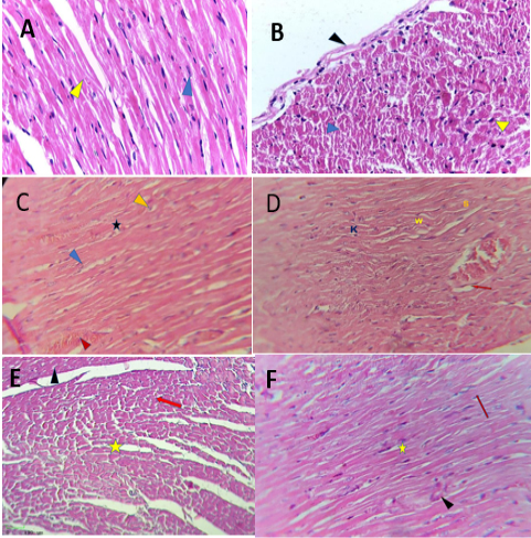
Histological section of the heart in control rats showed that composed of elongated myofibers surrounded by epicardium. Each muscle fibers contain oval nuclei with few dense connective tissues between cardiac fibers (Figure 1A, B). Heart tissue of rats treated with SAL showed disarrangement of some myocadiac fibers with degenerated of nuclei, infiltration of inflammatory cells in the connective tissue between cardiac fibers, congestion of blood vessels was observed in the SAL group. As other section explained, degenerated with wavy some cardiac muscle, irregular sarcolemma with loss of muscle striation and karyohexis nuclei, also observed degeneration of the endothelial lining of blood vessels with pericardium spaces (Figure 1C, D).
Table 3: Effect of ALA and salbutamol on lipid profile.
Parameter
Control group
Sulbtamol group
Sulbtamol + AlA group
LSD
Total Cholesterol (mg/dl)
65.7± 2.62b
78.55 ±6.27a
61.6 ±1.4b
12.8
Triglyceride(mg/dl)
33.5 ±4.55b
99±2.16a
37±2.95b
53.4
LDL-C(mg/dl)
31.5± 2.64b
40.25± 1.70a
32.25±2.87b
8.0
HDL-C(mg/dl)
20.75± 2.62ab
12.5 1.68±b
20.34± 2.49a
10.62
VLDL-C(mg/dl)
22± 2.16a
22.8± 0.832a
22.6±0.91a
7.86
Values are shown as mean ± SD, with n = 6/group. Different letters indicate significant differences at (p < 0.05) based on LSD post hoc test.
While histological section of the treated heart with (SAL₊ALA) showed clear structure of the myocardium which appear as cross striated myofibers. Few hemorrhages between cardiac fibers, clear epicardium layer with empty intercellular spaces and mild congested blood vessels, it also noted besides inflammatory cellular infiltration between the cardiac fibers (Figure 1E, F).
DISCUSSION
Troponin I and CK-MB are released into the plasma, and their level rises when there is cardiac muscles damage. The results of the present study revealed a significant decrease in cT-I and CK-MB concentration in the groups treated with ALA when compared with the salbutamol group these results are in agreement with . According to the findings, catecholamine-induced oxidative stress causes beta-adrenoreceptor hyperstimulation, which leads to a complex functional and structural myocardial injury that causes cellular damage. Additionally, researchers discovered that oxidative stress could inhibit sarcolemma calcium transport, resulting in calcium overload and cardiac dysfunction. This could lead to in-situ troponin degradation and the release of low molecular weight fragments into the circulation during a period of Ca2+ overload (Ahmed and Masoud, 2024).
Because alpha lipoic acid has strong antioxidant properties, oxidative stress reduction could be one of the ways that post-reperfusion arrhythmias are decreased (Zheng etal., 2024). Alpha lipoic acid has cardioprotective effects on ischemia-reperfusion injury through a mechanism that involves activation of aldehyde dehydrogenase 2. Alpha-lipoic acid raises the sulfane sulfur content. Through scavenging ROS and activating the K-ATP channel, sulfane sulfur should be converted to H2S, shielding the heart from after-reperfusion arrhythmias ; .
According to Serbinova et al. (1992) rats hearts fed lipoic acid provided protection from ischemia-reperfusion damage in an isolated perfused Langendorff heart system. Lipoic acid also preserved vitamin E in heart tissue, enhanced post-ischemic left ventricular functional recovery, and reduced lactate dehydrogenase leakage and lipid peroxidation .
Our results supported earlier studies that showed ALA supplementation was beneficial in lowering triglycerides, total cholesterol, LDL and HDL levels in individuals with metabolic disorders, but had no effect on VLDL levels. ALA may act on rising blood and liver triglycerides by suppressing liver lipogenic gene expressions, reduce hepatic triglyceride production, and promote the removal of triglyceride-rich lipoproteins animal models currently provide evidence of a lipid-lowering response to ALA therapy ; . ALA may act on rising blood and liver triglycerides by suppressing liver lipogenic gene expressions, reduce hepatic triglyceride production, and promote the removal of triglyceride-rich lipoproteins . Total cholesterol and LDL was decreased by ALA most likely through the raising lipoprotein lipase activity, starting the liver’s synthesis of LDL receptors, which increases the uptake of cholesterol back into the hepatic system or increases the synthesis of apolipoprotein A component and raises serum adiponectin, which increases the β-oxidation of FFAs .
There may be a connection between ALA’s effects on glucose consumption and lipid metabolism. When fructose is administered to rats, ALA improves insulin activity, insulin sensitivity, and glucose elimination. Reducing the levels of lipids in the blood to normal may lead to ineffective control of the major enzymes involved in lipid metabolism. One possible explanation for the decrease in plasma TG concentrations is the decreased availability of precursor FFAs and the improved peripheral tissue clearance from elevated LPL activity (Salehi et al., 2019). HDL-C plasma concentrations may have increased in ALA-treated rats because of enhanced synthesis of HDL components and delayed clearance. The ATP-binding cassette transporter A1, which is essential for the efflux of cholesterol from macrophages to HDL particles, is activated by lipoic acid. Also, elevated lipoic acid levels can promote the synthesis of ApoA-I, a significant HDL protein component that is necessary for the development of mature HDL particles. Lipoic acid may promote cholesterol mobilization and efflux by activating signaling cascades through receptors such as scavenger receptor class B type I. The preservation of normal phospholipid concentrations in tissues and plasma may be due to ALA antioxidant properties : . The absence of significant differences in VLDL could be that the VLDL metabolic pathway may not be affected by ALA at this dose.
The results of microscopic examination of cardiac section of rats treated with SAL showed more effects on the heart tissue, including degenerated cardio fiber, degenerated nuclei and irregular with infiltration of inflammatory cells congestion of blood vessels, abnormal sarcolemma with karyohexise nuclei. Also observe wavy cardiac fibers with few of fiber striation. SAl is used blockers of β1 receptors and it acts on the heart by competing with the site of the β1receptors on the heart tissue. SAL could slow down the strength of cardiac contraction with reduce oxygen consumption, also reduce the volume of blood flow . Histological section of the heart treated (SAL and ALA) showed normal branching muscle fibers, with normal elongated nuclei and mild congested blood vessels, associated with the antioxidant activity of AlA, its cardiovascular protective properties also block pro-inflammatory genes and proteins, including cytokines, TNF-α, IL-1β-, and IL-6. To control adipogenesis, activate transcription factors and peroxisome proliferative-activated receptors ; ; .
CONCLUSION AND RECOMMENDATIONS
The study concluded that alpha lipoic acid has ameliorative effects in cardiac biomarker, lipid profile and cardiac tissue against myocardial ischemia induced salbutamol in experimental animals. These findings suggest a potential cardioprotective effect of ALA, but further studies are needed.
ACKNOWLEDGMENT
The authors would like to thank the faculty and staff of Basrah University College of Veterinary Medicine’s animal house for their support and advice.
NOVELTY STATEMENT
The novelty of this study lies in demonstrating the potential therapeutic effects of alpha lipoic acid on salbutamol-induced myocardial ischemia. The study offers valuable insights by systematically exploring its impact on cardiac markers and lipid profiles in experimental animals. The key finding is the significant improvement in cardiac markers and lipid profile and decreasing harmful elements while boosting protective HDL levels. This suggests that ALA could play an ameliorative role in managing myocardial ischemia, advancing research in cardiovascular protection.
AUTHOR’S CONTRIBUTION
The work provided here was a collaborative effort by all authors. In addition to writing the final publication, each author had an impact on the study’s idea and design, data collecting, analysis, and interpretation.
Ethical approval
Under IACUC ethical permission, this investigation was conducted in 2024 on animal 25/37 at the College of Veterinary Medicine, University of Basrah, Iraq.
Generative AI and AI-assisted technology statement
The authors declare that no Genrative AI was used in the creation of this manuscript.
Conflict of interest
The authors have declared no conflict of interest.
Figures
Figure 1:.
(A): Cardiac section of rat from the control group showing a normal structure of cardiac fibers (blue), nuclei located centrally (yellow). (B): Transverse section of cardiac muscle for the control group showing epicardium (black) transverse of muscle fibers (blue), normal oval nuclei (yellow). (C): Histological section of the treated heart with SAL showing disarrangement of some myocadiac fibers (blue) with infiltration of inflammatory cells (black)degenerated nuclei (yellow) and congestion of blood vessels(red). (D): Showing degenerated with wavy (W) some cardiac muscle, irregular sarcolemma(S), karyohexis nuclei (K), and congestion of blood vessels with congestion of endothelial lining sheet (red). (E): Histological section of the treated heart with (SALâ‚ŠALA) showing longitudinal and cross section myocardia fibers (red arow), empty intercellular spaces (yellow star) with regular epicardium (black arow). (F): Showing elongated and branching cardiac fibers (red arow) normal empty intercellular spaces (yellow star) mild congested blood vessels (black arow) H and E stain(X40).
References
Abd-Halim SAS, Ghafar NA, Jubri Z, Das S (2018). Induction of myocardial infarction in experimental animals: A review. J. Clin. Diagn. Res., 12(11). https://doi.org/10.7860/JCDR/2018/36997.12221[CrossRef]
Afsheen N, Khalil-ur-Rehman KUR, Jahan N, Khan KM, Zia MA (2017). Salbutamol: A substituent of isoproterenol to establish an experimental animal model to induce myocardial infarction.
Ahmed AA, Masoud RA (2014). Cardioprotective potential of basil oil and vitamin E against oxidative stress in experimental myocardial infarction induced by epinephrine in rats. Al-Azhar Assiut Med. J., 12(4): 204.
Ahsan F, Mahmood T, Usmani S, Bagga P, Shamim A, Tiwari R, Siddiqui MH (2020). A conglomeration of preclinical models related to myocardial infarction. Braz. J. Pharma. Sci., 56: e18365. https://doi.org/10.1590/s2175-97902019000418365[CrossRef]
Akbari M, Ostadmohammadi V, Lankarani KB, Tabrizi R, Kolahdooz F, Khatibi SR, Asemi Z (2018). The effects of alpha-lipoic acid supplementation on glucose control and lipid profiles among patients with metabolic diseases: A systematic review and meta-analysis of randomized controlled trials. Metabolism, 87: 56-69. https://doi.org/10.1016/j.metabol.2018.07.002[CrossRef]
Akhtar A, Abbas SA, Zaidi SHM, Sohail A, Alam MI, Raza L, Alam MI (2023). The acute effects of the use of salbutamol and ipratropium on the heart rates of patients with obstructive airway disease. Cureus, 15(10). https://doi.org/10.7759/cureus.46409[CrossRef]
Alves NE, Valdes S, Silveira C, Duarte-Martino H, Milagro FI, Moreno-Aliaga MJ, Ribeiro S (2012). Studies on mechanistic role of natural bioactive compounds in the management of obesity an overview. TONUTRAJ, 5(1): 193-206. https://doi.org/10.2174/1876396001205010193[CrossRef]
Aodah AH, Devi S, Alkholifi FK, Yusufoglu HS, Foudah AI, Alam A (2023). Effects of taraxerol on oxidative and inflammatory mediators in isoproterenol-induced cardiotoxicity in an animal model. Molecules, 28(10): 4089. https://doi.org/10.3390/molecules28104089[CrossRef]
Capece U, Moffa S, Improta I, Di Giuseppe G, Nista EC, Cefalo CM, Mezza T (2022). Alpha-lipoic acid and glucose metabolism: A comprehensive update on biochemical and therapeutic features. Nutrients, 15(1): 18. https://doi.org/10.3390/nu15010018[CrossRef]
Iciek M, Bilska-Wilkosz A, Kozdrowicki M, Górny M (2023). Reactive sulfur species in human diseases. Antioxid. Redox Signal., 39(13-15): 1000-1023. https://doi.org/10.1089/ars.2023.0261[CrossRef]
Janssen PM, Elnakish MT (2019). Modeling heart failure in animal models for novel drug discovery and development. Expert opin. Drug discov., 14(4): 355-363. https://doi.org/10.1080/17460441.2019.1582636[CrossRef]
Lee Y, Lim JW, Kim H (2022). α-lipoic acid inhibits cerulein/resistin-induced expression of interleukin-6 by activating peroxisome proliferator-activated receptor-γ in pancreatic acinar cells. Mol. Med. Rep., 26(2): 264. https://doi.org/10.3892/mmr.2022.12780[CrossRef]
Lewis GF, Rader DJ (2005). New insights into the regulation of HDL metabolism and reverse cholesterol transport. Circ. Res., 96(12): 1221-1232. https://doi.org/10.1161/01.RES.0000170946.56981.5c[CrossRef]
Marques L, Vale N (2022). Salbutamol in the management of asthma: A review. Int. J. Mol. Sci., 23(22): 14207. https://doi.org/10.3390/ijms232214207[CrossRef]
Motazedian S, Ghaffarpasand F, Mojtahedi K, Asadi N (2010). Terbutaline versus salbutamol for suppression of preterm labor: A randomized clinical trial. Ann. Saudi Med., 30(5): 370–375. https://doi.org/10.4103/0256-4947.67079[CrossRef]
Salehi B, Berkay YY, Antika G, Boyunegmez TT, Fawzi MM, Lobine D, Sharifi-Rad J (2019). Insights on the use of α-lipoic acid for therapeutic purposes. Biomolecules, 9(8): 356. https://doi.org/10.3390/biom9080356[CrossRef]
Seo EY, Ha AW, Kim WK (2012). α-Lipoic acid reduced weight gain and improved the lipid profile in rats fed with high fat diet. Nutr. Res. Pract., 6(3): 195–200. https://doi.org/10.4162/nrp.2012.6.3.195[CrossRef]
Cremer DR, Rabeler R, Roberts A, Lynch B (2006). Long-term safety of α-lipoic acid (ALA) consumption: A 2-year study. Regulat. Toxicol. Pharmacol., 46(3): 193-201. https://doi.org/10.1016/j.yrtph.2006.06.003[CrossRef]
Serbinova E, Reznick SKAZ, Packer L (1992). Thioctic acid protects against ischemia-reperfusion injury in the isolated perfused Langendorff heart. Free Radic. Res. Commun., 17(1): 49-58. https://doi.org/10.3109/10715769209061088[CrossRef]
Star B (2023). Mitochondrial dysfunction in sepsis: Identifying mechanisms and novel therapies. [Thesis fully internal (DIV), University of Groningen]. University of Groningen.
Suryavanshi SV, Kulkarni YA (2017). NF-κβ: A potential target in the management of vascular complications of diabetes. Front. Pharmacol., 8: 798. https://doi.org/10.3389/fphar.2017.00798[CrossRef]
Superti F, Russo R (2024). Alpha-lipoic acid: Biological mechanisms and health benefits. Antioxidants, 13(10): 1228. https://doi.org/10.3390/antiox13101228[CrossRef]
Tharwat M, Marzok M (2024). Implementation of cardiac troponin I as a sensitive biomarker for myocardial injury in animals. J. Appl. Vet. Sci., 9(4): 10-16.
Thirunavukkarasu V, Nandhini AA, Anuradha CV (2004). Effect of α‐lipoic acid on lipid profile in rats fed a high‐fructose diet. J. Diab. Res., 5(3): 195-200. https://doi.org/10.1080/15438600490486778[CrossRef]
Yamauchi T, Kamon J, Minokoshi YA, Ito Y, Waki H, Uchida S, Kadowaki T (2002). Adiponectin stimulates glucose utilization and fatty-acid oxidation by activating AMP-activated protein kinase. Nat. Med., 8(11): 1288-1295. https://doi.org/10.1038/nm788[CrossRef]
Zhang Y, Han P, Wu N, He B, Lu Y, Li S, Li Y (2011). Amelioration of lipid abnormalities by α‐lipoic acid through antioxidative and anti-inflammatory effects. Obesity, 19(8): 1647-1653. https://doi.org/10.1038/oby.2011.121[CrossRef]
Zheng H, Xu Y, Liehn EA, Rusu M (2024). Vitamin C as scavenger of reactive oxygen species during healing after myocardial infarction. Int. J. Mol. Sci., 25(6): 3114. https://doi.org/10.3390/ijms25063114[CrossRef]