
Our study was performed to determine if alpha lipoic acid (ALA) may increase sperm quality in manganese-treated and healthy adult rats. A total of 24 adult rats, randomly assigned to four groups, were administered the following treatments for 56 days. G1: control, animals received distilled water orally. G2: healthy rats were administered 600 mg/kg B.W of manganese in the form of MnCl2 every day via gavage. G3: healthy rats were administered 40 mg/kg B.W of ALA every day via gavage. G4: received 600 mg/kg of MnCl2 and 40 mg/kg B.W. of ALA orally, every day via gavage. Blood samples were collected on days 0, 28, and 56. The results revealed a significant (p≥0.05) reduction in testosterone, glutathione peroxidase, total antioxidant capacity, and epididymis sperm quality, a significant increase(P<0.05) in oxidative stress in the G2 group, while, there was a significant (P<0.05) boost in luteinizing hormone, follicle-stimulating hormone, malondialdehyde, abnormalities and dead sperms percentage in G2. On the other hand, a significant (P< 0.05) increase in testosterone, improvement of epididymis sperm quality, and a decrease in the biomarker of oxidative stress in G3. The histological examination of the testes revealed several pathological abnormalities in the testicular tissue of the G2 group. In conclusion of this study, we can see that ALA’s ability to restore all passive effects caused by MnCl2 and improve epididymis sperm quality has been demonstrated in G4 groups.
Research Article · Volume 57, Issue 2, 5 April 2025 · Open Access
Ameliorative Effect of Alpha Lipoic Acid on Manganese-Induced Testicular Dysfunction
· ·
Affiliations & Notes ⌄
- 1Medical College, Ibn Sina University of Medical and Pharmaceutical Sciences, Baghdad, Iraq 2Medical College, Al Iraqia University, Baghdad, Iraq 3Al-Muthanna University, Samawah, Iraq * Corresponding author: Wassan.mh.husain@ibnsina.edu.iq
Article Info ⌄
- Journal:
- Pakistan Journal of Zoology
- Published:
- 5 April 2025

From the article
- A significant decrease in testosterone concentration was registered during the administration of Mn for 56 days, this evidence may be a result of the effect of Mn on cholesterol levels, and by the way testosterone synthesis.
- Therefore, ALA reduced the negative effect of Mn in reproductive function of male rats.
- This explains why testosterone concentration decrease in rats exposed to ALA and Mn compared with rat’s exposure only to Mn.
- Unlike, AlA which showed a good change in testicular tissue and improved spermatogenesis.
Selected from the article's own text.
Abstract
Introduction
Alpha-lipoic acid (ALA), repeatedly referred to as thioctic acid and 1,2-dithiolane-3-pentanoic acid, is a naturally occurring hydrophilic and hydrophobic chemical that is found in large quantities in the cytoplasm and cellular membranes of both plants and animals . It acts as a cofactor for numerous key mitochondrial enzymes related to energy metabolism. ALA has received a lot of attention for its possible health advantages, including its antioxidant qualities and participation in several metabolic processes . It has been utilized as a pet food as well as a nutritional supplement, multivitamin formula, and anti-aging supplement . Due to its inadequate natural occurrence in the human diet, the body naturally synthesizes it in the liver, heart, and testicles to produce the necessary alpha-lipoic acid . By interacting with vitamin C and glutathione, which then recycles vitamin E, it also defends cellular membranes . In addition to being a powerful antioxidant, it has the potential to chelate metals . ALA physically eliminates blood glucose and transforms energy into ATP . Its therapeutic potential has been examined and supports its use in treating conditions including cancer, AIDS, vascular disease, inflammations, diabetes, hypertension, neurodegenerative illnesses, and diabetic polyneuropathies .
Manganese, is a vital trace mineral, that has a variety of functions in the body’s physiological processes. While Mn is essential for regular growth, development, and metabolism, overexposure to Mn can have adverse health consequences . Concern over the dangers of Mn to people and animals has been raised by the developing vanadium titanium and steel industries . Therefore, the health of both humans and animals is now greatly threatened by Mn pollution. Therefore, scholars from all around the world have recently expressed great worry about the toxicity of Mn. Additionally, the Mn exposure resulted in inflammatory harm to the liver and cerebrum , as well as apoptotic damage to the spleen . More recently, literature has emerged that offers contradictory findings about the detrimental effects of Mn on male reproductive function. And because there is restricted clinical research exploring the impact of ALA on the reproductive well-being of males. Therefore, the major objective of this study was to investigate the effect of Mn and ALA on the epididymis sperm quality of adult male rats and their ability to improve it.
Materials and Methods
Chemicals
Alpha lipoic acid (ALA) was obained from NOW FOODs 395s.Glen Elly Rd. Blooming dale, IT 60108, USA while, MnCl2 were obtained from Mscclesfield-chestnre (UK), testosterone, luteinizing hormone (LH) and follicle-stimulating hormone (FSH) kits were obtained from Monobind Inc (USA). Glutathione peroxidase (GPx), malondialdehyde (MDA)and total antioxidant capacity (TAC), microplate assay kits were purchased from Cohesion Biosciences.
Experimental design
Twenty-four adult rats, four months old, weighing 350±40 g were housed as six rats per cage and were placed in the room for ten days for acclimation at 22-25 oC with air change continually, with a light_ dark cycle of 12:12h per day. Animals in our study followed the ARRIVE criteria (https://arriveguidelines.org/). The experimental animals were randomly assigned to four groups and administered the following treatments for 56 days: G1: received distilled water orally. G2: healthy rats were administered 600 mg/kg B.W of Mn in the form of MnCl2 every day via gavage. G3: healthy rats were administered 40 mg/kg B.W of ALA every day via gavage. G4: received oral administration of 600 mg/kg MnCl2 and 40 mg/kg of ALA every day via gavage. MnCl2 dose was calculated according to Goudarzi et al. (2020) (median dose was selected from 6 graduated doses as administrative dose in our study) and ALA dose was calculated according to Behnamifar et al. (2003).
Blood samples and biochemical analysis
For serum collection, animals were anesthetized by intramuscular injection of ketamine and xylazine (90mg /40mg), and blood samples were collected in a gel tube at zero, 28, and 56 days of the experiment via cardiac puncture. Serum sample prepared by centrifugation of coagulated blood at 2500 rpm for 15 min. Testosterone LH, FSH, MDA, TAC and GPX were assessed by ELISA kit according to kit manufacturer’s instructions.
Sperm collection and histological preparation
On day 56, the rats were anesthetized and sperm samples were collected from the left caudal epididymis via crushed in by microsurgical scissor (1 ml of ham’s solution) to release spermatozoa for testing. The epididymal sperm suspension was held at 37°C in a humid environment of 5% CO2. A single drop of the epididymis sperm suspension was observed under a microscope to measure the sperm quantity and charcerteristics such as motility, abnormality, and dead sperm following the method of Husain and Ali (2022). After animals were slaughtered, the right testes were fixed in formalin and processed for the histological section cutting. The histological alterations in testes were recorded after staining with hematoxylin and eosin .
Statistical analysis
In the current study, the data were statistically analyzed for comparison between means and variance by Tukey’s, using Graph Pad Prism version 8.0.2 was and one-way and two-way ANOVA (Walters et al., 2021).
Results
Effect of ALA on hormone level
Figure 1A shows a significant decline (P ≥ 0.05) of testosterone hormone concentration during 56 days of experiment in G2 administrated rats as compared with zero time and other administrated groups, whereas rats in G3 showed significant elevation (P < 0.05) of testosterone concentration in all periods of experiment except at zero time and as a compared with all other expermintal rats. On the other hand, non-significant difference was observed in testosterone concentration of the control and G4 groups. A decrease in testosterone was observed after 56 days compared with zero day in G2.
Figure 1B shows significant elevation (P ≤ 0.05) of FSH level in G2 as compared with other groups and zero time of the same animals. In contrast, G3 showed a significant (P ≥ 0.05) decline in FSH as compared with zero time and other administrated groups. The G4 group showed significant elevation (P ≤ 0.05) in FSH within 56 days of the experiment when compared with the control group at the same time of the experiment.
Figure 1C shows a significant elevation (P ≤ 0.05) of LH level in G2 administrated animals as compared with other groups. Unlike animals in G3 and G4, there was a significant decline (P ≤ 0.05) in LH as a compared with G2. However, G4 group has a non-significant difference when compared with animals in the control group.
Effect of ALA on antioxidants and enzymes
Figure 2A shows a concentration of serum GPX during the study period. In a comparison with the control group, the G2 group showed a significant decrease (P ≤ 0.05) in GPX. Whereas a significant elevation (P ≥ 0.05) was seen in G3 and G4 as compared with G1. Within the same group, G4 showed a non-significant difference in GPX as compared with zero time of study.
Figure 2B shows MDA levels. Animals in G2 groups showed a significantly elevation (P≥0.05) of MDA in all experimental groups compared with zero time, as well as when compared with other animals an all study groups. Animals in G3 on the other hand showed a significant decrease (P ≤0.05) compared with control and all time of study in the same group.
Serum level of total antioxidant capacity (TAC) is shown in Figure 2C. Rats in G2 group showed significant decrease (P ≤ 0.05) in TAC as compared with all other groups and in 56 days register a significant decrease (P ≤ 0.05) in TAC as compared with zero and 28 days of experiment. A significant elevation (P ≥ 0.05) of TAC was rigistered in G3 and G4 as compared with control and G2 group.
Effect of ALA on sperms quality
Effect of ALA on sperm concentration, motility, abnormality, and dead sperm percent of all groups in current study are illustrated in Figure 3. Sperm concentration and motility showed a significant elevation (P≥0.05) in G3 animals as opposed to observation of animals in G2 group. Furthermore, in the G4 group, the index rises significantly (P≥0.05) in sperm concentration and motility as compared with the G2 group and non-significant difference as compared with the control group. Sperm abnormality and dead percent showed significantly elevation (P≥0.05) in animals of G2 administrated animals when a compared with control group whereas showed a significant decrease (P≤0.05) in sperm abnormality in G3 groups as a compared with control group.
Effect of ALA on histological structure of testes
Histological tissue sections of testes in the untreated group exhibited substantially normal distribution of germinal cells (Fig. 4A), which is similar to the structure of testes of the G4 group (Fig. 4D). In G2 group (Fig. 4B), the majority of the seminiferous tubules displayed severe atrophy. The tubules appeared lengthened and distorted, occasionally seemed smaller in size in comparison with the control group. The tubules were surrounded by thick edematous interstitial tissue. Unlike the other groups, the histological examination of testicular section from G3 (Fig. 4C) demonstrated that the most of the seminiferous tubules were filled with sperms, and the phases of spermatogenesis were visible, including sub-capsular congestion of blood vessels sinuses and connections between tubules and the interstitial space.
Discussion
The current study found that changes in reproductive hormones concentration, sperm motility, sperm concentration, and sperm normality test with alteration of antioxidant markers during the administration period of Mn and ALA. While Mn is essential for regular growth, development, and metabolism, overexposure to Mn can have adverse health consequences . A significant decrease in testosterone concentration was registered during the administration of Mn for 56 days, this evidence may be a result of the effect of Mn on cholesterol levels, and by the way testosterone synthesis. Some studies indicate that excessive Mn intake can cause liver dysfunction and impaired cholesterol production ; these may be correlated with the decrease in the production of testosterone. Anyhow, Mn is known to cause neurotoxicity, notably in dopaminergic circuits in the brain , the alteration of these pathways may affect the hypothalamic release of gonadotropin-releasing hormone (GnRH), which regulates the pituitary gland’s output of luteinizing hormone. However, LH increases testosterone synthesis in the testes, hence changes in GnRH/LH signaling might result in lower testosterone levels. On the other hand, low testosterone levels often cause positive feedback on the hypothalamus and pituitary gland axis , increase the production of GnRH then stimulate the production of LH and FSH. By the way, Mn disrupts this feedback loop and causes dysregulation of testosterone secretion and lower testosterone levels. This finding corroborates the ideas of Chandel and Jain (2017), who suggested that MnCl2 exposure at high dosages exhibited a considerable negative influence on male reproductive outcomes.
Our results demonstrate decrease of sperm concentration and sperm motility with elevation in sperm abnormality and dead sperm percentag in G2 group. This may be due to hormonale distraction and inflamation in testes after expoture to highy concentration of Mn. Inflammation can compromise the blood-testis barrier, raise oxidative stress, and impede spermatogenesis. However, the inflammation over time in the testes can lead to the generation of defective sperm, compromising male fertility . Moreover, Mn preferentially accumulates in mitochondria, where it impairs oxidative phosphorylation and enhances the formation of reactive oxygen species (ROS) . Excessive ROS generation causes the oxidation of membrane polyunsaturated fatty acids, resulting in an abundance of lipid peroxidation products leading to an increase of sperm abnormality.
Concerning the effect of Mn on the antioxidant system, the current study showed an elevation of MDA, decrement in TAC, and GPX. This result pointed out that Mn exposure has been inhibiting the activity of antioxidant enzymes such as GPX , which normally help neutralize ROS and protect cells from oxidative damage. Additionally, Mn exposure can generate ROS, such as radicals made up of superoxide (O2•−) and hydroxyl radicals (•OH), these ROS can cause lipid peroxidation, which is a reactive process that produces products from lipid peroxidation like MDA. MDA is a measure of oxidative damage to cell membranes that indicates the degree of lipid peroxidation .
In regard to the histological study of the testicular tissue of rats after administration to Mn confirmed a decrease in the level of testosterone. The presence of the necrotic area and some endemic areas in the histological section of testes in the G2 group indicate the presence of inflammation .
On the other hand, the use of ALA demonstrates the elevation of testosterone concentration may be due to the elimination of oxidative stress and improved testicular function . Therefore, ALA reduced the negative effect of Mn in reproductive function of male rats. Overall, ALA supplementation may improve the metabolism of lipids and cholesterol levels as well as LDL cholesterol and triglyceride levels . This explains why testosterone concentration decrease in rats exposed to ALA and Mn compared with rat’s exposure only to Mn. In alternative pathway, the previous studies indicate that ALA has metal-chelating properties . Additionally, chelation treatment is now the most successful technique for managing metal toxicity. Therefore, ALA might theoretically have lowered Mn levels through chelation mechanisms, perhaps alleviating the Mn harm effect in the testis.
Regarding to ROS state, ALA has a unique chemical composition that neutralizes various ROS, such as superoxide radicals (O2•−), radicals made up of hydroxyl (•OH), and radicals that are peroxyl (ROO•). The sulfur-containing thiol structure in ALA interacts with ROS, producing stable molecules and avoiding further oxidant damage to biological substances such as proteins, lipids, and DNA . This finding agrees with a histological section of testes in rats treated with ALA alone and ALA plus Mn.
Conclusions and Recommendations
Based on the preceding findings, you can conclude that exposure of adult male rats to Mn showed negative indicators in epididymis sperm quality and male reproductive hormones, as well as an increase in oxidative stress, whereas administration of ALA improves the effect of Mn on male rats’ reproductive parameters and oxidative stress. This result was confirmed with histological observation of testicular tissue which demonstrates the atrophic necrosis, and thickness of edematous interstitial tissue surrounding the seminiferous tubule, which affect the germinal cell and sperm formation in rats receiving Mn. Unlike, AlA which showed a good change in testicular tissue and improved spermatogenesis.
Declarations
Acknowledgment
I would like to thank everyone who helped me with this study.
Funding
The study received no external funding.
Ethical statement
This study, with IACUC. No. 2579 on September 10th, 2023, was conducted in the Animal House at the Specialized Veterinary Studies Laboratory, Baqubah, Diyala Province, Iraq.
Statement of conflict of interest
The authors have declared no conflict of interest.
Figures
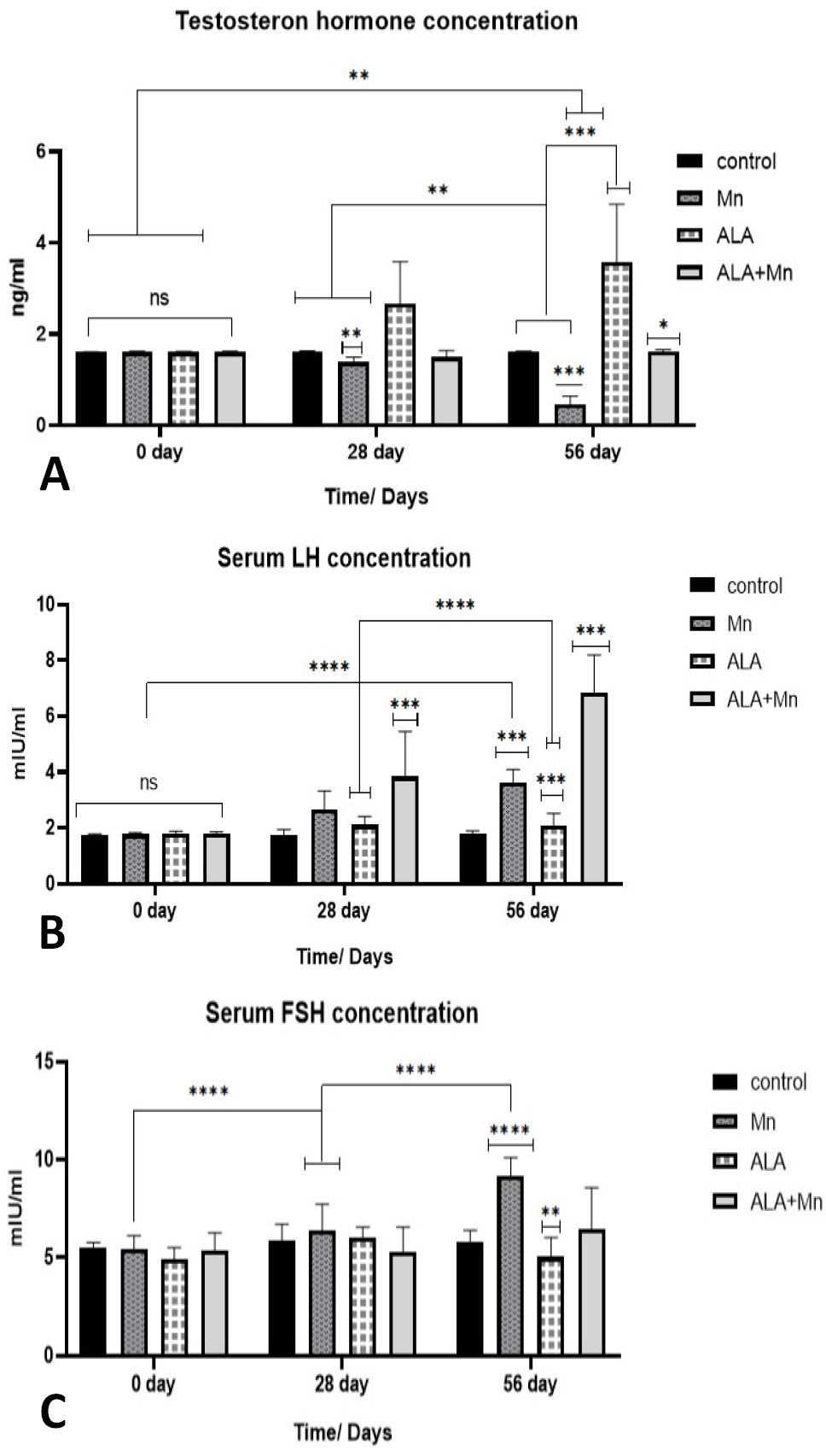
Effect of Mn, ALA, and Mn +ALA on testosterone concentration (A), LH (B), and FSH (C) concentration (mIU/ml) in rats at on day 0,28, and 56) the experiment. Values represented as M ±SE, n = 6 rats. ns: (P>0.05), ** (P<0.05), ***(P<0.001), ****(P<0.0001).
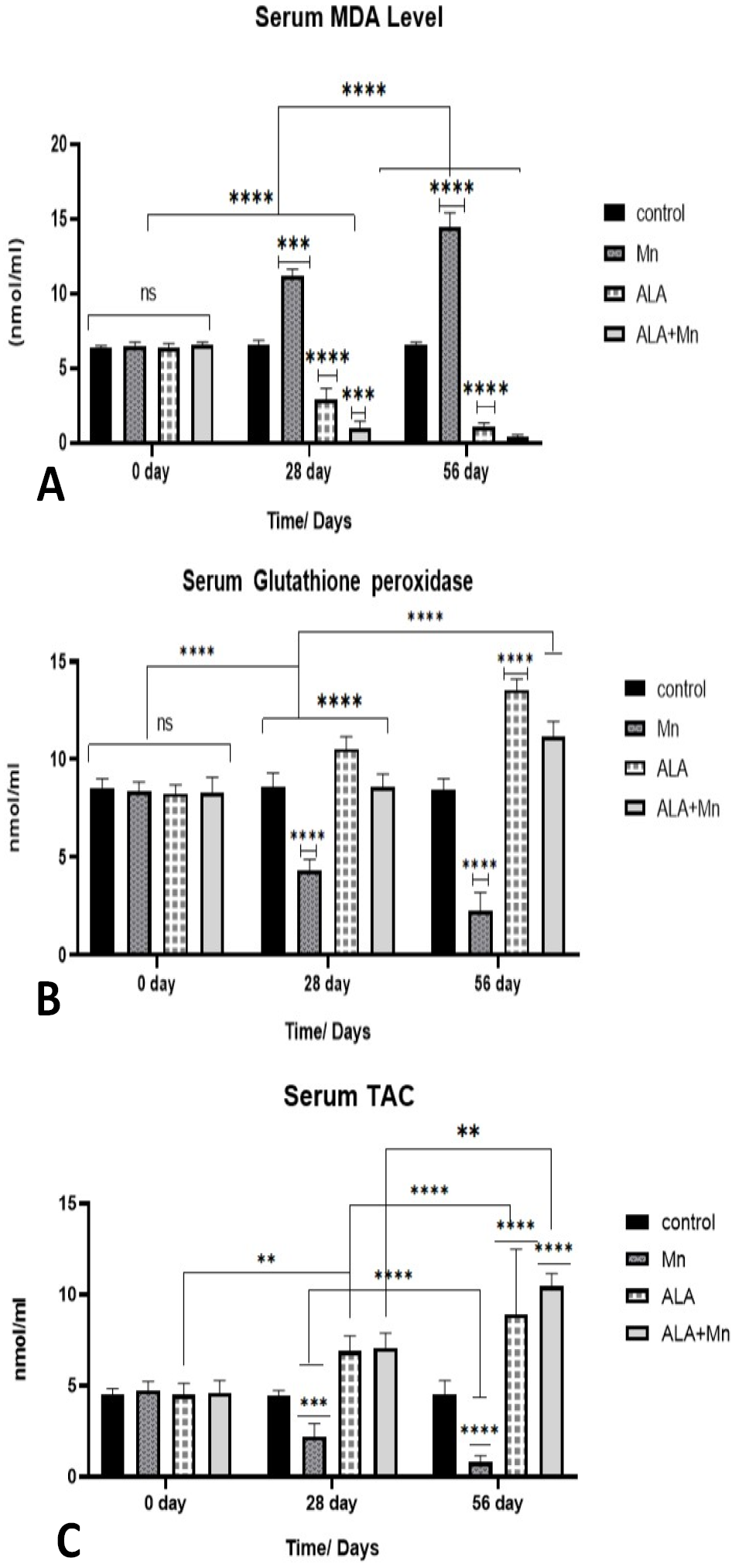
The effect of Mn, ALA, and Mn +ALA on MDA (A), serum glutathione peroxidase (B) and total antioxidant capacity (TAC) (C) (nmol/ml) in rats on days 0, 28, and 56 days of experiment. Values represented as M ±SE, n = 6 rats. ns: (P>0.05), ** (P<0.05), ***(P<0.001), **** (P<0.0001).
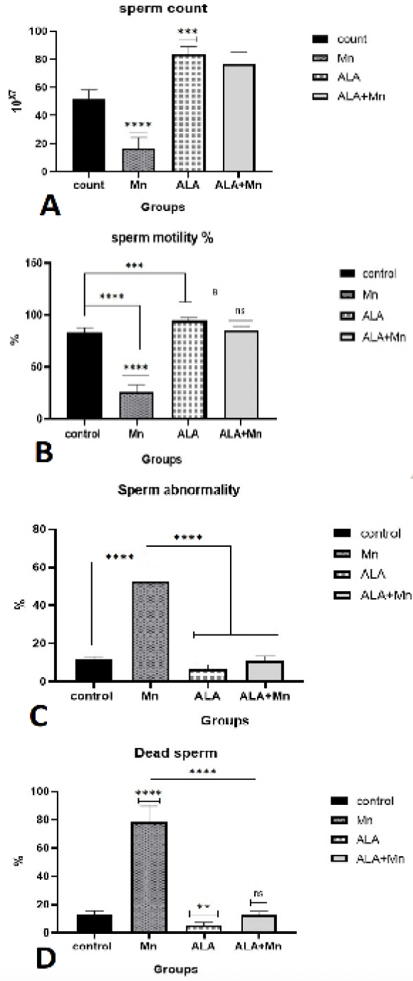
Effect of Mn, ALA and Mn +ALA on sperm count (A), sperm motility (B), sperm abnormality (C) and dead sperm (%) (D) in adult male rats at 56 day of experiment. Values represented as M ±SE, n = 6 rats. ns: (P>0.05), ** (P<0.05), *** (P<0.001), **** (P<0.0001).
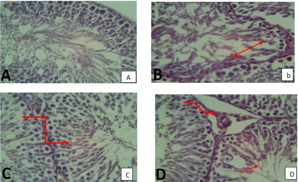
Histopathological section of testicular tissue, (A) section of testes from control group, showed normal structure and germinal cell. (B) section of testes from rats treated with (Mn), ( ) demonstrate the severe atrophic necrosis. (C) section of testes from rats treated with (ALA) showed ( ) a seminiferous tubule are filled with sperms, and the phases of spermatogenesis are visible. (D) section of testes from rats treated with (Mn + ALA) showed regular structure of testes and the seminiferous tubule filled with sperm () and milled congestion of blood vessel () (100 X).
References
- Attia, M., Ebtessam, A.E., Randa, M.Z. and Amal, A.E., 2020. An overview of the antioxidant effects of ascorbic acid and alpha lipoic acid (in liposomal forms) as adjuvant in cancer treatment. Antioxidants, 9: 359. https://doi.org/10.3390/antiox9050359[CrossRef]
- Ávila, D.S., João, B.T.R., Yousef, T., Ana, P.M.D.S., Abel, S.A., Bowman, B. and Michael, A., 2023. Manganese neurotoxicity. In: Handbook of neurotoxicity. Springer. pp. 2305–2329. https://doi.org/10.1007/978-3-031-15080-7_3[CrossRef]
- Baj, J., Wojciech, F., Aleksandra, B., Beata, K., Michał, F., Alicja, F., Grzegorz, T., Piero, P., Grzegorz, B. and Elżbieta, R-B., 2023. Consequences of disturbing manganese homeostasis. Int. J. mol. Sci., 24: 14959. https://doi.org/10.3390/ijms241914959[CrossRef]
- Behnamifar, A., Shaban, R., Mohammad, A., Karimi, T. and Mohsen, S., 2003. Effects of dietary alpha-lipoic acid supplementation on the seminal parameters and fertility potential in aging broiler breeder roosters. 100: 1221-1238.
- Bjørklund, G., Mariia, S., Roman, L., Monica, B., Massimiliano, P., Ioan, S., Oksana, S., Kateryna, S. and Salvatore, C., 2022. Natural compounds and products from an anti-aging perspective. Molecules, 27: 7084. https://doi.org/10.3390/molecules27207084[CrossRef]
- Chandel, M. and Jain, G.C., 2017. Effects of manganese exposure on testicular histomorphology, serum hormones level and biochemical marker parameters in Wistar rats. J. biol. Sci. Med., 3: 1–14. https://doi.org/10.3390/oxygen2010001[CrossRef]
- Cilio, S., Monica, R., Gianluca, V., Benito, F.M., Gaetano, G., Federico, C., Gianpiero, F., Erika, D.Z. and Felice, C., 2022. Beneficial effects of antioxidants in male infertility management: A narrative review. Oxygen, 2: 1–11.
- Corradi, P.F., Renato, B.C. and Loren, W.G., 2016. Physiology of the hypothalamic pituitary gonadal axis in the male. Urol. Clin., 43: 151–162. https://doi.org/10.1016/j.ucl.2016.01.001[CrossRef]
- Deore, M.S., Saba, N., Anoop, K. and Flora, S.J.S., 2021. Alpha-lipoic acid protects co-exposure to lead and zinc oxide nanoparticles induced neuro, immuno and male reproductive toxicity in rats. Front. Pharmacol. 12: 626238. https://doi.org/10.3389/fphar.2021.626238[CrossRef]
- Dutta, S., Pallav, S., Petr, S. and Shubhadeep, R., 2021. Oxidative stress, testicular inflammatory pathways, and male reproduction. Int. J. mol. Sci., 22: 10043. https://doi.org/10.3390/ijms221810043[CrossRef]
- Fenga, C., Silvia, G., Valentina, D.S., Luca, F. and Massimo, L., 2017. Immunological effects of occupational exposure to lead. Mol. Med. Rep., 15: 3355–3360. https://doi.org/10.3892/mmr.2017.6381[CrossRef]
- Gomes-Silva, A.P., Paloma, C.M., Laís, N.S., Marcella, D.S.A.S. and Juliana, E.P., 2023. Exposure to manganese during sertoli cell formation and proliferation disturbs early testicular development in rats. Reprod. Toxicol., 120: 108447. https://doi.org/10.1016/j.reprotox.2023.108447[CrossRef]
- Goudarzi, M., Mojtaba, H.K., Alireza, M., Mojtaba, K. and Esrafil, M., 2020. Protective effect of alpha-lipoic acid on Di- (2-Ethylhexyl ) phthalate-induced testicular toxicity in mice. https://doi.org/10.1007/s11356-020-07817-1[CrossRef]
- Han, Y., Jiali, C., Di, A., Ying, H. and Zhenwu, T., 2022. Occurrence, potential release and health risks of heavy metals in popular take-out food containers from China. Environ. Res., 206: 112265. https://doi.org/10.1016/j.envres.2021.112265[CrossRef]
- Husain, W.M., Araak, J.K. and Ibrahim, O.M.S., 2019. Effect of different doses from ZnONPS on the pituitary-testes axis function in adult male rats. Adv. Anim. Vet. Sci., 7. https://doi.org/10.17582/journal.aavs/2019/7.7.550.556[Full text][CrossRef]
- Jan, A.T., Mudsser, A., Kehkashan, S., Arif, A., Inho, C. and Qazi, M.R.H., 2015. Heavy metals and human health: Mechanistic insight into toxicity and counter defense system of antioxidants. Int. J. mol. Sci., 16: 29592–29630. https://doi.org/10.3390/ijms161226183[CrossRef]
- Khan, H., Thakur, G.S., Randhir, S.D. and Mohamed, M.A.D., 2022. α-lipoic acid, an organosulfur biomolecule a novel therapeutic agent for neurodegenerative disorders: An mechanistic perspective. Neurochem. Res., 47: 1853–1864. https://doi.org/10.1007/s11064-022-03598-w[CrossRef]
- Kurutas, E.B., 2015. The importance of antioxidants which play the role in cellular response against oxidative/nitrosative stress: Current state. Nutr. J., 15: 1–22. https://doi.org/10.1186/s12937-016-0186-5[CrossRef]
- Li, L. and Yang, X., 2018. The essential element manganese, oxidative stress, and metabolic diseases: Links and interactions. Oxid. Med. Cell. Longev., 5: 7580707. https://doi.org/10.1155/2018/7580707[CrossRef]
- Liu, Y., Meijin, Y., Jiawen, C., Ye, D., Xiaohua, T. and Zuozhong, Z., 2021. Heat shock proteins took part in oxidative stress-mediated inflammatory injury via NF-ΚB pathway in excess manganese-treated chicken livers. Ecotoxicol. environ. Saf., 226: 112833. https://doi.org/10.1016/j.ecoenv.2021.112833[CrossRef]
- Majdalawieh, A.F., Sarah, D. and Sarah, M.Y., 2020. Effects of sesamin on fatty acid and cholesterol metabolism, macrophage cholesterol homeostasis and serum lipid profile: A comprehensive review. Eur. J. Pharmacol., 885: 173417. https://doi.org/10.1016/j.ejphar.2020.173417[CrossRef]
- Miao, Z., Zhang, K., Bao, R., Li, J., Tang, Y. and Teng, X., 2021. The mechanism of manganese-caused inflammatory damage: Th1/Th2 imbalance and heat shock proteins mediated inflammation via activating NF-ΚB pathway in chicken nervous system in vivo and in vitro. Environ. Sci. Pollut. Res., 28: 44361–437. https://doi.org/10.1007/s11356-021-13782-0[CrossRef]
- Nyarko-Danquah, I., Edward, P., Alexis, D., Karam, F., Soliman, A., Michael, A. and Eunsook, L., 2020. Manganese accumulation in the brain via various transporters and its neurotoxicity mechanisms. Molecules, 25: 5880. https://doi.org/10.3390/molecules25245880[CrossRef]
- Prathima, P., Venkaiah, K., Pavani, R., Daveedu, T., Munikumar, M., Gobinath, M. and Valli, M., 2017. α -lipoic acid inhibits oxidative stress in testis and attenuates testicular toxicity in rats exposed to carbimazole during embryonic period. Toxicol. Rep., 4: 373–381. https://doi.org/10.1016/j.toxrep.2017.06.009[CrossRef]
- Segat, B.B., Lucas, B.M., Rodrigo, C., Roberta, C., Hugo, T., Alexandra, L., Adolfo, H. and Christiane, F., 2023. Scavenging of reactive species probed by EPR and ex-vivo nanomolar reduction of lipid peroxidation of manganese complexes. J. Inorgan. Biochem., 239: 112060. https://doi.org/10.1016/j.jinorgbio.2022.112060[CrossRef]
- Wesselink, E., Koekkoek, W.A.C., Grefte, S., Witkamp, R.F. and Van Zanten, A.R.H., 2019. Feeding mitochondria: Potential role of nutritional components to improve critical illness convalescence. Clin. Nutr., 38: 982–995. https://doi.org/10.1016/j.clnu.2018.08.032[CrossRef]
- Husain, W.M. and Ali, Z.S., 2022. The effective role of phitofert ® supplement in male reproductive hormones and epididymal sperm quality in carbon tetrachloride (CCL4) treated rats. Adv. Anim. Vet. Sci., 10: 292. https://doi.org/10.17582/journal.aavs/2022/10.2.292.297[Full text][CrossRef]
- Wu, L., Yanqi, L., Ze, Y., Yanhong, W., Wei, L., Guoqiang, Z. and Li, W., 2023. Blood manganese and non-alcoholic fatty liver disease in a high manganese exposure area in China. J. Hlth. Popul. Nutr., 42: 118. https://doi.org/10.1186/s41043-023-00467-2[CrossRef]
- Zhang, Y., Lixia, C., Ruimeng, S., Ruijuan, L., Ting, D., Yuhan, L., Xinming, Z., Rongtian, S. and Yanfei, Q., 2022. Multienzymatic antioxidant activity of manganese-based nanoparticles for protection against oxidative cell damage. ACS Biomater. Sci. Eng., 8: 638–648. https://doi.org/10.1021/acsbiomaterials.1c01286[CrossRef]
- Zhu, Y., Shu, L. and Xiaohua, T., 2016. The involvement of the mitochondrial pathway in manganese-induced apoptosis of chicken splenic lymphocytes. Chemosphere, 153: 462–470. https://doi.org/10.1016/j.chemosphere.2016.03.081[CrossRef]
Services
Article usage statistics
Loading…