
Microplastics (MPs) are free radicals, and exposure towards them can cause hepatotoxicity by increasing serum glutamic-pyruvic transaminase (SGPT) and serum glutamic-oxaloacetic transaminase (SGOT) enzymes in the blood, leading to liver cell damage. Aiming to find natural antioxidants for minimizing the negative impacts by MPs, this study investigated pineapple due to its rich contents of the aforementioned nutrients such as bromelain, flavonoids, and vitamin C, to prevent and mitigate hepatotoxicity. Employing a true experiment with a post-test control group design, 15 rats Rattus norvegicus were selected as samples and divided into five treatments: normal treatment, positive control with 4 200 µg d–1 oral-induced PET-MPs (sizes: < 0.2 mm), and three treatments of pineapple extract (PE) at 87.5 mg kg–1 BW, 175 mg kg–1 BW, and 350 mg kg–1 BW. The statistical analysis utilized ANOVA and regression correlation. The results showed decreases in SGPT levels, SGOT levels, and liver cell damage percentages in all samples receiving PE. The optimum dosage was 350 mg kg–1 BW with suppression levels of 41 %, 42 %, and 76 %, respectively, compared to the control. It is therefore confirmed that PE has the ability to act as an antioxidant and anti-inflammatory agent. This research will be further developed by comparing the efficacy of PE to other natural antioxidants, followed by formulating it in the form of effervescent tablets for public consumption in order to achieve the Sustainable Development Goals, especially the Goal 3: Good health and well-being.
Special Issue: Novel Advances in Agricultural Science and Technology for Sustainable Farming in Tropical Region · Volume 39, Issue 1, 10 October 2025 · Open Access
Effectiveness of Pineapple Extract in Improving Microplastic Hepatotoxicity in Male White Rats of Wistar Strain
· · · · · · · · ·
Affiliations & Notes ⌄
- 1Department of Clinical Pathology, University of Muhammadiyah Malang - UMM, Malang 65145, East Java, Indonesia
- 2Department of Agriculture, Graduate Program, UMM, Malang 65144
- 3Department of Internal Medicine, UMM
- 4Department of Chemical Engineering, Diponegoro University, Semarang 50275, Central Java, Indonesia
- 5Department of Biology and Biotechnology, The Hashemite University, Zarqa 13133, Jordan
- 6UMM Faculty of Medicine Students. , *Correspondence | Diah Hermayanti, Department of Clinical Pathology, Medical Faculty, University of Muhammadiyah Malang, Jl. Bendungan Sutami No.188, Malang 65145, East Java, Indonesia
- 7Email: diahhermayanti_umm@yahoo.com
Article Info ⌄
- Journal:
- Sarhad Journal of Agriculture
- Published:
- 10 October 2025
From the article
- Further, MPs disrupt redox balance, leading to oxidative damage in the liver.
- Treatments with ethanolic fruit extract of pineapple at 200 mg kg–1 and 400 mg kg–1 significantly reduce liver biomarker enzymes.
- Administration of PE is able not only to reduce SGPT and SGOT levels but also to decrease the percentage of liver cell damage due to hepatotoxicity in MPs-induced R. novergicus.
- Although not as effective as the other treatments in reducing SGPT levels, the discrepancy is not statistically significant.
Selected from the article's own text.
Abstract
Introduction
Plastic pollution is a global health issue, and it is estimated that by 2050, approximately 12 × 109 t of plastic waste will be found worldwide . Humans can be exposed to microplastics (MPs) and even nanoplastics (NPs) mainly through diet or inhalation. Human exposure to MPs has increased due to the discovery of MPs in human-consumed seafood, drinking water, and food packaging . In Indonesia, MPs have been found in potato plantations, i.e., soil, organic fertilizer, irrigation water, potato seeds, potato production, and potato by-products , b, 2025). The same research team has discovered MPs contamination in not only 33 brands of edible oil and various Indonesian traditional snacks ; , but also grass field soil, main feed, supplementary feed, drinking water, goat feces, and goat milk at goat farms as well as commercial Red Palm Oil . MP pollutants are also found in salt , sugarcane (Saccharum officinarum L.) , Caridean shrimp (Caridea Dana, 1852) farming ponds , paddy germination , and assorted seeds of crops among them are paddy (Oryza sativa L.), peanut (Arachis hypogaea L.), soybean (Glycine max Merr.), corn (Zea mays L.), mungbean (Vigna radiate (L.) R. Wilczek), and palm oil (Elaeisguineensis Jacq.).
The small size of MPs allows these materials to easily pass-through biological membranes . This exposure impacts human health , as it has been reported to cause organ toxicity, neurotoxicity, and toxicity to the reproductive and developmental systems ; ; . MP accumulation in the liver can trigger the excessive production of reactive oxygen species (ROS), causing oxidative stress in hepatocytes and hepatotoxicity ; Li et al., 2023a). Studies by Banihashemi et al. (2022), Inaku et al. (2024, 2025), and Zou et al. (2023) have confirmed that the administration of MPs can increase serum glutamic-oxaloacetic transaminase (SGOT) and serum glutamic-pyruvic transaminase (SGPT) levels.
Oxidative damage caused by free radicals in the human body can be decreased, even prevented, by the body’s antioxidant defense system through free radical scavenging, metal chelation, and neutralization of reactive species through enzymatic activity. Consuming antioxidants from food is a way to maintain antioxidant levels in the body ; . Antioxidants limit the expression and activity of free radical-generating enzymes such as xanthine oxidase (XO) and NAD(P)H oxidase or boost the expression and activity of antioxidants such as glutathione peroxidase (GPx), catalase (CAT), and superoxide dismutase (SOD) to control the levels of reactive species ; .
Pineapple (Ananas comosus L.) is rich in antioxidants among them are bromelain, flavonoids, and vitamin C. Bromelain from whole pineapple is of medium antioxidant activity at an IC50 value of 3 624 μg mL–1 ; . This study aims to determine the optimal dose of PE in minimizing the impact of MPs on experimental animals, specifically male white rats of the Wistar strain (Rattus norvegicus (Berkenhout, 1769)). In the next stage, research involving human subjects is required to achieve the Sustainable Development Goals (SDGs), particularly the Goal 3: Good health and well-being .
Materials and Methods
Conducted in the Pharmacology Laboratory of the Faculty of Medicine, University of Muhammadiyah Malang, campus II (S 7°57’27.1332” and E 112°36’50.7312”) between February 10, 2025 and March 23, 2025 at constant temperature (28 °C ± 2 °C),
humidity (55 % ± 10 %), and 8 h light/16 h dark cycle (lights on at 09:00 and off at 17:00), the study was carried out by the description of ethical approval No. E.5.a/019/KEPK-UMM/II/2024 issued by the Health Research Ethics Commission, Faculty of Medicine, University of Muhammadiyah Malang.
Experimental animal
In this study, male R. norvegicus were selected as the experimental animals. Each of 15 rats was placed in a designated glass cage (300 mm × 300 mm × 300 mm), and all cages were arranged in a randomized block design based on the rats’ weight at the start of the study . Each treatment underwent three repeats as advised by Arifin and Zahiruddin (2017) by the provisions of “sample size calculation in animal studies”.
Once acclimatized for 1 wk, the rats were divided into five treatments, namely the normal treatment (N), the MPs positive control treatment (C), and the treatment groups (P1, P2, and P3) with MPs administration at 4 200 µg d–1, referring to the previous research with modification ; ; Li et al., 2023b). Pineapple Extract (PE) was applied at the doses of 87.5 mg kg–1 BW (P1), 175 mg kg–1 BW (P2), and 350 mg kg–1 BW (P3) .
During the 35 d research period (Li et al., 2023b), all treatments were fed with the standard BR1 feed (PT. Japfa Comfeed Indonesia) of 20 g d–1, while drinking water was provided ad libitum. Every day, the remaining feed, drinking water, and feces were recorded for MPs content observation ; .
Pineapple extract (PE)
Pineapple samples were obtained from pineapple plantations in Blitar area, East Java (S 7°59’45.15” and E 112°11’5.3628”), harvested in November 2024. PE was prepared in Materia Medika Laboratory, Batu, East Java, Indonesia (S 7°51’55.386” and E 112°31’8.238).
An amount of 1 kg of fresh pineapple flesh was cut into small pieces. Once pureed with a blender (Philips 5000 series HR2223/70, China), the juice was dissolved into a mush with a bit of 95 % ethanol (food grade, PT. Gael Vada Indonesia). Next, 3 000 mL of ethanol solvent was stirred in (Vertical ribbon mixer, China) until homogeneous. The result was covered with aluminum foil and stored at a temperature of 4 °C for 24 h (PLR 386 Thermo Scientific, Lab. refrigerator). The filtrate was then separated from its pulp (Merck-millipore EZ-stream vacuum filtration Pump, Germany) and evaporated at a temperature of 40 °C (RE100-S LED digital rotary evaporator, China), speed of 10 rpm to 20 rpm (1 rpm = 1/60 Hz), and suction pressure of 2 atm (1 atm = 101.325 Pascal) to obtain a concentrated extract liquid. Once no more liquid dripped into the collection flask, indicating that the extraction was complete, the evaporation process was finished .
Microplastics
MPs used in this study were of the polyethylene terephthalate (PET) type made from used PET plastic bottles and pellets. Ground into powder in a rice mill, the material was then filtered using an 80-mesh sieve (equivalent to 0.2 mm) supplied by the Research and Community Service Institute of Universitas Islam Madura (Coordinates: 7°9’20.1708” S and 113°27’27.9504” E).
Unlike several previous studies on in vivo MPs ; Li et al., 2023b; , this study found hindrance in obtaining MPs in micrometer (µm) or nanometer (nm) sizes due to limited equipment availability . While it is a weakness, the situation also serves as a novelty point of this study, as the danger of MPs, even in mm sizes, particularly in R. norvegicus, can be demonstrated.
SGPT and SGOT examination
A blood sample was taken from the ventricle of the heart of each sample, left to rest for 1 h, and then centrifuged to obtain the serum. The serum was then run through spectrophotometry (DR6000 UV-Vis, Germany) to examine SGPT and SGOT activities .
Liver histology examination
The liver of each sample was placed in 10 % neutral formalin preservative and then prepared in sections using paraffin, stained with Hematoxylin. Each preparation was observed through five fields of view to calculate the number of normal cells and the number of damaged ones, encompassing necrosis, fatty degeneration, and hydropic degeneration. The results were further processed to obtain the percentage of damaged cells in each field of view and the average rate of cell damage from each treatment . SGOT and SGPT examination and liver histology observation were carried out in the Central Laboratory for Research and Diagnostics of “Satwa Sehat”, Malang, Indonesia (S 7°57’43.8408” and E 112 36’8.1036”). All samples were analyzed with a blind system.
Statistical analysis
The results of each double sample were presented as the mean and standard deviation. Statistical analysis was conducted by the Shapiro-Wilk test for data normality and the Levene test for data homogeneity ; . Significant differences between the treatments and the control were determined by one-way analysis of variance (ANOVA, P < 0.05), followed by the Tukey (honestly significant difference - HSD) test, using SPSS 20.0. Origin 9.5 was used to create and modify all the graphs .
A correlation test was performed post-ANOVA to determine the relationship between PE and the observed variables, namely SGPT, SGOT, and liver cell damage ; . Next, a linearity graph was calculated between the PE dose and the observed variables to prove the existence of linearity .
Result and Discussion
Analysis of variance
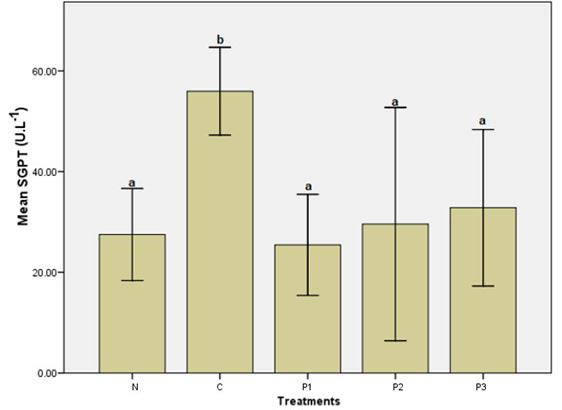
Serum glutamic pyruvic transaminase (SGPT): Rates of SGPT after MPs exposure at 4 200 µg d–1in five treatments are listed in Figure 1. Meanwhile, supporting data (Sd, SEM, 95 % confidence interval for mean, and minimum and maximum) are listed in Supplementary Table 1A.
Figure 1 indicates that the MPs’ introduction has induced SGPT production, with the highest rate observed in C (55.95 µL µL-1), which is significantly different from the rates in the other treatments. SGPT rates in treatments N, P1, P2, and P3 are not substantially different from each other, and they are lower compared to C at 51 %, 54 %, 47 %, and 41 %, respectively. It can be inferred that the PE antioxidant is capable of reducing SGPT after MPs come into contact.
Serum glutamic oxaloacetic transaminase (SGOT)
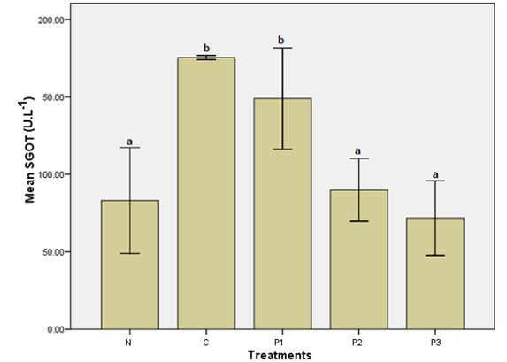
Rates of SGOT after MPs exposure at 4 200 µg d–1 in five treatments are as detailed in Figure 2. Meanwhile, supporting data (Sd, SEM, 95% confidence interval for mean, and minimum and maximum) are listed in Supplementary Table 1B.
PE has shown noteworthy effects on SGOT rates. While not significantly different in treatments N, P2, and P3, they are lower than the other two. Treatment C, although not substantially different from P1, has the highest rate of SGOT of all it is 52 %, 49 %, and 58 % higher compared to treatments N, P2, and P3, respectively. The gaps are statistically significant. A considerable decrease in SGOT rates is noted in P2 and P3, at 43 % and 54 %, compared to P1.
Liver cell damage
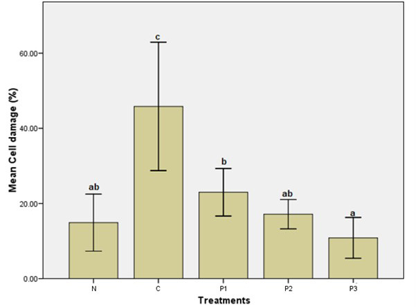
Rates of liver cell damage after MPs exposure at 4 200 µg d–1 in five treatments are depicted in the form of a bar chart in Figure 3. Meanwhile, supporting data (Sd, SEM, 95 % confidence interval for mean, and minimum and maximum) are listed in Supplementary Table 1C.
The experiment has highlighted the sizeable impact of PE antioxidant on liver cell damage rates in the rats. Treatment C is significantly higher than the other treatments, which are 67 % (N), 50 % (P1), 62 %
(P2), and 76 % (P3) lower, respectively. P3 records the lowest damage rates, while those in N, P1, and P2 are not significantly different from each other. A remarkable result is observed in the significant 53 %
decrease in P3 compared to P1. However, further analysis has proven that the damage rate gaps in treatments P3, P2, and N are not statistically different.
Liver histology examination
To support the findings presented in Figures 1, 2, and especially Figure 3, liver histology examination was also performed, and the results are shown in Figure 4.
The liver cell damage histology in Figure 4 comprises cell degeneration and necrosis. In C treatment, where the samples were exposed to MPs without receiving PE treatment (Figure 4B), the damaged cells appear to be the most severe. Fewer damaged cells occur in P1 (Figure 4C), and a further decrease is visible in P2 (Figure 4D). Maximum cell regeneration is evident in P3.
Regression and correlation analysis
Following up on the aforementioned ANOVA test, regression and correlation analyses were conducted to determine the correlation between the effect of PE antioxidant and the variables used to validate the data in Figures 1, 2, and 3. Correlation coefficient value, R square, and regression equation are specified in Table 2, while linearity graphs are presented in Figure 5A, B, C.
Table 2: Regression and correlation analysis on the observed variables and PE antioxidant.
| Observed variables | Correlation coefficient | P value (P > 0.05) | R square (%) | Regression equation Y= EP, X= Observed variables |
| SGPT | -0.469 | 0.062 | 22.0 | Y = 43.095 - 0.047 X |
| SGOT | -0.875 | 0.000 | 76.6 | Y = 168.43 - 0.307 X |
| Liver cell damage | -0.849 | 0.000 | 72.1 | Y = 37.81 - 0.090 X |
Discussion
MPs in experimental models, such as cells, organs, and animals, are toxic because they cause oxidative stress ; . Excessive reactive oxygen species (ROS) production and free radical formation can lead to oxidative stress, resulting in damage to cells, tissues, and organs ; . Cellular oxidative stress is defined as the imbalance between ROS production and antioxidants ; . This imbalance can lead to the malfunction or structural modification of major cellular molecules, such as lipids, proteins, and DNA (Sadasivam et al., 2022). Lipid peroxidation, resulting from an oxidative attack on the unsaturated acyl chains of lipids by free radicals, can modify membrane structure and facilitate the lateral diffusion of lipids, thereby significantly
reducing the stretching modules of membranes and increasing permeability . In experimental rats, MPs induce hepatotoxicity, characterized by elevated levels of SGPT and SGOT enzymes, as well as a decrease in the expression of oxidative stress-related proteins, including sirtuin 3 (SIRT3) and superoxide dismutase-2 (SOD2) . Meanwhile, nanometer-sized plastics (NPs) in large yellow croaker (Larimichthyscrocea Richardson, 1846), a carnivorous marine fish, have altered the fatty acid composition and fish muscle texture by enhancing oxidative stress and disrupting lipid metabolism, indicating that PS - NPs can induce liver lipid deposition by inhibiting lipolysis . The level of SGPT and alkaline phosphatase (ALP) can increase 2 to 3 times in hepatocellular, cholestatic, or mixed hepatic damage. In addition to SGPT and SGOT, elevated levels of ALT and AST in the blood are indicative of hepatocyte necrosis and inflammation. The rise of AST is often regarded as less than that of ALT in viral hepatitis, but both are clinically relevant in detecting acute hepatic damage . Further, MPs disrupt redox balance, leading to oxidative damage in the liver. This exposure also disrupts hepatic lipid metabolism by stimulating lipid synthesis while inhibiting catabolism, ultimately leading to the development of fatty liver . Moreover, MPs can induce liver fibrosis by activating the Wnt/β-catenin signaling pathway . Although the size of MPs in this research is on the mm scale, despite being measured in micrometer (µm) or nanometer (nm), the results exhibited in Figures 1-4 indicate their negative impacts on the liver of R. norvegicus.
The phytochemical constituents of pineapple are saponins, tannins, steroids, flavonoids, terpenoids, naphthoquinone, inulin, alkaloids, phenols, and amino acids. Additionally, the fruit also contains phytosterols, cardiac glycosides, proteins, and polyphenols. Bromelain and vitamin C are also identified in the fruit of the pineapple ; . The significant amount of polyphenols in pineapple fruit extract and juice indicates their high antioxidant activity, which has the potential to be used in preventive medication and the treatment of various diseases (Jovanović et al., 2018). The pineapple crown produces 0.26 % dried crude bromelain with a total protein content of 44.10 % and an IC50 value of 3 624 μg mL–1, equivalent to 1 590.18 μg mL–1 of total protein . Treatments with ethanolic fruit extract of pineapple at 200 mg kg–1 and 400 mg kg–1 significantly reduce liver biomarker enzymes. Histopathological reports revealed that administration of paracetamol caused degeneration of fatty cysts, infiltration of lymphocytes, proliferation of Kupffer cells, and congestion of liver sinusoids. Upon treatment with the ethanolic fruit extract of pineapple, normal hepatic globular architecture, reduced lymphatic infiltration, and normal Kupffer cell proliferation were observed, suggesting that the ethanolic fruit extract of pineapple protects the liver from adverse conditions .
Table 2 indicates negative coefficient correlation values in all observed variables. Referring to Praptiningsih (2022) and Prasad (2022), it is inferred that the higher the PE dosage, the lower the rates of observed variables (SGPT, SGOT, and liver cell damage) will be. R-squared values of SGPT= 22.0 %, SGOT= 76.6 %, and cell damages= 72.1 % represent the effect of PE administration at 22.0 %, 76.6 %, and 72.1 % towards the values of respective observed variables.
The linearity graphs in Figure 5A-C show that all regression lines between the independent variable and each of the three dependent variables are related to the bottom right, indicating relevance between PE antioxidant administration and SGPT, SGOT, and liver cell damage rates ; . The higher PE dosage significantly affects the lower rates of observed variables, and vice versa.
Conclusions
Administration of PE is able not only to reduce SGPT and SGOT levels but also to decrease the percentage of liver cell damage due to hepatotoxicity in MPs-induced R. novergicus. The optimum result was recorded by P3 receiving PE at 350 mg kg–1 BW with the lowest SGOT level and liver cell damage percentage. Although not as effective as the other treatments in reducing SGPT levels, the discrepancy is not statistically significant. It is conclusive that PE is feasible for treating and preventing liver diseases in R. norwegicus.
MPs–induced at 4 200 µg d–1 were of ground polyethylene terephthalate (PET) and filtered with an 80-mesh sieve to a size of 0.2 mm. While larger than micrometers (µm) or nanometers (nm) in size, its existence brought disconcert to the samples’ bodily systems. It is therefore evident that MPs, even in millimeter size, can be hazardous to health.
Recommendations
This research will be further developed by comparing the efficacy of PE to other natural antioxidants against MPs exposure, especially at higher doses. Referring to the findings of Zou and You (2024), which surveyed 109 countries, indicating that Indonesian people consumed MPs at a rate of approximately 15 g MPs mo–1, the exposure dose of MPs in this study is relatively low. The dangers of MPs in Indonesian society must be prevented to achieve the Sustainable Development Goals, especially the Goal 3: Good health and well-being. Once a positive result is determined for the appropriate dosage of PE for human consumption, formulating PE in the form of effervescent tablets should be feasible for public use.
Acknowledgments
The authors express their gratitude to the Research and Community Service Institute, University of Muhammadiyah Malang (LPPM-UMM), which has funded this research under the letter of consent E.3a/368.10-RPK-UMM/2024, dated October 23, 2024. Heartfelt thanks are also extended to Mardiana Sri Susanti (Aura Statistics Consultant), Trias Agung Pakati (Mayantara School), and Dewi Mariyam and staff (Central Laboratory for Research and Diagnostics of “Satwa Sehat”) for preparing this manuscript. Additionally, Surya Andhi Angga Putra, Resky Adelia Saputri, and Miftha Churochman have assisted in the in vivo test.
Novelty Statement
The authors have conducted a search on Google Scholar for an unspecified period. As of July 15, 2025, no scientific papers on the ability of PE to mitigate the effects of PET-MPs on the liver of R. norvegicus have been found. Two studies by Inaku et al. (2024, 2025) investigated the liver and kidneys of R. norvegicus exposed to PE manufactured by a chemical manufacturer for 14 d. However, the studies above did not include antioxidant tests. Based on the data, the authors declare that the manuscript submitted to the Sarhad Journal of Agriculture is novel.
Author’s Contribution
Diah Hermayanti and Roy Hendroko Setyobudi: Conceptualized and designed the study, elaborated the intellectual content, performed literature search, materials research, manuscript preparation, and manuscript revision.
Meddy Setiawan: Research supervision and grant funding.
Suherman Suherman, Mohammed Ali Wedyan, and Muhannad Illayan Saleem Massadeh: Manuscript review.
Rika Permatasari Sanusi, Rona Nisrina, Nurul Izzah Dwi Faradinah, and Hajar Tsabita: investigator.
All authors have read and approved the final manuscript.
Supplementary material
There is supplementary material associated with this article, i.e., Tables 1A, 1B, and 1C. The supplementary material is listed below (after the list of references).
Generative AI and AI-assisted technology statement
The authors stated that they didn’t use generative AI and AI-assisted technology in preparing this manuscript.
Conflict of interest
The authors have declared no conflict of interest.
Supplementary Table 1: Mean, standard deviation, standard error of SGPT, SGOT, and cell damage.
|
| N | Mean | Std. deviation | Std. error | 95% confidence interval for mean | Minimum | Maximum | |
| Lower bound | Upper bound | |||||||
| A: SGPT | ||||||||
| N | 3 | 27.5033 | 3.67525 | 2.12190 | 18.3735 | 36.6332 | 25.06 | 31.73 |
| PC | 3 | 55.9500 | 3.50287 | 2.02238 | 47.2484 | 64.6516 | 53.76 | 59.99 |
| P1 | 3 | 25.4367 | 4.03956 | 2.33224 | 15.4018 | 35.4715 | 20.99 | 28.88 |
| P2 | 3 | 29.5733 | 9.32125 | 5.38163 | 6.4181 | 52.7286 | 19.46 | 37.82 |
| P3 | 3 | 32.8200 | 6.24875 | 3.60772 | 17.2972 | 48.3428 | 26.45 | 38.94 |
| Total | 15 | 34.2567 | 12.50729 | 3.22937 | 27.3304 | 41.1830 | 19.46 | 59.99 |
| B: SGOT | ||||||||
| N | 3 | 83.1133 | 13.76243 | 7.94574 | 48.9256 | 117.3011 | 73.79 | 98.92 |
| PC | 3 | 175.3800 | 0.53777 | 0.31048 | 174.0441 | 176.7159 | 175.04 | 176.00 |
| P1 | 3 | 148.9133 | 13.14159 | 7.58730 | 116.2678 | 181.5589 | 136.87 | 162.93 |
| P2 | 3 | 89.9033 | 8.15480 | 4.70818 | 69.6457 | 110.1610 | 80.67 | 96.12 |
| P3 | 3 | 71.7200 | 9.69039 | 5.59475 | 47.6477 | 95.7923 | 64.08 | 82.62 |
| Total | 15 | 113.8060 | 43.07060 | 11.12078 | 89.9543 | 137.6577 | 64.08 | 176.00 |
| C: Cell damage (%) | ||||||||
| N | 3 | 14.9000 | 3.05043 | 1.76116 | 7.3223 | 22.4777 | 12.56 | 18.35 |
| PC | 3 | 45.8467 | 6.87954 | 3.97191 | 28.7569 | 62.9364 | 39.60 | 53.22 |
| P1 | 3 | 22.9933 | 2.54700 | 1.47051 | 16.6662 | 29.3204 | 20.93 | 25.84 |
| P2 | 3 | 17.1433 | 1.57119 | .90713 | 13.2403 | 21.0464 | 15.33 | 18.10 |
| P3 | 3 | 10.8367 | 2.18793 | 1.26320 | 5.4015 | 16.2718 | 9.29 | 13.34 |
| Total | 15 | 22.3440 | 13.21195 | 3.41131 | 15.0275 | 29.6605 | 9.29 | 53.22 |
Figures
SGPT rates in MPs-administered R. novergicus.
SGOT rates in MPs-administered R. novergicus.
Liver cell damage rates in MPs-administered R. novergicus.
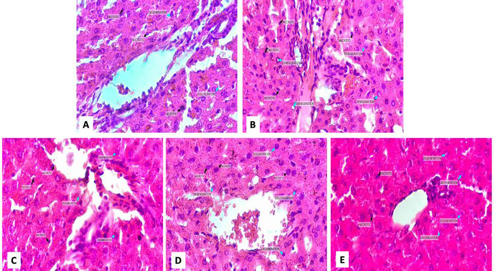
Histology images of the liver (A= normal treatment; B=control treatment; C=P1; D=P2; E=P3 (Hematoxylin-eosin, 400×).
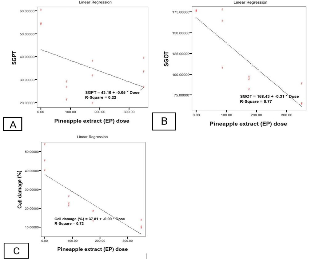
Linearity graph on PE antioxidant and SGPT (A), SGOT (B), cell damage (C).
References
- Arifin, W.N. and W.M. Zahiruddin. 2017. Sample size calculation in animal studies using resource equation approach. Malays. J. Med. Sci., 24(5): 101–105. https://doi.org/10.21315/mjms2017.24.5.11[CrossRef]
- Ayoka, T.O., B.O. Ezema, C.N. Eze and C.O. Nnadi. 2022. Antioxidants for the prevention and treatment of non-communicable diseases. J. Explor. Res. Pharmacol., 7(3): 179–189. https://doi.org/10.14218/JERP.2022.00028[CrossRef]
- Azeh, E.G., N.F. Nwalo and B.N. Nkeh-Chungag. 2022. Free radicals, oxidative stress-related diseases and antioxidant supplementation. Altern. Ther. Health Med., 28(1): 114.
- Azizah, M., 2018. Anti-inflammatory effect test of pineapple (Ananas comosus L.) fruit flesh extract on male white mice induced by egg albumin. J. Ilmiah Bakti Farmasi, 3(1): 41–46.
- Banihashemi, A., S. Soltanian, A. Gholamhosseini and M. Banaee. 2022. Effect of microplastics on Yersinia ruckeri infection in rainbow trout (Oncorhynchus mykiss). Environ. Sci. Pollut. Res., 29: 11939–11950. https://doi.org/10.1007/s11356-021-16517-3[CrossRef]
- Baty, F., C. Ritz, S. Charles, M. Brutsche, J.P. Flandrois, and M.L. Delignette-Muller. 2015. A toolbox for nonlinear regression in R: The package nlstools. J. Stat. Softw. 66(5): 1–21. https://doi.org/10.18637/jss.v066.i05[CrossRef]
- Chaudhary, P., P. Janmeda, A.O. Docea, B. Yeskaliyeva, A.F.A. Razis, B. Modu, D. Calina and J. Sharifi-Rad. 2023. Oxidative stress, free radicals and antioxidants: potential crosstalk in the pathophysiology of human diseases. Front. Chem. Sec. Med. Pharma. Chem., 10: 1–24. https://doi.org/10.3389/fchem.2023.1158198[CrossRef]
- Chiang, C.C., H. Yeh, R.F. Shiu, W.C. Chin and T.H. Yen. 2024. Impact of microplastics and nanoplastics on liver health: Current understanding and future research directions. World J. Gastroenterol., 30(9): 1011–1017. https://doi.org/10.3748/wjg.v30.i9.1011[CrossRef]
- Damat, D., R.H. Setyobudi, I. Iswahyudi, S. Suherman, Y. Sincihu, H.A. Manshur, A. Anekawati, S.K. Wahono, M. Anwar, M.P. Garfansa, R.A. Saputri and S.A. Amani. 2025. Determination and quantification of microplastics in cooking oil sold in Indonesia. Sarhad J. Agric., 39 (Special issue 1): 198-207. https://dx.doi.org/10.17582/journal.sja/2023/39/s1.198.207[Full text][CrossRef]
- Damat, D., R.H. Setyobudi, J.S. Utomo, Z.Vincēviča-Gaile, A. Tain and D.D. Siskawardani. 2021. The characteristics and predicted of glycemic index of rice analogue from modified arrowroot starch (Maranta arundinaceae L.). Jordan J. Biol. Sci., 14(3): 389–393. https://doi.org/10.54319/jjbs/140302[CrossRef]
- Das, A., 2023. The emerging role of microplastics in systemic toxicity: Involvement of reactive oxygen species (ROS). Sci. Total Environ., 895: 165076. https://doi.org/10.1016/j.scitotenv.2023.165076[CrossRef]
- Dewi, N.D.M.A., N.I. Wiratmini and S.K. Sudirga. 2021. Histology of mice (Mus musculus L.) liver and kidney induced by carbon tetrachloride (CCl4) after being given soursop leaf extract (Annona muricata L.). J. Biol. Udayana, 26(1): 21–31. https://doi.org/10.24843/JBIOUNUD.2022.v26.i01.p03[CrossRef]
- Ekalaturrahmah, Y.A.C., M.T. Shidqi, M.P. Garfansa, M.S. Sholeh, F.K. Hartati, A. Sutanto, I. Iswahyudi and A.K. Putri. 2025. Identification and quantification of microplastics in water, sediment, and shrimp collected from shrimp farming ponds. Bioresour. Technol. Rep., 29(102064). https://doi.org/10.1016/j.biteb.2025.102064[CrossRef]
- Garfansa, M.P., R.H. Setyobudi, I. Iswahyudi, S. Anwar, D. Damat, T. Liwang, M.S. Susanti, D. Hermayanti, M. Setiawani, T.D.N. Subchi, D. Mariyam, Y.M. Ananda, S.K. Wahono, A. Basir, M. Musrif and A.S. Khan. 2024. Potential impact microplastic polyethylene terephthalate on mice. Sarhad J. Agric., 39(1): 47-60. https://doi.org/10.17582/journal.sja/2023/39/s1.47.60[Full text][CrossRef]
- Hector, A., 2021. The new statistics with R: An introduction for biologists. Oxford University Press. https://doi.org/10.1093/oso/9780198798170.001.0001[CrossRef]
- Hendroko, R., A. Wahyudi, S.K. Wahono, G.A. Praptiningsih, Salafudin, Salundik and T. Liwang. 2013. Bio-refinery study in the crude jatropha oil process: Co-digestion sludge of crude jatropha oil and capsule husk Jatropha curcas Linn. as biogas feedstocks. Int. J. Technol., 4(3): 202–208. https://doi.org/10.14716/ijtech.v4i3.115[CrossRef]
- Hermayanti, D., R.H. Setyobudi, S. Anwar, M.P. Garfansa, I. Iswahyudi, M. Setiawan, T. Liwang, T.D.N. Subchi, L. Zalizar and P.G. Adinurani. 2024. The effect of polyethylene terephthalate microplastics on the growth of mice. Bio Web Conf., 104(00005): 1–17. https://doi.org/10.1051/bioconf/202410400005[CrossRef]
- Inaku, A.H.R., H. Hadiyanto, H.R Abdurachim, T. Bariroh, and S. Rachmawati. 2025. In vivo test of acute exposure of polyethylene microplastics on kidney and liver of Rattus norvegicus Wistar strain rats. Ecol. Eng. Environ. Tech., 26(2): 164–177. https://doi.org/10.12912/27197050/196654[CrossRef]
- Inaku, A.H.R., H. Hadiyanto, H.R. Abdurachim, T. Bariroh and H.H.A. Matin. 2024. Kidney and liver disorders due to microplastic exposure: Chronic in vivo study in male white rats. J. Kesehatan Masyarakat, 20(1): 120–130. https://doi.org/10.15294/kemas.v20i1.48781[CrossRef]
- Iswahyudi, I., W. Widodo, W. Warkoyo1, R.H. Setyobudi, D. Damat, D. Roeswitawati, S. Anwar, T.D.N. Subchi, I.R. Utarid, M.P. Garfansa, M.S. Sholeh, I. Ekawati, R. Tonda, W.A. Mujianti, D.R.A. Sukma, S.U. Lestari and C. Anam. 2024. Effect of high-density polyethylene, polyvinyl chloride and low-density polyethylene microplastics on seeding of paddy. Sarhad J. Agric., 39 (Special issue 1): 61–70. https://doi.org/10.17582/journal.sja/2023/39/s1.61.70[Full text][CrossRef]
- Ji, L.L. and D. Yeo. 2021. Oxidative stress: An evolving definition. Fac. Rev., 10(13). https://doi.org/10.12703/r/10-13[CrossRef]
- Jones, L., A. Barnett and D. Vagenas. 2025. Linear regression reporting practices for health researchers, a cross-sectional meta-research study. PLoS One, 20(3): e0305150. https://doi.org/10.1371/journal.pone.0305150[CrossRef]
- Jovanovic, M., M. Milutinovic, M. Kostic, B. Miladinovic, N. Kitic, S. Brankovic and D. Kitic. 2018. Antioxidant capacity of pineapple (Ananas comosus (L.) Merr.) extracts and juice. Lek. Sirov., 38: 27–30. https://doi.org/10.5937/leksir1838027J[CrossRef]
- Kıran, T.R, O. Onder and K.A. Bay. 2023. Oxidative stress and antioxidants in health and disease. J. Lab. Med., 47(1): 1–11. https://doi.org/10.1515/labmed-2022-0108[CrossRef]
- Kumar, D., Suchitra, J. Mundlia, S.K. Yadav, D. Yadav, N. Aggarwal, H. Chopra, V. Kumar and M.A. Kama. 2025. Anticancer potential of pineapple and its bioactive compound bromelain. Curr. Pharm. Des., 31(6): 461–483. https://doi.org/10.2174/0113816128303910240713180835[CrossRef]
- Lai, W., D. Xu, J. Li, Z. Wang, Y. Ding, X. Wang, X, Li, N. Xu, K. Mai and Q. Ai. 2021. Dietary polystyrene nanoplastics exposure alters liver lipid metabolism and muscle nutritional quality in carnivorous marine fish large yellow croaker (Larimichthys crocea). J. Hazard. Mater., 5(419): 126454. https://doi.org/10.1016/j.jhazmat.2021.126454[CrossRef]
- Lamichhane, G., A. Acharya, R. Marahatha, B. Modi, R. Paudel, A. Adhikari, B.K. Raut, S. Aryal and N. Parajuli. 2023. Microplastics in environment: Global concern, challenges, and controlling measures. Int. J. Environ. Sci. Technol., 20: 4673–4694. https://doi.org/10.1007/s13762-022-04261-1[CrossRef]
- Li, S., Y. Ma, S. Ye, S. Tang, N. Liang, Y. Liang and F. Xiao. 2021. Polystyrene microplastics trigger hepatocyte apoptosis and abnormal glycolytic flux via ROS-driven calcium overload. J. Hazard. Mater., 417: 126025. https://doi.org/10.1016/j.jhazmat.2021.126025[CrossRef]
- Li, X., L. Feng, Q. Kuang, X. Wang, J. Yang, X. Niu, L. Gao, L. Huang, P. Luo and L. Li. 2024. Microplastics cause hepatotoxicity in diabetic mice by disrupting glucolipid metabolism via PP2A/AMPK/HNF4A and promoting fibrosis via the Wnt/β-catenin pathway. Environ. Toxicol., 39(2): 1018–1030. https://doi.org/10.1002/tox.24034[CrossRef]
- Li, Y., L. Tao, Q. Wang, F. Wang, G. Li and M. Song. 2023a. Potential health impact of microplastics: A review of environmental distribution, human exposure, and toxic effects. J. Environ. Health, 1(4): 249–257. https://doi.org/10.1021/envhealth.3c00052[CrossRef]
- Li, Z., Y. Yang, X. Chen, Y. He, N. Bolan, J. Rinklebe, S.S. Lam, W. Peng and C. Sonne. 2023b. A discussion of microplastics in soil and risks for ecosystems and food chains. Chemosphere, 313: 137637. https://doi.org/10.1016/j.chemosphere.2022.137637[CrossRef]
- Losada-Barreiro, S., Z. Sezgin-Bayindir, F. Paiva-Martins and C. Bravo-Díaz. 2022. Biochemistry of antioxidants: Mechanisms and pharmaceutical applications. Biomedicines, 10(12): 3051. https://doi.org/10.3390/biomedicines10123051[CrossRef]
- Poljsak, B., V. Kovaˇc and I. Milisav. 2021. Antioxidants, food, processing and health. Antioxidants, 10(433): 1–12. https://doi.org/10.3390/antiox10030433[CrossRef]
- Praptiningsih, G.A., 2016. Design and analysis of agro trial data: Manual and SPSS. Plantaxia, Yogyakarta, Indonesia.
- Praptiningsih, G.A., 2022.Agrotechnology applied statistics. Deepublish, Yogyakarta.
- Praptiningsih, G.A., R.H. Setyobudi, A. Nindita, S.K. Wahono, M. Maizirwan, A. Sasmito, Y.A. Nugroho and T. Liwang. 2015. Chaterization of Jatropha curcas Linn. capsule husk as feedstock for anaerobic digestion. Energy Proc., 65: 264–273. https://doi.org/10.1016/j.egypro.2015.01.046[CrossRef]
- Prasad, S., 2022. Correlation and regression. In: Elementary statistical methods. Springer, Singapore. https://doi.org/10.1007/978-981-19-0596-4_5[CrossRef]
- Puspitasari, D., I. Lestari, C.K. Rahayuningsih and J. Christyaningsih. 2022. The effectiveness of Moringa oleifera leaf extract on hepatotoxic case reviewing from cadmium, SGOT and SGPT blood levels in white rats (Rattus norvegicus) induced with cadmium (Cd). J. Teknokes, 15(3): 137–146. https://doi.org/10.35882/teknokes.v15i3.278[CrossRef]
- Putri L.O., H. Diah and S. Fathiyah. 2013. The effect of giving pineapple extract (Ananas comosus L. Merr). Saintika Medika, 9(1): 25–32.
- Putri, E.B.P., A. Syafiuddin, S.A. Aini, I. Iswahyudi and M.P. Garfansa. 2023. Identification and quantification of microplastics in seawater and sea salt collected from sea salt ponds. Desalin. Water Treat 300: 130–135. https://doi.org/10.5004/dwt.2023.29719[CrossRef]
- Raptopoulou, C., P.A. Palasantza, M. Mitrakas, K. Kalaitzidou, A. Tolkou and A. Zouboulis. 2016. Statistical variation of nutrient concentrations and biological removal efficiency of a wastewater treatment plant. Water Util. J., 14: 5–17.
- Saati, E.A., R.H. Setyobudi, I. Iswahyudi, R. Asiyah, M. Taufiq, M. Churochman, M. Aula, S.K. Wahono, A. Hidayati, M.P. Garfansa, F.K. Hartati, A.D. Puspitarini, G.S.P. Diksa, and S.A. Amani 2025. Number and characteristics of microplastics contamination in commercial red palm oils. Food Hum.. 5: 100737. https://doi.org/10.1016/j.foohum.2025.100737[CrossRef]
- Sadasivam, N., Y.J. Kim, K. Radhakrishnan and D.K. Kim. 2022. Oxidative stress, genomic integrity, and liver diseases. Molecules, 27(3159): 1–18. https://doi.org/10.3390/molecules27103159[CrossRef]
- Samad, M.A., M.A. Kaleem, M.N. Sharif and A.N.U. Hud. 2018. Hepatoprotective activity of ethanolic fruit extract of A. comosus in paracetamol induced liver toxicity in rats. Int. J. Appl. Pharm. Sci. Res., 3(4): 67–71.
- Saptarini, N.M., D. Rahayu and I.E. Herawati. 2019. Antioxidant activity of crude bromelain of pineapple (Ananas comosus (L.) Merr) crown from Subang district, Indonesia. J. Pharm. Bioallied. Sci., 11(Suppl 4): S551–S555. https://doi.org/10.4103/jpbs.JPBS_200_19[CrossRef]
- Scheuermann, L., K. Petersen and E. Heseltine. 2022. Dietary and inhalation exposure to nano- and microplastic particles and potential implications for human health. World Health Organisation.
- Setyobudi, R.H. and S. Anwar. 2024. Microplactics debris: A call for research. International Conference on Advances in Seed Storage Techiques, Dept. of Agronomy, The University of Agriculture Peshawar, Pakistan, 25th – 26th April, 2024.
- Setyobudi, R.H., D. Hermayanti, S. Husen, M.P. Garfansa, T.D.N. Subchi, S. Suherman, S. Anwar, I. Iswahyudi,Y. Sincihu, A. Anekawati, M.S. Susanti, M. Anwar, T.A. Pakarti and S.A.A. Putra. 2025. Bioremediation using microorganisms and earthworms in reducing the impact of microplastics on potato cultivation. Sarhad J. Agric., 39(Special issue 1): 171-184. https://dx.doi.org/10.17582/journal.sja/2023/39/s1.171.184[Full text][CrossRef]
- Setyobudi, R.H., I. Iswahyudi, D. Damat, M.P. Garfansa, D. Hermayanti, S.D. Ramadani, Z. Vincevica-Gaile, L.R. Nurfadilah, E.S. Savitri and Z. Meylanzharie. 2024a. Presence of microplastics in water, soil, organic fertilizer, and potato plants on potato plantations. Environ. Qual. Manage., 34(2): e70001. https://doi.org/10.1002/tqem.70001[CrossRef]
- Setyobudi, R.H., S. Anwar, I. Iswahyudi, S. Husen, D. Damat, M.P. Garfansa, P.G. Adinurani, M. Mel, T. Liwang, R. Aprilianti, T.D.N. Subchi, M. Setiawan, D. Hermayanti, D. Mariyam, B.A. Prahardika, Z. Vincevica-Gaile, S.K. Wahono, T.N. Punjungsari, A. Fauzi, I. Andini, N.R. Malihah, I. Ekawati, D.D. Sulistyoningrum and Y.A.C. Ekalaturrahmah 2024b. Identification and quantification of microplastics contamination in potato from Malang Raya, Indonesia. Bio Web Conf., 104(00036): 1–15. https://doi.org/10.1051/bioconf/202410400036[CrossRef]
- Setyobudi, R.H., S. Anwar, M.P. Garfansa, T. Liwang, I. Iswahyudi, D. Damat, E.S. Savitri, S.K. Wahono, L. Latipun, P.G. Adinurani, T.D.N. Subchi, M. Setiawan, D. Hermayanti, D. Mariyam, A. Fauzi, Z. Vincevica-Gaile, M. Churochman, D. Sulistyoningrum, A.R. Farzana and I.O. Dewi. 2024c. Microplastic debris in palm cooking oil: A call for research. Bio Web of Conf., 104(00037): 1–17. https://doi.org/10.1051/bioconf/202410400037[CrossRef]
- Sharma, A., L. Kumar, M. Malhotra, A.P. Singh and A.P. Si. 2024. Ananas comosus (Pineapple): A comprehensive review of its medicinal properties, phytochemical composition, and pharmacological activities. J. Drug Deliv. Ther., 14(5): 148–157. https://doi.org/10.22270/jddt.v14i5.6557[CrossRef]
- Sincihu, Y., S. Morina, N.P. Sudewi, T.M. Mulyasari, P.T. Ningrum, Steven and D.A.L. Dewi. 2023. Identification of abundance of microplastic particles in cane sugar in Indonesia. Commun. Med. Publ. Health Indones. J., 3(3): 184–190. https://doi.org/10.37148/comphijournal.v3i3.123[CrossRef]
- Sun, H., N. Chen, X. Yang, Y. Xia and D. Wu. 2021. Effects induced by polyethylene microplastics oral exposure on colon mucin release, inflammation, gut microflora composition and metabolism in mice. Ecotoxicol. Environ. Saf., 220: 112340. https://doi.org/10.1016/j.ecoenv.2021.112340[CrossRef]
- Thakur, S., V. Kumar, R. Das, V. Sharma and D.K. Mehta. 2024. Biomarkers of hepatic toxicity: An overview. Curr. Ther. Res. Clin. Exp., 26(100): 10073. https://doi.org/10.1016/j.curtheres.2024.100737[CrossRef]
- United Nations, 2025. The 17th goal. UN-Department of Economic and Social Affairs Sustainable Development.
- Wahyudi, A., R.H. Setyobudi, I. Iswahyudi, M.P. Garfansa, E.S. Savitri, B.A. Prahardika, P. Patmawati, R.D. Putri, D.R.A. Sukma, N.S. Putra, E. Yunita and S.K. Wahono. 2025. Determination and quantification of microplastics in goat farms. Bioresour. Technol. Rep., 31(102199): 1–9. https://doi.org/10.1016/j.biteb.2025.102199[CrossRef]
- Yang, H., M, Zhou, H, Li, T. Wei, C. Tang, Y. Zhou and X. Long. 2020. Effects of low-level lipid peroxidation on the permeability of nitroaromatic molecules across a membrane: A computational study. ACS Omega, 5(10): 4798–4806. https://doi.org/10.1021/acsomega.9b03462[CrossRef]
- Zhao, X. and F. You. 2024. Microplastic human dietary uptake from 1990 to 2018 grew across 109 major developing and industrialized countries but can be halved by plastic debris removal. Environ. Sci. Technol., 58 (42): 19091–19091. https://doi.org/10.1021/acs.est.4c10354[CrossRef]
- Zou, H., H. Qu, Y. Bian, J. Sun, T. Wang, Y. Ma, Y. Yuan, J. Gu, J. Bian and Z. Liu. 2023. Polystyrene microplastics induce oxidative stress in mouse hepatocytes in relation to their size. Int. J. Mol. Sci., 17, 24(8): 7382. https://doi.org/10.3390/ijms24087382[CrossRef]
Services
Article usage statistics
Loading…