The cornea, which is the structure of the eye that is primarily exposed to the environment, is particularly susceptible to injury which may be a partial or a complete absence of a portion of corneal tissue from burns, abrasions, inadequate tear production, infections, various medical conditions, and refractive procedures. Such wounds frequently result from such injuries, which start the tissue’s healing process. Therefore, corneal wound healing is both a fundamental scientific issue and a major therapeutic challenge.
A single layer of basal cells and four to six layers of stratified squamous epithelial cells make up the corneal epithelium . With the assistance of Limbal stem cell multiplication, immigration, and specialization these layers are regularly shed and replaced to maintain corneal homeostasis . Adult limbal epithelial stem cells, which are situated at corneo-scleral junction’s periphery, are necessary for corneal epithelial regeneration . Continuous cells turn over maintain a constant structure and thickness which prevents corneal transparency loss.
In the case of corneal wounds, sheets of epithelial cells start to migrate to close the defect, after that, epithelial cells start to differentiate and stratify. Although it has been conclusively demonstrated that corneal epithelial stem cells play a role in wound healing, the role of stromal and endothelial stem cells in this process is still up for debate . healing of the cornea is a very complicated process starting by cell death then progenitor cell migration, proliferation, myofibroblast differentiation, and modification of the extracellular matrix. Regardless of the type of injury, the inflammatory process and formation of new blood capillaries are usually associated with a fibrotic process in the cornea .
The regenerative and reparative abilities of stem cell therapies have increased in interest during the recent years. As previously mentioned, the inflammatory response is crucial to the healing and fibrosis of corneal wounds. Only MSCs among the other stem cell types have the capacity to modulate the immune system. The ability of MSCs derived from various tissues to cure corneal wounds and restoration of corneal transparency has been studied . The therapeutic impact of the transplanted MSCs may be related to the suppression of angiogenesis and inflammation rather than the MSCs’ epithelial differentiation Recent researches suggests that EVs which derived from stem cells have the same effects on target tissues that their parental stem cells do.
Stem cell-derived EVs are being investigated for their potential in regenerative medicine and tissue repair. For example, in the treatment of cardiovascular and neurological illnesses . Due to the fact that EVs produced from MSCs have both regenerative and anti-inflammatory effects the area of research became increasingly interested in how MSC-derived EVs affect corneal wound healing and regeneration.
The cargos of EVs, which includes micro RNAs, certain proteins, lipids, and metabolites, control how they behave. Immunomodulatory/anti-inflammatory, antiangiogenic, and antiapoptotic capabilities of MSCs-derived EVs are anticipated to interact during the healing of corneal wounds in a way that favorably shifts the fibrotic process to a regenerative pathway. However, it is still unclear how exactly MSCs and their EVs promote corneal epithelial and stromal wound healing as well as corneal regeneration.
In this study we use New Zealand White rabbits albino rabbit as an experimental model which has the large cornea which acts approximately 30% of the globe , dimensions of the cornea is 14mm vertically & 15mm horizontally, its thickness is 407_20 µm The normal cornea in rabbits is composed of 4 layers, the external epithelial layer with average thickness is 30:40 µm, stroma the thickest part of cornea which is composed of parallel collagen bundles, Descemet membrane’s thickness is 7:8 µm which increases in old ages to 15 µm, and the inner layer endothelium as a single layer of the cell which have sodium ATPase pump responsible for keeping the cornea clear. Two types of corneal ulcers were induced in this experiment, central and peripheral ulcers. Usually thermal, mechanical, and chemical injuries, chronic diseases, or genetic defects found near to the limbus lead to Limbal Epithelial Stem Cells loss which causes limbal stem cell deficiency leading to retardation in the corneal healing process and allowing ingrowth of conjunctiva and neovascularization, and corneal opacity . Cultivated Limbal Epithelial Stem Cells or an autologous or allogeneic limbal graft is typically used to repair limbal stem cell deficiency in clinical situations.
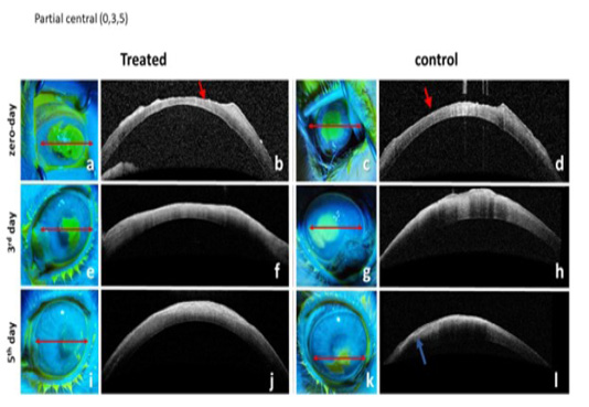
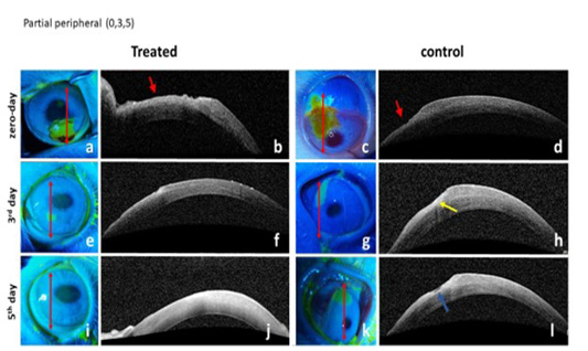
Lyophilized xeno-Mesenchymal Stem Cell Extracellular Vesicle which were isolated from canine MSCs were used for the treatment of both central & peripheral corneal ulcers. EVs promoted not only corneal epithelial cell migration and proliferation but also epithelial wound closure. Both treated groups at the 5th day of corneal wound induction appeared fluorescein -ve by fluorescein test that indicates the epithelial wound closure andAS-OCT images showed the appearence of a well-developed epithelial layer and a homogenous intact stroma unlike the control groups at the 5th day appear fluorescein +ve and AS-OCT image that showed irregular plaques of epithelium and hyperreflective lesion with different densities affecting the whole corneal thickness with some lesions appear as stromal fibrosis and thin stromal layer with sub epithelial cyst.
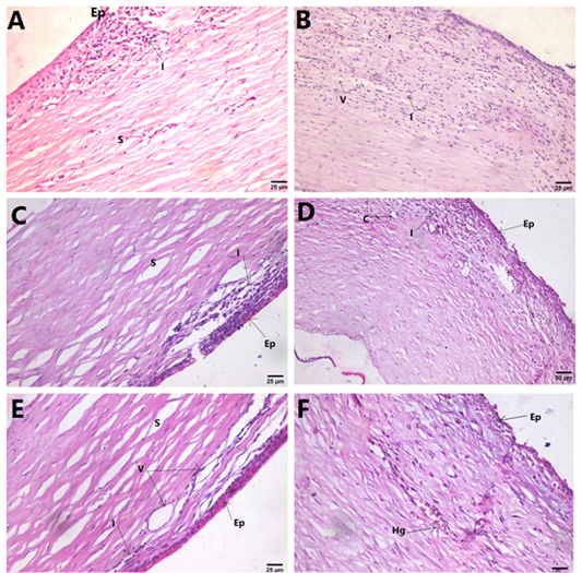
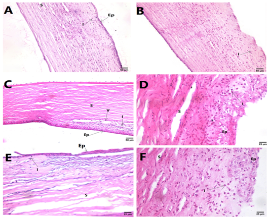
Histopathological examination by H&E staining for evaluation of the quality of healing for 3 successive weeks showed that both treated groups had a complete layer of epithelium appearing within 1week which increased in the number of rows in the 2nd and 3rd weeks and stromal edema decreased by time unlike control groups which had weak fragmented epithelium and sub-epithelial hemorrhage and vascularization.
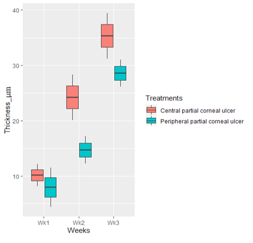
Statistical analysis of epithelial layer thickness revealed that time (weeks) and treatment of various corneal ulcers did have a statistically significant effect on average corneal ulcer epithelial thickness, which was indicated by the progressively increased corneal epithelial thickness across the testing weeks, Central partial corneal ulcer cases revealed significant higher thickness over time compared to peripheral cases.
This finding offers preliminary evidence that Xenogenous EVs have the ability to heal corneal wounds even if in peripheral corneal ulcer with deficiency of limbal stem cell, EVs was able to stimulate the remnant limbal stem cell and accelerate corneal defect healing than control group.