Research Article · Volume 14, Issue 7, 18 July 2026 · Open Access
Comparative Effects of β-Tricalcium Phosphate and Mesenchymal Stem Cell–Derived Extracellular Vesicles on Ilial Fracture Healing in a Mongrel Dog Model
· · · · ·
Affiliations & Notes ⌄
1Department of Surgery, Anesthesiology and Radiology, Faculty of Veterinary Medicine, Suez Canal University, 4.5 Ring Road, Ismailia, 41522, Egypt
2Department of Cytology and Histology, Faculty of Veterinary Medicine, Suez Canal University, 4.5 Ring Road, Ismailia, 41522, Egypt
3Department of Surgery and Theriogenology, Faculty of Veterinary Medicine, King Salman International University, Ras Sudr 46612, Egypt. *Correspondence | Mahmoud F. Ahmed, Department of Surgery, Anesthesiology and Radiology, Faculty of Veterinary Medicine, Suez Canal University, 4.5 Ring Road, Ismailia, 41522, Egypt
Nevertheless, marked differences were observed among the control, β-TCP, and MSC-EVs groups with respect to radiographic progression, biomechanical integrity, and histological bone quality, indicating distinct regenerative capacities associated with each treatment modality.
Notably, fractures treated with MSC-EVs exhibited significantly higher radiographic density from one to three months postoperatively compared with the control group, indicating accelerated and more effective bone regeneration.
In conclusion, this study establishes the regenerative efficacy of MSC-EVs in treating ilial body fractures in mongrel dogs, demonstrating accelerated osteogenesis, enhanced angiogenesis, and more complete structural remodeling compared to β-TCP alone.
Overall, these findings suggest that MSC-EVs are a promising therapeutic strategy and should be tested in clinical scenarios in future studies to evaluate their potential for optimizing fracture repair, improving bone quality, and restoring biomechanical integrity in veterinary orthopedics.
Selected from the article's own text.
Abstract
This randomized controlled study compared how β-tricalcium phosphate (β-TCP) and mesenchymal stem cell-derived extracellular vesicles (MSC-EVs) affected healing of surgically created ilial fractures in mongrel dogs. Nine healthy adult male mongrel dogs were randomly allocated into three groups (n = 3 per group). Standardized ilial shaft complete transverse fracture were surgically created and stabilized using dynamic compression plates (DCP). The control group received fixation alone, whereas the β-TCP and MSC-EVs groups received local augmentation at the fracture site. Postoperative lameness scoring and radiographic examinations were performed at regular intervals for 3 months. At the end of the experimental period, biomechanical tensile strength testing, histological examination, and histomorphometric analysis using BoneJ software were conducted. At 90 days, lameness scores did not differ significantly between groups (P> 0.05). The MSC-EVs group demonstrated significantly higher tensile strength compared with the control group (P < 0.05). Radiographic analysis revealed a significantly higher mean gray value at the fracture region of interest only in the MSC-EVs group compared with the control group at the end of the study (P < 0.05). Histomorphometric analysis showed significantly increased trabecular thickness and bone area percentage in both treatment groups relative to the control group (P < 0.05). In conclusion, both treatments improved bone healing compared to fixation alone, but MSC-EVs produced better results. However, MSC-EVs produced superior radiographic, histological, and biomechanical outcomes, underscoring their promising potential as an advanced therapeutic strategy for improving fracture management in orthopedic practice.
Pelvic fractures rank among the most common traumatic injuries in dogs treated by small animal veterinarians . Ilial body fractures account for roughly 18% to 46% of pelvic fractures in small animals . Ilial fractures are commonly oblique, causing displacement that disrupts hip alignment, limits weight-bearing, narrows the pelvic canal, and damages the lumbosacral nerve trunk ; . Additional intra-abdominal injuries, including bladder trauma, may also occur .
Veterinarians typically stabilize iliac body fractures by applying bone plates to the lateral surface of the ilium . Bone plates are unique among implants in that they can be contoured to precisely match the lateral surface of the ilium; once fixed, they effectively maintain reduction while restoring the bone’s normal curvature to prevent pelvic canal narrowing . Among available options, dynamic compression plates (DCP) are the most used for this purpose ; .Contemporary strategies for managing iliac fractures emphasize precise anatomical reduction, shortened hospitalization, expedited healing, and rapid restoration of function ; . In high-risk scenarios, intraoperative evaluation by the veterinary surgeon may indicate the need for adjunctive bone healing promoters, such as in open fractures or high-energy traumatic fractures where standard fixation alone may not ensure bone union .
β-tricalcium phosphate (β-TCP), a widely adopted bioceramic, serves as an effective synthetic bone graft substitute owing to its biocompatibility, osteoconductivity and osteoinductivity, with cell-mediated resorption facilitating defect regeneration ; . It promotes reliable bone formation with low infection risk and minimal morbidity, aligning with goals of integration, augmentation, and replacement .
The use of mesenchymal stem cells (MSCs) in canine fracture repair represents a promising advancement in orthopedics, offering potential benefits including accelerated bone regeneration, shorter recovery periods, and improved functional outcomes . Unfortunately, widespread clinical use is limited by the lack of standardized protocols for their isolation, characterization, and administration . Concerns remain regarding safety, efficacy, and long-term stability, as well as potential risks such as immune reactions, tumor formation, or unwanted differentiation ; . Current evidence indicates that transplanted stem cells aid tissue repair primarily through paracrine effects, releasing growth factors, cytokines, chemokines, and extracellular vesicles (EVs) . These signals regulate resident cell activity and modify the local microenvironment, with EVs considered the key contributors to the therapeutic effects of stem cells by delivering functional proteins, lipids, and nucleic acids that modulate, repair, and regenerate damaged tissues . By recapitulating the paracrine signaling of MSCs, EV-based strategies harness comparable regenerative potential while mitigating the inherent risks of cell therapy, including uncontrolled differentiation, tumorigenicity, and immune rejection, thereby offering a safer and more standardized alternative for clinical translation .
We hypothesized that adding β-TCP or MSC-EVs to DCP fixation would improve healing compared to DCP fixation alone, with β-TCP enhancing structural bone formation and MSC-EVs promoting faster early healing through angiogenesis, inflammation modulation, and osteogenesis. Accordingly, this study aimed to evaluate and compare the bone-healing efficacy of β-TCP and MSC-EVs using clinical lameness scoring, radiographic assessment, biomechanical tensile strength testing, and histopathological analysis.
MATERIALS AND METHODS
Study design
The study was carried out at the Department of Surgery, Anesthesiology and Radiology, Faculty of Veterinary Medicine, Suez Canal University. The minimum sample size was calculated using G*Power version 3.1.9.2 . Nine clinically healthy male mongrel dogs, aged 15–20 months and weighing 17–20 kg, were randomly allocated into three groups using a sealed white envelope randomization procedure: Control group (n = 3), induced ilial body fractures stabilized with DCP alone; β-TCP group (n= 3), induced ilial fracture stabilized by DCP with application of β-TCP; and MSC-EVs group (n = 3), induced ilial fracture stabilized by DCP with application of MSC-EVs. The dogs were maintained on a standard diet in accordance with NRC nutritional standards for dogs, with free access to water . The dogs were housed in the animal house for 14 days before the experiment to allow for acclimatization and a complete health assessment, including musculoskeletal examination, to exclude any conditions that might interfere with the study.
Animal preparation and anesthesia
Prior to surgery, food and water were withheld for 10 and 2 hours, respectively. Prophylactic cefotaxime (Cefotax®, EIPICO, Tenth of Ramadan, Egypt) was administered preoperatively at a dose of 50 mg/kg intramuscularly (IM). The dogs received IM injections of acepromazine (Calmivet®, Vetoquinol, France) at a dose of 0.05 mg/kg and atropine sulfate (Memphis Pharmaceutical, Egypt) at a dose of 0.04 mg/kg, followed by an IM injection of nalbuphine HCl (Nalufin®, Amoun Pharmaceutical Co., Al Qalyubia, Egypt) at a dose of 1 mg/kg. General anesthesia was induced using an intravenous (IV) injection of propofol (Diprivan®, AstraZeneca, UK) at 2.2 mg/kg and maintained with 2% isoflurane (IsoFlo®, Zoetis, USA) in oxygen . During surgery, lactated Ringer’s solution was administered IV at a rate of 10 mL/kg/hour. The surgical site, extending from the dorsal midline to the stifle joint and from a point 10 cm cranial to the iliac crest to the tail head caudally, was clipped and aseptically prepared for surgery using povidone iodine (Nile Co. for pharmaceutical and chemical industries, Cairo, Egypt).
Experimental ilial body fracture and repair
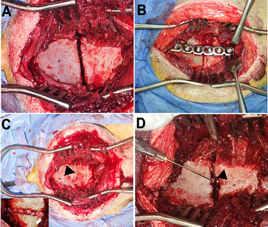
Surgical exposure was performed as described previously . The same surgeon performed all the surgical procedures. Briefly, dogs were placed in a lateral recumbent position. A skin incision was made from the cranial extent of the right iliac crest to 1–2 cm caudal to the greater trochanter, centered over the ventral third of the iliac wing. The subcutaneous tissue and gluteal fat were incised to expose the intermuscular septum between the middle gluteal muscle and the long head of the tensor fasciae latae muscle. The incision continued to separate the tensor fasciae latae from the middle gluteal muscle cranially and from the superficial gluteal muscle caudally. Dissection was performed to further separate these muscles and expose the ventral border of the ilium. The iliolumbar vessel was isolated and ligated, and the deep and middle gluteal muscles were reflected from the lateral surface of the ilium. A complete transverse fracture was created at the right ilial shaft using an oscillating orthopedic bone saw (System 8 Precision, Stryker, USA) equipped with blades measuring 90–105 mm in length, 20–25 mm in width, and 1.27–1.37 mm in thickness, with continuous saline irrigation applied throughout the procedure. After fracture reduction , a six-hole DCP was contoured to match the normal curvature of the lateral ilial surface using a plate bender and secured with three 3.5 mm Ø cortical screws in the cranial fragment and three in the caudal fragment (Control group). In the β-TCP group, β-TCP granules (Regen Store, UK) were placed into the fracture line prior to fixation with the DCP. In the MSC-EVs group, 6×10⁵ MSC-Evs (Almimas Pharma, distributed by BioHeal, Egypt) were applied to the fracture line (Figure 1).
The muscles and subcutaneous were sutured in a simple continuous pattern using size 0 absorbable polyglycolic acid sutures (EGYSORB®, Taisier Group, Cairo, Egypt). The skin was routinely closed with size 1 polypropylene sutures (EGYSORB®) in a simple interrupted pattern.
Postoperative management
All dogs were maintained on cage rest throughout the postoperative period. The surgical site was inspected and dressed twice daily. Prophylactic antibiotic therapy consisted of intramuscular cefotaxime (50 mg/kg every 8 h) administered for 7 days. Postoperative analgesia included intramuscular meloxicam at a dose of 0.2 mg/kg (Mobitil®, MUP, Cairo, Egypt), administered once daily for 5 days. Skin sutures were removed on the 10th postoperative day. Dogs were continuously monitored until the end of the experimental period (90 days). An experienced veterinarian unaware of treatment assignments assessed pain using the short-form Glasgow Composite Pain Scale (CMPS-SF) . Pain scores were recorded during the first postoperative week. Rescue analgesia (nalbuphine hydrochloride, 1 mg/kg IM) was administered when the pain score reached ≥6, based on the dog’s overall clinical condition.
Postoperative evaluation
Daily clinical examinations were performed for the operated dogs during the first postoperative week and weekly thereafter throughout the study period (90 days). The posture and gait of each dog were recorded postoperatively on days 1, 3, 7, 14, 21, 30, 60, and 90. Lameness was evaluated by two experienced veterinary surgeons who were blinded to the treatment groups. Assessment was performed according to using a scoring system ranging from 0 to 4, where 0= no lameness, 1= subtle weight-bearing lameness, 2= obvious weight-bearing lameness, 3= intermittent non-weight-bearing lameness, and 4= consistent non-weight-bearing lameness.
Postoperative radiographic examinations of the operated ilium were performed immediately after surgery and subsequently at 14, 30, 60, and 90 days postoperatively to monitor healing at the fracture line using digital X-ray imaging (Control-X system, Control-X Medical Zrt. (cPlc), Dunakeszi, Hungary). Sequential radiographs were analyzed using Fiji in ImageJ for assessment of mean gray value at the fracture site (region of interest) as previously described .
By the end of the experiment (90 days), dogs were euthanized with an overdose of sodium pentobarbital IV . Fresh ilial specimens were harvested and carefully cleared of surrounding soft tissues. Each sample was longitudinally sectioned into standardized bone strips measuring approximately 1 cm in width and 5 cm in length. To prevent damage at the gripping sites during testing, both ends of each bone strip were embedded in rubber tubes filled with non-shrink epoxy mortar, which was allowed to harden and form rigid supportive caps. The prepared specimens were then centrally aligned and mounted in a 60-ton hydraulic universal testing machine (Universal Material Tensile/Bend Tester; model PT-1100-600, Perfect International Instruments Co., Ltd., Guangdong Province, China; maximum test force: 600 kN). Tensile loading was applied at a constant displacement rate of 0.01 mm/s until failure .
Histological examination and histomorphometric analysis
Iliac bone specimens were harvested from all experimental groups. Sections were subjected to standard histological processing . Tissues were fixed in 10% neutral-buffered formalin for 72 h to preserve morphological integrity, followed by decalcification in 10% ethylenediaminetetraacetic acid (EDTA; pH 7.4) for 4–6 weeks at 20-25˚C with regular solution changes. Following decalcification, samples were dehydrated through graded alcohols, cleared in xylene, and embedded in paraffin wax. Serial coronal sections of 5 µm thickness were obtained using a rotary microtome and mounted on glass slides. For general morphological evaluation, sections were stained with hematoxylin and eosin (H and E) following standard protocols . Histological evaluation was performed using a high-resolution light microscope (Olympus BX53, Olympus Corporation, Tokyo, Japan). Digital images were acquired at both low and high magnifications using a DP74 digital camera (Olympus Corporation, Tokyo, Japan). Both cortical (compact) and trabecular regions of the iliac bone were assessed.
A separate set of iliac bone samples from each dog was prepared to evaluate bone mineralization and collagen deposition. After fixation in 10% neutral-buffered formalin, samples used for von Kossa staining were not decalcified, embedded in methyl methacrylate, sectioned, and stained with silver nitrate to identify mineralized areas. For Masson’s trichrome staining, sections were deparaffinized, rehydrated, and treated with Bouin’s solution to enhance collagen visualization. Nuclei were stained with Weigert’s iron hematoxylin, followed by staining of cytoplasm and muscle fibers with Biebrich scarlet–acid fuchsin. Collagen was then selectively highlighted with aniline blue after differentiation. Finally, sections were dehydrated, cleared, and mounted for microscopic evaluation. For image analysis, three non-overlapping fields per region were acquired at standardized magnification from (i) cortical and (ii) trabecular bone (TB) within the region of interest. Using ImageJ (v1.53) with manual thresholding, the black/brown signal was segmented, and the mineralized area (%) was calculated as (black-positive pixels/total field area) × 100 for each field, then averaged per dog and region. Group means were compared using one-way ANOVA followed by Tukey’s post hoc test (GraphPad Prism v9). Statistical significance was set at P < 0.05.
Quantitative histomorphometric analysis was carried out using the BoneJ plugin integrated within ImageJ software (Version 1.53, National Institutes of Health, Bethesda, MD, USA), as previously described . The following parameters were quantitatively measured: trabecular thickness (µm), defined as the average width of trabeculae; bone area, expressed as a percentage of the total field; and vascular density, calculated as the number of blood vessels per square micrometer (vessels/µm²).
Statistical analysis
Statistical analysis was performed using SPSS version 29 (IBM SPSS Statistics for Windows, version 29.0, Armonk, NY: IBM Corp). Data normality was assessed using the Shapiro–Wilk test. Normally distributed variables, such as mean gray value, and tensile strength, were analyzed using one-way analysis of variance (ANOVA) followed by Tukey’s post hoc test. Between-group comparisons for non-normally distributed data were performed using the Kruskal–Wallis test with Bonferroni correction. Spearman’s rho correlation coefficients were used to quantify associations between treatment groups and healing-related parameters.
RESULTS
Clinical evaluation
All dogs recovered from surgery without wound discharge, infection, or unusual inflammation. Surgical wounds healed uneventfully within 10 days postoperatively. All dogs exhibited intermittent non-weight-bearing lameness (score 3) on the first postoperative day, with one dog in the control group displaying consistent non-weight bearing lameness (score 4). Lameness improved gradually in all groups after surgery. By postoperative day 21, dogs showed subtle weight-bearing lameness (score 1), and by day 60, no lameness was detected in any operated dogs (score 0), indicating complete functional recovery following fracture intervention. Kruskal–Wallis analysis revealed no statistically significant differences in lameness scores among the experimental groups at any time point (P > 0.05) (Table 1). However, within-group temporal analysis demonstrated a significant improvement in lameness scores over time in all groups (Control: χ² (7) = 20.407, P = 0.005; β-TCP: χ² (7) = 20.402, P = 0.005; MSC-EVs: χ² (7) = 20.818, P = 0.004). Post-hoc comparisons showed that lameness scores at 60 and 90 days were significantly lower than those recorded at 1–3 days and 7 days (e.g., Control: 60 days vs. 1-day, adjusted P = 0.043; β-TCP: adjusted P = 0.032).
Table 1: Lameness scores in experimental groups over time: Mean ranks and statistical comparison using the Kruskal–Wallis test.
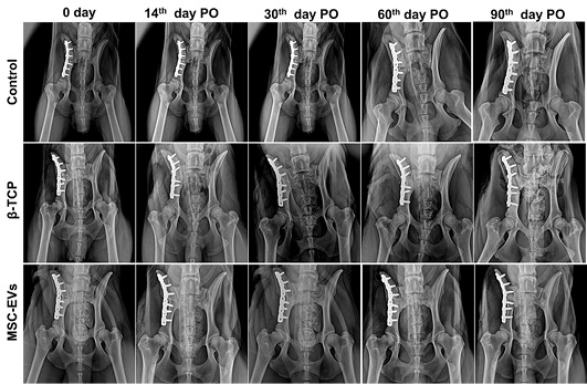
Sequential radiographic examinations of the ilium demonstrated a gradual increase in radiopacity at the fracture site, indicating ongoing callus formation and bone remodeling (Figure 2). Quantitative radiographic analysis of the mean gray (radiopacity) value at the fracture site showed an increase from day 0 to 90 postoperatively (Table 2). Throughout the follow-up period, the DCP and cortical screws remained stable, with no evidence of implant failure or displacement. Fracture alignment was well maintained, and the fracture site was initially visible as an area of low radiodensity, which gradually diminished over time as healing advanced.
Table 2: Descriptive statistics of mean gray values in the region of interest across experimental groups over time.
Time
Groups
Mean
SE
Minimum
Maximum
Day 0
Control
141.116b
3.793
119.69
150.76
β-TCP
168.15a
9.214
150.76
227
MSC-Evs
132.264b
7.111
111.67
161.67
14th day PO
Control
157.916a
3.049
150.29
175.88
β-TCP
166.325a
8.301
136
210.5
MSC-Evs
155.276a
10.091
118.21
203.76
30th day PO
Control
162.579b
3.096
149.25
171.25
β-TCP
177.962ab
5.633
165
215.05
MSC-Evs
195.088a
8.796
152
220.5
60th day PO
Control
160.81b
2.758
149.43
170.23
β-TCP
178.602ab
5.114
157.83
200.89
MSC-Evs
201.228a
13.377
145.79
270.24
90th day PO
Control
174.65b
4.647
157.5
196.38
β-TCP
189.547ab
5.012
180.81
223.76
MSC-Evs
203.084a
8.206
170.8
250.21
Means with different superscript letters within the same column at each time point are significantly different (P < 0.01). β-TCP, Beta tri-calcium phosphate; MSC-EVs, Mesenchymal stem cell-derived exosomal vesicles; PO, postoperatively.
Tensile strength evaluation of ilial bone segments
Tensile strength was lowest in controls (33.02 ± 1.14 kN) and highest with MSC-EVs (44.36 ± 0.72 kN). The MSC-EVs group had significantly greater tensile strength compared with both the Control and β-TCP groups (P < 0.01). The β-TCP group showed higher tensile strength than the Control group; however, this difference was not statistically significant (P > 0.01) (Table 3).
Table 3: Tensile strength (kN) of ilial bone segments at the end of the experiment across experimental groups.
Group
Mean
SE
Minimum
Maximum
Control
33.0233b
1.14333
30.9
34.82
β-TCP
40.8833a
0.83475
39.24
41.96
MSC-Evs
44.3633a
0.72066
43.17
45.66
Means with different superscript letters are significantly different (P < 0.01). β-TCP, Beta tri-calcium phosphate; MSC-EVs, Mesenchymal stem cell-derived exosomal vesicles.
Histological findings
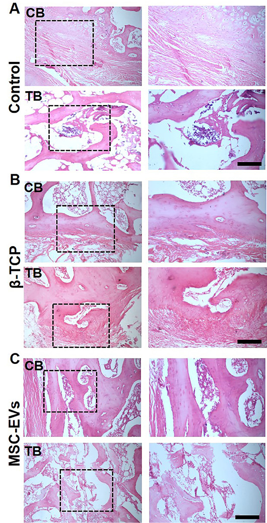
Bone healing patterns differed markedly between groups histologically (Tables 4 and 5). Histological analysis of compact bone without promotors showed mainly disorganized woven bone at the fracture site. The matrix exhibited randomly oriented collagen fibers and numerous embedded osteocytes. The Haversian system remained poorly organized, with only partial reestablishment of concentric lamellae and vascular channels. Periosteal bone apposition was evident, marked by active osteoblasts lining the outer cortical surfaces and focal periosteal thickening. Osteoclasts were observed at the endosteal border, reflecting ongoing bone remodeling. The medullary region contained fibrovascular granulation tissue, with limited evidence of marrow restoration (Figure 3A). In trabecular bone, newly formed woven trabeculae were irregular in size and poorly interconnected. The marrow spaces were partially filled with fibrous tissue, suggesting a prolonged or incomplete endochondral ossification process. Angiogenic activity was moderate, and osteoblastic lining was sparse, indicating a delayed transition toward lamellar bone (Figure 3A).
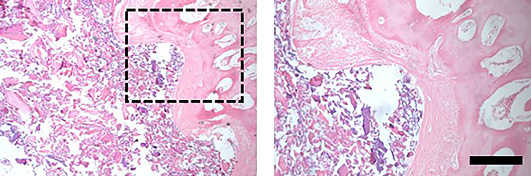
In one dog, spontaneous healing was incomplete, with mainly immature, disorganized bones. The defect area showed persistent fibrous tissue, little mature bone, limited blood vessel formation, and signs of chronic inflammation, indicating delayed bone healing (Figure 5).
Histological sections revealed extensive osteoconductive integration of β-TCP granules within the defect. These appeared as eosinophilic, acellular crystalline remnants, surrounded by newly deposited bone matrix. Bone formation was initiated directly on the surface of the scaffold, resulting in composite areas of woven and early
Table 4: Comparative histological features of ilial healing in compact and trabecular regions across experimental groups.
Parameter
Control
β-TCP
MSC-EVs
Compact Bone
Bone Type
Predominantly woven bone
Woven + early lamellar bone
Mature lamellar bone
Osteon (Haversian) Structure
Poorly formed or absent
Partially restored + early remodeling
Well-developed
Osteoblast Activity
Moderate + lining new bone
High + around β-TCP particles
High + reduced as remodeling completes
Osteoclast Presence
Present (active remodeling)
Present (resorbing β-TCP and bone)
Present at remodeling fronts
Periosteal Response
Mild periosteal thickening
Enhanced periosteal bone formation
Restored periosteum with mature bone
Fibrosis/Inflammation
Moderate
Mild to moderate, especially near particles
Absent
Trabecular Bone
Trabecular Pattern
Irregular, disorganized woven trabeculae
Trabeculae integrated with scaffold, moderate continuity
Table 5: Histomorphometric analysis (Mean ± SD) of trabecular thickness, vascular density, and bone area (%) in experimental groups.
Group
Trabecular thickness (µm)
Vascular density (vessels/µm²)
Bone area (% of the total field)
Control
87 ± 5.83
10.50 ± 1.87
26.33 ± 3.56
β-TCP
106.83 ± 5.34
14.66 ± 2.16
37.83 ± 2.99
MSC-Evs
138.34±4.76
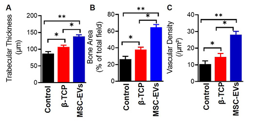
28.26±1.99
64.5±3.39
lamellar bone. Osteoblasts were abundant, particularly at the bone-scaffold interface. Although β-TCP facilitated matrix deposition, the Haversian system remained underdeveloped at this stage. Periosteal activity was enhanced compared to the control, and limited inflammatory infiltrates were observed (Figure 3B). The β-TCP supported the formation of thicker, more continuous trabecular zones. Osteoblasts and osteocytes were more numerous compared to controls, and marrow spaces exhibited improved vascularization with decreased fibrous content. Evidence of early lamellar bone formation was noted on trabecular surfaces, although scaffold remnants persisted, indicating that complete resorption and remodeling were still in progress (Figure 3B).
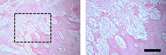
In one dog from the β-TCP group, bone formation was partially improved, with new bone growing along the scaffold surface. However, residual β-TCP particles remained and were surrounded by immature bone and multinucleated giant cells, suggesting incomplete material resorption. Although trabecular continuity was better than controls, some non-mineralized areas persisted, indicating uneven bone maturation (Figure 6).
Application of MSC-EVs induced the most advanced histological pattern of regeneration. The cortical bone at the repair site was composed of advanced and more mature lamellar bone, with clearly defined Haversian systems and concentric lamellae surrounding central canals. Osteocytes were well distributed within lacunae, and osteoblasts were sparsely but strategically positioned, suggesting a late phase of matrix remodeling. The periosteum and endosteum were structurally restored. No evidence of fibrous tissue or residual inflammation was observed (Figure 3C). In trabecular areas, MSC-EVs treatment resulted in the formation of highly organized lamellar trabeculae, showing advanced remodeling and mineralization. Trabecular thickness and interconnectivity exceeded all other groups. The marrow space exhibited extensive vascular networks, and marrow composition included adipocytes and hematopoietic cells, indicating successful regeneration of bone marrow microarchitecture. Multinucleated osteoclasts were occasionally seen at remodeling fronts, reflecting dynamic turnover (Figure 3C). Osteoclasts were identified in all groups; however, the pattern and extent of osteoclastic activity varied. In the Control group, activity was limited and associated with areas of incomplete remodeling. In the β-TCP group, osteoclasts were mainly observed around residual material particles. In contrast, the MSC-EVs group showed more organized and balanced remodeling activity, consistent with more advanced bone maturation.
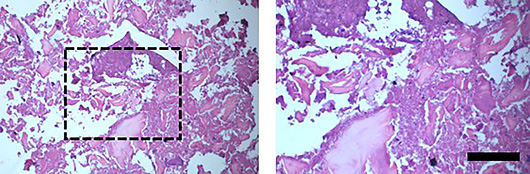
The MSC-Evs group exhibited the most consistent and mature bone regeneration. Histology revealed well-organized lamellar bone, restored Haversian systems, and normalized marrow architecture. However, even in this group, mild remodeling irregularities were noted in some sections, including excess osteoid seams, focal osteoclastic activity, or variable trabecular orientation, which may reflect ongoing dynamic turnover or species-specific healing kinetics. Notably, no signs of chronic inflammation, necrosis, or fibrous encapsulation were observed in this group (Figure 7).
The histomorphometric analysis was consistent with the histological findings, as the control group exhibited the lowest trabecular thickness, bone area, and vascularization percentage, whereas the MSC-EV group demonstrated the highest values (Figure 4).
Mineralized matrix quantification confirms superior regeneration with MSC-EVs
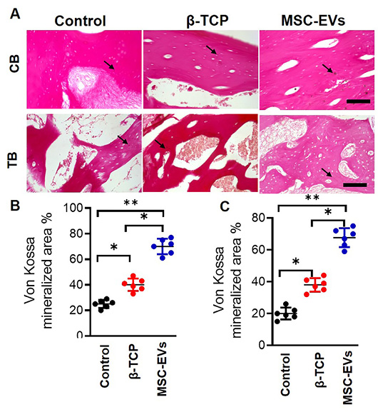
Von Kossa staining demonstrated a stepwise increase in mineral deposition across groups in both compact and trabecular compartments (Figure 8). In compact bone, the mineralized area was lowest in the Control group, increased with β-TCP, and was greatest with MSC-EVs. A similar pattern was observed in TB. Qualitatively, Control fields displayed sparse, discontinuous mineral foci, whereas β-TCP sections showed mineral nucleation along and between scaffold remnants. MSC-EVs sections showed the most continuous and mature mineralized network.
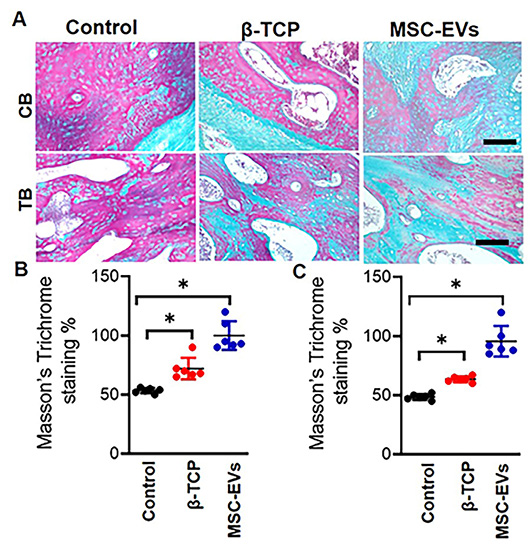
Masson’s trichrome staining revealed a graded increase in green collagen signal across groups (Figure 9). Control sections displayed thin, discontinuous green bands with wide non-stained gaps at the defect perimeter. β-TCP increased collagen deposition mainly along particle interfaces and peri-defect borders, producing partial bridges. MSC-EVs showed the most uniform and extensive collagen matrix, with continuous cortical spans and minimal interruptions. Quantitatively, the trichrome-positive (green) area increased stepwise from Control to β-TCP and MSC-EVs. In the trabecular compartment, Control fields contained scattered, thin green trabeculae with poor interconnection, whereas β-TCP increased green signal around scaffold remnants, yielding patchy bridges between struts. Meanwhile, MSC-EVs presented the most mature pattern, characterized by a dense, well-connected green network spanning the field.
DISCUSSION
This study directly compared β-TCP and MSC-EVs for repairing experimental ilial fractures in dogs. By integrating clinical outcomes with radiographic, biomechanical, and histopathological assessments. Both treatments improved healing compared to DCP alone, but MSC-EVs produced consistently better bone structure, mineralization, and strength.
In this study, all experimentally induced iliac fractures in dogs healed clinically without postoperative complications, consistent with the high success rates and favorable clinical outcomes reported following adequate surgical stabilization of ilial body fractures in dogs ; . Nevertheless, marked differences were observed among the control, β-TCP, and MSC-EVs groups with respect to radiographic progression, biomechanical integrity, and histological bone quality, indicating distinct regenerative capacities associated with each treatment modality.
Clinically, all dogs showed a consistent postoperative lameness pattern: partial weight-bearing within 48 hours, progressive improvement by the end of the first week, and full limb function restoration over time, with no significant differences in lameness scores among β-TCP, MSC-EVs, and control groups at any interval. This absence of intergroup differences indicates that early functional recovery was driven primarily by surgical stabilization and time, rather than the applied promotor. Similar patterns occur in femoral and pelvic fracture dog models, where rapid gait normalization often precedes or decouples from histological bone maturity ; .
Radiographic assessment using ImageJ-based relative bone density analysis demonstrated a progressive increase in radiopacity across all experimental groups, reflecting continuous mineral deposition and callus maturation during fracture healing. Increased radiographic density is a well-established indicator of advancing mineralization and osseous union . Notably, fractures treated with MSC-EVs exhibited significantly higher radiographic density from one to three months postoperatively compared with the control group, indicating accelerated and more effective bone regeneration. The superior radiographic outcomes observed in the MSC-EVs group align with growing evidence that MSC-EVs enhance osteogenesis primarily through paracrine mechanisms rather than direct cellular engraftment. MSC-EVs are known to promote osteogenic differentiation and extracellular matrix mineralization by delivering bioactive cargo that modulates key regenerative signaling pathways . In contrast, the moderate increase in radiographic density observed in the β-TCP group is consistent with its established role as an osteoconductive scaffold. While β-TCP provides a structural framework that supports new bone ingrowth, its relatively slow and sometimes incomplete resorption may delay remodeling and, in some cases, elicit localized foreign body reactions that compromise optimal regeneration .
Biomechanical evaluation through tensile strength testing corroborated the radiographic findings. The MSC-EVs group exhibited the highest tensile strength values, significantly exceeding those of the control group, indicating superior structural integrity and load-bearing capacity of the regenerated bone. Although β-TCP treatment resulted in improved tensile strength compared with controls, its biomechanical performance remained inferior to that of MSC-EVs. This finding suggests that while β-TCP contributes to early mineral deposition and mechanical stabilization, it lacks the biological cues required for complete osteoinduction and long-term remodeling . The modest mechanical improvement observed with β-TCP may be attributed to its ability to facilitate initial mineralization; however, insufficient biological integration can limit maturation and functional restoration of bone tissue . In contrast, the enhanced biomechanical competence observed in the MSC-EVs group could be attributed to the synergistic regenerative effects of MSC-EVs. These vesicles transport a diverse array of osteoinductive molecules, including osteogenic microRNAs , growth factors, and extracellular matrix-modulating proteins . Collectively, these bioactive components stimulate osteoblast differentiation, enhance angiogenesis, and modulate inflammatory responses, thereby promoting the formation of a more organized, mature, and biomechanically competent bone matrix .
Microscopic examination revealed important differences in bone quality and organization between treatments. In the control group, spontaneous fracture healing was characterized by the persistence of woven bone, irregularly arranged trabeculae, fibrous tissue infiltration, and incomplete restoration of the marrow cavity. These features are indicative of delayed remodeling and limited transition from woven to lamellar bone, reflecting suboptimal healing. This may be attributed to insufficient vascularization and inadequate osteogenic signaling, which impede complete and organized bone regeneration ; . Meanwhile, the application of β-TCP resulted in improved osteoconductive activity, as evidenced by increased osteoblast proliferation, thicker and more regularly arranged trabeculae, and enhanced neovascularization . These histological improvements indicate that β-TCP effectively provides a scaffold that supports cellular attachment and early bone formation . Nevertheless, the continued presence of residual β-TCP particles, multinucleated giant cells, and immature or poorly organized Haversian systems suggests ongoing material resorption and incomplete structural remodeling. This delayed maturation likely accounts for the moderate biomechanical performance observed in the β-TCP group, highlighting the limitations of osteoconductive scaffolds in the absence of strong biological signaling ; .
In contrast, the MSC-EVs group exhibited the most advanced and well-organized bone regeneration. Histological sections demonstrated extensive formation of mature lamellar bone with fully developed Haversian systems, reestablished periosteal and endosteal layers, restored marrow architecture, and minimal inflammatory infiltrates. Quantitative histomorphometric analysis further confirmed significantly greater bone area, trabecular thickness, vascular density, osteoblast counts, and lamellar bone proportion in this group compared with the other treatments. These findings strongly support the concept that MSC-EVs actively modulate the fracture healing microenvironment through the delivery of bioactive molecules including osteogenic microRNAs and growth factors that promote angiogenesis, osteoblast differentiation, and coordinated bone remodeling ; ; . Moreover, the strong correlations observed between MSC-EVs treatment and key histological indicators of bone maturity reinforce their pivotal role in accelerating and enhancing bone regeneration. Collectively, these histopathological outcomes demonstrate that MSC-EVs not only accelerate the rate of bone formation but also improve the structural quality and maturity of regenerated bone, offering a clear biological advantage over osteoconductive scaffolds alone.
This study has several limitations. The small sample size and short follow-up restricted evaluation to early and intermediate stages of fracture healing without assessing long-term remodeling or durability. The model involved simple fractures without critical-sized gaps, limiting the assessment of bone regeneration in larger defects. The MSC-EV dose was selected as a pilot and may not represent the optimal therapeutic dose. Additional limitations include the lack of detailed EV characterization, and potential baseline differences in fracture radiographs. A combined β-TCP + MSC-EV group was not included, so potential synergistic effects could not be assessed. Future studies with larger sample sizes, longer follow-up, dose-response analyses, comprehensive EV profiling, blinded assessment of histological sections, and combination treatment groups are needed to validate and expand these findings and better define their translational potential in clinical settings.
CONCLUSION AND RECOMMENDATIONS
In conclusion, this study establishes the regenerative efficacy of MSC-EVs in treating ilial body fractures in mongrel dogs, demonstrating accelerated osteogenesis, enhanced angiogenesis, and more complete structural remodeling compared to β-TCP alone. Overall, these findings suggest that MSC-EVs are a promising therapeutic strategy and should be tested in clinical scenarios in future studies to evaluate their potential for optimizing fracture repair, improving bone quality, and restoring biomechanical integrity in veterinary orthopedics.
ACKNOWLEDGEMENTS
The authors would like to express their sincere gratitude to Prof. Dr. Marwa Abd El Moneim Hassan, Faculty of Veterinary Medicine, Suez Canal University, for her valuable guidance and support in the statistical analysis of the study. The authors also extend their appreciation to Dr. Mohamed A. Hashem, Faculty of Veterinary Medicine, Suez Canal University, for his professional technical assistance.
NOVELTY STATEMENT
This study is the first to comparatively evaluate the bone-healing efficacy of β-tricalcium phosphate (β-TCP) and mesenchymal stem cell–derived extracellular vesicles (MSC-EVs) in experimentally induced ilial fractures in mongrel dogs using integrated clinical, radiographic, biomechanical, and histopathological assessments, providing new insights into the role of MSC-EVs as an emerging bone-healing promoter in veterinary orthopedics.
AUTHOR CONTRIBUTION
Saad L. Saad contributed to writing original draft, validation, methodology, investigation, software, visualization, formal analysis, data curation, and conceptualization. Mohammed H. Shekidef contributed to conceptualization, methodology, formal analysis, investigation, resources, data curation, writing original draft, writing review and editing, supervision, validation, project administration, and funding acquisition. Ibrahim H. Ahmed, and Mohamed H. El-Daharawy contributed to writing original drafts and writing review and editing, methodology, validation, supervision, investigation, and conceptualization. Elsayed Metwllay contributed to writing original draft and writing review and editing, methodology, formal analysis, data curation, investigation, visualization, resources, software, validation, and conceptualization. Mahmoud F. Ahmed contributed to writing original draft and writing review and editing, methodology, validation, supervision, investigation, resources, formal Analysis, visualization, and conceptualization.
Ethical approval statement
All experimental procedures were reviewed and approved by the Institutional Animal Use and Care Committee of the Faculty of Veterinary Medicine, Suez Canal University (Approval No. 2021005) and were carried out in compliance with the ARRIVE guidelines.
Data availability statement
The data that support the findings of this study are available on request from the corresponding author.
Generative AI and AI assisted technology statement
The authors declare that no generative AI and AI assisted technology was used in the creation of this manuscript.
Statement of conflict of interest
The authors have declared no conflict of interest.
Figures
Figure 1:.
Surgical induction and repair of ilial body fractures in dogs, showing (A) exposure of the iliac shaft, (B) fixation using a dynamic compression plate (DCP), (C) application of β-tricalcium phosphate (β-TCP) at the fracture line, and (D) application of MSC-EVs at the fracture line.
Figure 2:.
Sequential ventrodorsal radiographic assessment of operated ilial fractures in dogs, showing healing progression just after surgery (day 0), at 14 days, 30 days, 60 days, and 90 days postoperatively.
Figure 3:.
Histological evaluation of ilial bone regeneration across treatment groups. Representative HandE-stained sections of compact bone (CB) and trabecular bone (TB) from ilial fracture sites in the control, β-TCP, and MSC-EVs groups are shown. Left panels depict lowmagnification views with boxed regions, and right panels show higher-magnification views of the boxed areas.
Figure 4:.
Quantitative histomorphometric analyses include trabecular thickness (µm) (A), bone area as a percentage of the total tissue field in the defect region (B), and vascular density (number of vessels per µm²) within marrow spaces (C). Data are presented as mean ± SD, and statistical significance was determined using one-way ANOVA with Tukey’s post hoc test (*P < 0.05, **P < 0.01). Scale bars = 100 µm.
Figure 5:.
Histological features of healing in the control group. HandE-stained section of a dog ilial defect. The left image showed a low-magnification view with the region of interest indicated by a dashed box, while the right image showed a higher-magnification view of the boxed area. The defect exhibits irregular trabecular architecture, persistent fibrous connective tissue, absence of mature lamellar bone, residual fibrocartilaginous tissue, and poorly vascularized surrounding marrow spaces. Scale bar = 100 µm.
Figure 6:.
Histological features of β-TCP-mediated bone healing in HandE-stained sections. The left image showed a low-magnification view with the region of interest indicated by a dashed box, while the right image showed a higher-magnification view of the boxed area. Residual β-TCP particles were embedded within immature woven bone, and multinucleated giant cells were present near the particles, indicative of a mild foreign body reaction. Scale bar = 100 µm
Figure 7:.
Histological (HandE-stained section) features of the MSC-EV–treated iliac defect. The left image shows a low-magnification view with the region of interest indicated by a dashed box, while the right image shows a higher-magnification view of the boxed area. New bone formation with irregular trabecular architecture is observed with marked inflammatory cell infiltration. Scale bar = 100 µm.
Figure 8:.
Von Kossa assessment of mineralized matrix at the fracture site across experimental groups. (A) Representative von Kossa–stained sections of compact bone (CB) and trabecular bone (TB). Mineralized matrix appears black (arrows), and counterstain is red. Scale bars = 100 µm. (B and C) Quantification of mineralized area in compact bone (B) and trabecular bone (C) is expressed as a percentage of the field. Data are presented as mean ± SD. Statistical analysis was performed using one-way ANOVA with Tukey’s post hoc test (*P < 0.05, **P < 0.01). n = 6 section/3 animals.
Figure 9:.
Masson’s Trichrome assessment of matrix maturation at the fracture site across experimental groups. Representative Masson’s Trichrome–stained sections of compact bone (CB) and trabecular bone (TB). In CB, collagen-rich/mineralized matrix appears blue–teal, cytoplasm and osteoid appear red–magenta, and nuclei are dark. In TB, collagenous trabecular networks are highlighted in blue teal. Scale bars = 100 µm. Quantification of Masson’s Trichrome–positive area in compact bone (B) and trabecular bone (C) is expressed as a percentage of the tissue field. Data are presented as mean ± SD. Statistical analysis was performed using one-way ANOVA with Tukey’s post hoc test; *P < 0.05 indicates significant differences between groups. n = 6 section/3 animals.
References
Abed RA, Nazht HH, Omar RA (2022). Limb Gait Score of Femur Fracture Fixated by Intramedullary Pinning in Dogs. Arch Razi Inst., 77(3): 1027-1032.
Banu SA, Mamachan M, El-Husseiny HM, Golchin A, Sharun K (2025). Mesenchymal stem cell therapy in veterinary orthopaedics: Evidence from canine clinical medicine. Vet. Res. Commun., 49(5): 290. https://doi.org/10.1007/s11259-025-10843-4[CrossRef]
Böhme B, Laurent C, Milis O, Ponthot J-P, Balligand M (2022). Determination of canine long bone ultimate tensile strain by digital image correlation. J. Orthop. Res. Ther., 7: 1221.
Bohner M, Santoni BLG, Döbelin N (2020). β-tricalcium phosphate for bone substitution: Synthesis and properties. Acta Biomaterialia., 113: 23-41. https://doi.org/10.1016/j.actbio.2020.06.022[CrossRef]
Çatalkaya E, Yayla S, Altan S, Ersöz Kanay B, Saylak N, Hatipoğlu ŞA (2024). Retrospective study on pelvic fractures in cats and dogs (2020-2022). Kocatepe Vet. J., 17(1): 15-21. https://doi.org/10.30607/kvj.1370455[CrossRef]
Clarke KW, Trim CM, Hall LW (2014). Anaesthesia of the dog. In: Veterinary Anaesthesia, 11th ed by Clarke KW, Trim CM, Hall LW. W.B. Saunders. pp. 405-498.
Close B, Banister K, Baumans V, Bernoth EM, Bromage N, Bunyan J, Erhardt W, Flecknell P, Gregory N, Hackbarth H, Morton D, Warwick C (1997). Recommendations for euthanasia of experimental animals: Part 2. DGXT of the European Commission. Lab Anim., 31(1): 1-32. https://doi.org/10.1258/002367797780600297[CrossRef]
Council NR (2006). Nutrient requirements of dogs and cats. National Academies Press, Washington, DC.
Dias IE, Cardoso DF, Soares CS, Barros LC, Viegas CA, Carvalho PP, Dias IR (2021). Clinical application of mesenchymal stem cells therapy in musculoskeletal injuries in dogs-a review of the scientific literature. Open Vet J., 11(2): 188-202. https://doi.org/10.5455/OVJ.2021.v11.i2.2[CrossRef]
Domingos J, Caseiro R, Atayde L, Pinto PO, Mendonça C, Campos JM, Maurício AC (2017). Clinical application of macroporous ceramic to promote bone healing in veterinary clinical cases. Materials, Technologies and Clinical Applications by Baino F. IntechOpen.
Doube M, Kłosowski MM, Arganda-Carreras I, Cordelières FP, Dougherty RP, Jackson JS, Schmid B, Hutchinson JR, Shefelbine SJ (2010). BoneJ: Free and extensible bone image analysis in ImageJ. Bone. 47(6): 1076-1079. https://doi.org/10.1016/j.bone.2010.08.023[CrossRef]
Emami A, Arabpour Z, Izadi E (2025). Extracellular vesicles: Essential agents in critical bone defect repair and therapeutic enhancement. Mol. Biol. Rep., 52(1): 1-25. https://doi.org/10.1007/s11033-024-10209-0[CrossRef]
Fathy MZ, Ragab GH, Seif MM, Gadallah SM, Deeb S, Safwat NM (2018). Clinico-radiographic and histopathologic evaluation of iliac shaft fracture in dogs (an experimental study). Beni-Suef Univ. J. Basic Appl. Sci., 7(2): 165-170. https://doi.org/10.1016/j.bjbas.2017.09.001[CrossRef]
Faul F, Erdfelder E, Lang AG, Buchner A (2007). G*Power 3: A flexible statistical power analysis program for the social, behavioral, and biomedical sciences. Behav. Res. Methods, 39(2): 175-191. https://doi.org/10.3758/BF03193146[CrossRef]
Foo JB, Looi QH, How CW, Lee SH, Al-Masawa ME, Chong PP, Law JX (2021). Mesenchymal stem cell-derived exosomes and MicroRNAs in cartilage regeneration: Biogenesis, efficacy, miRNA enrichment and delivery. Pharmaceuticals, 14(11): 1093-1118. https://doi.org/10.3390/ph14111093[CrossRef]
Franch J, Díaz-Bertrana C, Lafuente P, Fontecha P, Durall I (2006). Beta-tricalcium phosphate as a synthetic cancellous bone graft in veterinary orthopaedics: A retrospective study of 13 clinical cases. Vet. Comp. Orthop. Traumatol., 19(4): 196-204. https://doi.org/10.1055/s-0038-1633001[CrossRef]
Garcia DC, Mingrone LE, Sá MJCd (2022). Evaluation of osseointegration and bone healing using pure-phase β - TCP ceramic implant in bone critical defects. A systematic review [Systematic Review]. Front. Vet. Sci., 9: 2022. https://doi.org/10.3389/fvets.2022.859920[CrossRef]
Geiger M, Blem G, Ludwig A (2016). Evaluation of ImageJ for relative bone density measurement and clinical application. J. Oral Health Craniofac Sci., 1(1): 12-21. https://doi.org/10.29328/journal.johcs.1001002[CrossRef]
Hammond G (2016). Imaging of fractures. In BSAVA Manual of Canine and Feline Fracture Repair and Management. British Small Animal Veterinary Association. https://doi.org/10.22233/9781910443279.6[CrossRef]
Hayashi K, Schulz KS, Fossum T (2018). Management of Specific Fractures. In Small Animal Surgery,5th ed by Fossum TW. Elsevier Health Sciences. pp. 1036–1134.
Ho Nguyen CT, Bui MH, Lam PH (2024). Residual calcified material volume of β-tricalcium phosphate with platelet-rich fibrin in unilateral alveolar bone graft. Maxillofac Plast Reconstr. Surg., 46(1): 7. https://doi.org/10.1186/s40902-024-00420-1[CrossRef]
Hoffberg JE, Koenigshof AM, Guiot LP (2016). Retrospective evaluation of concurrent intra-abdominal injuries in dogs with traumatic pelvic fractures: 83 cases (2008-2013). J. Vet. Emerg. Crit. Care (San Antonio)., 26(2): 288-294. https://doi.org/10.1111/vec.12430[CrossRef]
Ivanovska A, Wang M, Arshaghi TE, Shaw G, Alves J, Byrne A, Butterworth S, Chandler R, Cuddy L, Dunne J, Guerin S, Harry R, McAlindan A, Mullins RA, Barry F (2022). Manufacturing mesenchymal stromal cells for the treatment of osteoarthritis in canine patients: Challenges and recommendations. Front. Vet. Sci., 9: 897150. https://doi.org/10.3389/fvets.2022.897150[CrossRef]
Kim H-S, Shin TH (2025). Canine and feline mesenchymal stem cells: An integrative review of direct comparative studies by species and tissue source. J. Vet. Sci., 26(6). https://doi.org/10.4142/jvs.25163[CrossRef]
Layton C, Bancroft JD, Suvarna, SK (2019). Fixation of tissues. In: Bancroft’s Theory and Practice of Histological Techniques, 8th ed by Suvarna SK, Layton C, Bancroft JD. Elsevier. pp. 40-63. https://doi.org/10.1016/B978-0-7020-6864-5.00004-9[CrossRef]
Lewis DD, Stubbs WP, Neuwirth L, Bertrand SG, Parker RB, Stallings JT, Murphy ST (1997). Results of screw/wire/polymethylmethacrylate composite fixation for acetabular fracture repair in 14 dogs. Vet. Surg., 26(3): 223-234. https://doi.org/10.1111/j.1532-950X.1997.tb01489.x[CrossRef]
Lu H, Zhou Y, Ma Y, Xiao L, Ji W, Zhang Y, Wang X (2021). Current application of beta-tricalcium phosphate in bone repair and its mechanism to regulate osteogenesis. Front. Mater., 8: 1-16. https://doi.org/10.3389/fmats.2021.698915[CrossRef]
Łuczak JW, Palusińska M, Matak D, Pietrzak D, Nakielski P, Lewicki S, Grodzik M, Szymański Ł (2024). The future of bone repair: Emerging technologies and biomaterials in bone regeneration. Int. J. Mol. Sci., 25(23): 1-28. https://doi.org/10.3390/ijms252312766[CrossRef]
Moens NMM, DeCamp CE (2018). Fractures of the Pelvis. In Veterinary surgery: Small Animal, 2nd ed by Johnston SA, Tobias KM. Elsevier Health Sci., pp. 938-956.
Moi TSM, Minto BW, Macedo AP, Lucena DVF, Malta CAS, Dias LGGG (2022). Preliminary study on a novel dedicated plate for iliac fractures in dogs. PLoS One,17(8): e0269313 https://doi.org/10.1371/journal.pone.0269313.[CrossRef]
Neamat A, Gawish A, Gamal-Eldeen AM (2009). β-Tricalcium phosphate promotes cell proliferation, osteogenesis and bone regeneration in intrabony defects in dogs. Arch. Oral Biol., 54(12): 1083-1090. https://doi.org/10.1016/j.archoralbio.2009.09.003[CrossRef]
Petrovsky B, Knuth T, Aponte-Colón C, Hoefle W, Kraus K, Naiman J, Yuan L, Mochel JP, Zellner E (2021). Short-term outcomes of 59 dogs treated for ilial body fractures with locking or non-locking plates. Vet. Surg., 50(5): 1076-1086. https://doi.org/10.1111/vsu.13656[CrossRef]
Phelps SM (2023). The immunomodulatory role of canine mesenchymal stem cell-derived extracellular vesicles. Master’s thesis, Oregon State University, United States.
Reid J, Nolan A, Hughes J, Lascelles D, Pawson P, Scott E (2007). Development of the short-form Glasgow Composite Measure Pain Scale (CMPS-SF) and derivation of an analgesic intervention score. Anim. Welf., 16(S1): 97-104. https://doi.org/10.1017/S096272860003178X[CrossRef]
Saba E, Sandhu MA, Pelagalli A (2024). Canine mesenchymal stromal cell exosomes: State of the art characterization, functional analysis and applications in various diseases. Vet. Sci., 11(5): 187.
Schindelin J, Arganda-Carreras I, Frise E, Kaynig V, Longair M, Pietzsch T, Preibisch S, Rueden C, Saalfeld S, Schmid B, Tinevez J-Y, White DJ, Hartenstein V, Eliceiri K, Tomancak P, Cardona A (2012). Fiji: An open-source platform for biological-image analysis. Nat. Methods, 9(7): 676-682. https://doi.org/10.1038/nmeth.2019[CrossRef]
Schulze F, Lang A, Schoon J, Wassilew GI, Reichert J (2023). Scaffold guided bone regeneration for the treatment of large segmental defects in long bones. Biomedicines, 11(2): 1-30. https://doi.org/10.3390/biomedicines11020325[CrossRef]
Song Y, Liang F, Tian W, Rayhill E, Ye L, Tian X (2025). Optimizing therapeutic outcomes: Preconditioning strategies for MSC-derived extracellular vesicles. Front. Pharmacol., 16: 1509418. https://doi.org/10.3389/fphar.2025.1509418[CrossRef]
Szponder T, Wessely-Szponder J, Sobczyńska-Rak A, Żylińska B, Radzki RP, Polkowska I (2018). Application of platelet-rich plasma and tricalcium phosphate in the treatment of comminuted fractures in animals. In Vivo., 32(6): 1449-1455. https://doi.org/10.21873/invivo.11398[CrossRef]
Zhong D, Wang CG, Yin K, Liao Q, Zhou X, Liu AS, Kong LY (2014). In vivo ossification of a scaffold combining β-tricalcium phosphate and platelet-rich plasma. Exp. Ther. Med., 8(5): 1381-1388. https://doi.org/10.3892/etm.2014.1969[CrossRef]
Zhou H, Qi Y-X, Zhu C-H, Li A, Pei DD (2023). Mesenchymal stem cell-derived extracellular vesicles for treatment of bone loss within periodontitis in pre-clinical animal models: A meta-analysis. BMC Oral Health., 23(1): 701. https://doi.org/10.1186/s12903-023-03398-w[CrossRef]
Comparative Effects of β-Tricalcium Phosphate and Mesenchymal Stem Cell–Derived Extracellular Vesicles on Ilial Fracture Healing in a Mongrel Dog Model · ResearchersLinks