
Stem cells derived from Wharton’s jelly of human umbilical cord (WJMSCs) are considered as the potential therapeutic agents for regeneration and are a valuable source for stem cell banking. Current investigation shows metabolic disorders are not only responsible for genetic problems and certain disease risk factors in adult life but also have adverse effects on perinatal environment. This study was designed for the comparative analysis of viability and healing properties of GDM affected WJMSCs (GDM-WJMSCs) with Normal controls (N- WJMSCs). Mesenchymal stromal cells were isolated from Wharton’s jelly of human umbilical cords from normal and gestational diabetic mothers. Cell viability was tested by trypan blue exclusion method and wound healing potential by scratch assay method. Healing of the scratch was observed at different time intervals (0h, 6h, 24h, 48h). The images were analyzed by Image J software. Morphological analysis was done under inverted microscope. Results showed that with each passaging stage the viability of GDM cells remained normal whereas Wound healing potential of GDM-WJMSCs was decreased as compared to controls. Moreover, there were few morphological changes observed between the two groups. In conclusion GDM affects wound healing potential of WJMSCs and morphology of the cells to some extent whereas the viability of cells remains unaffected by GDM.
Research Article · Volume 54, Issue 2, 9 January 2022 · Open Access
A Comparative Analysis of Wound Healing Potential, Cell Viability and Morphology of Normal and Gestational Diabetic Affected Wharton’s Jelly Derived Mesenchymal Stem Cells
· · · · · · ·
Affiliations & Notes ⌄
- 1Department of Anatomy, Faculty of Basic Health Science, Ziauddin University, Karachi. 2Dr. Zafar H. Zaidi Center for Proteomics, University of Karachi. 3Department of Research, Ziauddin University, Karachi. 4Department of Pharmacology, Faculty of Basic Health Science, Ziauddin University, Karachi. , * Corresponding author: drnabilarasheed@gmail.com
- 2nabila.rasheed@zu.edu.pk
Article Info ⌄
- Journal:
- Pakistan Journal of Zoology
- Published:
- 9 January 2022

From the article
- In this study we assessed the wound healing potential, cell viability and morphological aspects of Wharton’s jelly derived mesenchymal stem cells of GDM affected and non-affected mothers.
- Our results showed that cells viability with different passaging (P0, P1, P2) was increased in both group of WJMSCs Table II.
- Our results show that there was significantly decrease in the healing potential in GDM WJMSCs compared to control Table III.
- Pre conditioning of the cells to enhance their survival rate, reduced apoptosis and improved propagation should be employed for using these GDMWJMSCs for therapeutics as well as for stem cells banking.
Selected from the article's own text.
Abstract
INTRODUCTION
Regenerative medicine is a branch of medicine linked with emerging treatments that restore or substitute injured, unhealthy, or damaged cells, tissues, or organs to regenerate or establish function and structure. Innovation and investigation in tissue engineering, bio-imaging, cells and on different types of stem cells are vital for the evolution of regenerative medicine, as discovered in many recent case studies and clinical trials (Van Rhijn et al., 2018; .
Stem cells are present as an endogenous system of renewal and repairing in human body. Two most inimitable properties of stem cells are self-renewal, and differentiation ability into several cell lines under suitable conditions . In different conditions like myeloid disorders, WJMSCs have been used successfully, and more than 100 ongoing clinical trials are evaluating their efficacy in the treatment of a variety of other conditions including Alzheimer disease, stroke, cerebral palsy, autism, Broncho pulmonary dysplasia, cartilage injury, osteoarthitis, full thickness burns, hearing loss and inborn metabolic disorders like diabetes etc. .
Stem cells have been classified on the basis of the tissue source of origin, as hematopoietic stem cells (HSCs) and Mesenchymal stem cells (MSCs). Mesenchymal Stem cells can be isolated from bone marrow, adipose tissue, Umbilical cord, Wharton’s jelly, placenta, dental tissues, skin and from other different sources.
The outstanding stemness potential and uses of mesenchymal stem cells in different clinical conditions have flickered attention in the possible effects of several syndromes and diseases on the stem cells. Self-renewal and differentiation capacities of mesenchymal stem cells are strongly exaggerated by metabolic diseases .
Gestational diabetes mellitus (GDM) is a temporary metabolic disorder occurs due to the glucose imbalance and insulin insensitivity during pregnancy period ; . Literature reports GDM have several cellular and transcriptomic effects on Wharton’s jelly derived MSCs (Amrithaj et al., 2012). These effects result in the alteration of proliferation and differentiation ability of WJMSCs. In GDM, the function of p16Ink4a protein, a major protein that regulate certain cell division steps are altered , which leads to inhibit the function of cyclin-dependent kinase and arrest the cell cycle at different steps, causes the proliferation ability of WJMSCs to be decrease in many organs .
In this study, we compared the basic properties of WJMSCs obtained from gestational diabetes mellitus (GDM) patients (GDM- WJMSCs) and normal pregnant women (NWJMSCs) including the morphological analysis, cell viability and wound healing ability of WJMSCs. Our study aims to evaluate the effects of gestational diabetes on healing capacity and viability of WJMSCs, for the better understanding and utilization of these MSCs for regenerative medicines and stem cells banking.
MATERIALS AND METHODS
Procurement of human umblical cord tissue
This was an experimental study performed at Multi-Disciplinary Research Lab Ziau Uddin University and at Dr. Zafar H. Zaidi Center of Proteomics (University of Karachi). This study was approved by Ethical Research Committee, Ziauddin University.
After taking consent from patients, umbilical cords were obtained from normal and gestational diabetic mothers of full term caesarian sections, aged between 18 to 30yr (primary gravida). The diagnosis of GDM was based on the International Association of Diabetes and Pregnancy Study Groups Consensus and Panel recommendation, (OGTT at 20 weeks, controlled GDM patients by diet and exercise). All hepatitis B and C positive mothers, a pre-pregnancy diagnosis of diabetes any complicated pregnancies with conditions of fetal anomaly/ IUGR, PIH, preeclampsia, ruptured membranes/placenta previa were excluded for the study.
Isolation and expansion of WJMSCs
The umbilical cord samples were obtained at the time of cesarean delivery from GDM patients and healthy pregnant women. The cord was rinsed several times with sterilized PBS solution and sliced into 3-mm-thick pieces. The vessels and amnion were removed from cord segments. Wharton’s jelly tissues were minced with scissors. Trypsin was added for tissue digestion and cells harvesting. The tissue pieces were cultured in Dulbecco’s modified eagle medium (DMEM) containing Pen/Strep, antifungal drug, sodium pyruvate, L glutamine and10% fetal bovine serum (FBS) and cultured in 5 % CO2 incubator at 37 °C for further cell proliferation.
Subculturing
When the cell confluency reached 80%, cells were sub-cultured into two T25 cm flasks. Medium were aspirated prior cells washing with sterile PBS. 2-3 mL of 0.25% trypsin was added to the cells and incubated at 37 °C for 4-5 min. After incubation complete medium was added to stop the trypsin reaction and the suspended cells were transferred into 15 mL falcon tube, centrifuged at 300 X g for 8 min and were seeded in sterile tissue culture treated flasks each containing 9 ml fresh complete DMEM.
RNA isolation and quantification
Total RNA was isolated from the isolated cells in 30µl reaction volume using TRIZOL method. Prior to RNA isolation, pipettes, glassware and bench top were sanitized by RNAse Erase spray. Cells were trypsinized and the cell pellet was resuspended in 1 mL Trizol reagent. Chloroform (200 μL per mL Trizol) was then added to the mixture and incubated at 25 ˚C for 15-20 min. Phase separation was performed by centrifuging the mixture at 10,000 X g for 30 min. Two phases appeared; lower organic phase and upper transparent aqueous phase and in between these two phases, a pellet of protein and DNA was present. Upper aqueous phase was proceeded for RNA extraction by transferring to a sterile tube. RNA was then precipitated by chilled isopropanol with centrifugation at 10000 X g for 10 min at 4 ˚C. RNA was re-hydrated by 75% ethanol and pelleted at 1000 X g for 15 min followed by air drying. The dried pellet 19 was then re-suspended in 30μL sterile, nuclease free water and stored at -80 ˚C till further use. The isolated RNA was quantified with MultiScan Sky Spectrophotometer.
cDNA synthesis
One microgram RNA was reverse transcribed using Revert Aid First Strand cDNA synthesis kit according to manufacturer’s protocol. Reaction mixture contained 1 μg RNA, 1 μL random hexamer and the volume was made up to 12 μL with DEPC treated sterile water. This mixture was first incubated at 70C for 5 min, and then placed on ice for 1 min. 4 μL 5X reaction buffer, 2 μL dNTPs and 1μL RNase out (20u/μL) were added in the tube and incubated at 25 °C for 5 min. After incubation, 1 μL of Reverse Transcriptase enzyme was added, microfuged and then incubated at 25 °C for 10 min followed by two other incubations, first at 42 °C for 60 min and second at 70 °C for 10 min. After incubation, the cDNA mixture tubes were incubated at 4˚C for 10 min, chilled on ice and were either used immediately for PCR or stored at -20 oC till further use.
PCR amplification of stem cells markers
RT-PCR of WJMSCs cells were performed for the analysis of expression of stem cells markers (CD 90, CD 73, CD 105) and GAPDH was used as an internal standard. Sequence, annealing temperature, and product sizes of each primer are enlisted in Table I.
Primers reconstitution
Primers for each gene was designed using the primer3 design program at http://frodo.wi.mit.edu/primer3/, and purchased from Z. The primers were reconstituted in 10 mM Tris-HCl/EDTA (TE) buffer (pH 8). 100 μM primer stock was prepared from master primer vials and was further diluted to 10 μM in TE buffer, pH 8.0. The Primer sequences are listed in Table I.
Table I. List of WJMSCs markers and primers sequence.
| Marker/gene | Primer sequences (5’-3’) | Product size | Annealing temperature |
| CD 90 | Forward TTGGATGAGGAGTGGTTGGG Reverse TTGGTTGTGGCTGAGAATGC | 181 | 56˚C |
| CD 73 | Forward GCTCGTTCACCAAGGTTCAGC Reverse TCTCGATCAGTCCTTCCACACC | 198 | 57˚C |
| CD 105 | Forward TCCATTGTGACCTTCAGCCT Reverse CTTGGATGCCTGGAGAGTCA | 176 | 57˚C |
| GAPDH | ForwardCCAGAACATCATCCCTGCCT ReverseCCTGCTTCACCACCTTCTTG | 185 | 58˚C |
For 25 μL PCR reaction, 1μg of cDNA was amplified by using Go Taq® Green Master Mix 2X (Promega, USA) according to manufacturer’s instructions. The reaction mixture contained 12.5 μL Master mix, 0.5 μL of each primer, and 1 μg of cDNA. Reaction volume (25 μL) was maintained by adding sterile, nuclease free water. The mixture was subjected to centrifugation and then placed in thermal cycler. PCR reaction was carried out in Thermocycler machine. GAPDH was used as internal standard in all experiments. Reaction started with the initial denaturation at 95°C for 5min, followed by 40 cycles of denaturation, annealing, and extension at 95°C, 58°C and 72°C, respectively and final extension at 72°C for 10 min. The amplified PCR products were checked on 1% agarose gel and then stored at -20°C.
Morphological observation of stem cells
Cells isolated from the normal and GDM patient’s cord were analyzed for morphological changes under inverted microscope (Floid cell image system) at different passages stages (p0, p1, p2) at 10x magnification power.
Cell viability assay
Cell viability was evaluated by Trypan Blue dye exclusion method. Cell suspension were mixed with the dye at 1:1 ratio and then visually examined under inverted microscope. A viable cell showed a clear cytoplasm whereas a nonviable cells took the dye and had a blue cytoplasm.
Percentage of viable cells were calculated by using the following formula
% viable cells = (Number of viable cells ÷ Number of total cells)] × 100.
In-vitro wound healing assay
In vitro scratch assay was used to analyze the wound healing potential of normal and GDM isolated WJ-MSCS. Control and GDM MSCs were grown in 6-well plate. Horizontal and vertical scratches was made though sterile 10µL tip on layer of confluent cells placed on culture plate. Cells were imaged at 0 h followed by incubation at 37°C in 5% CO2 incubator. Further images were taken at 6, 12 and 24 h. Cellular migration and closure of the wounded area was analyzed by Image J software used for image analysis. (Version: 1.50 b). Following formula was used to calculate the wound closure:
% of wound closure = [(At = 0h−At = 0h) / At = 0h] × 100%
Where, At= 0h is the area of wound measured immediately after scratching. At= Δh is the area of wound measured 06, 12, 24 or 48 h after scratching.
Statiscal analysis
SPSS program (version 20) was used for analysis of data. All numerical values were presented as mean± SEM. The mean and SEM of the treatment group was generated by independent sample t test. The significant difference between the groups was considered significant at set P-value < 0.05.
RESULTS
Characterization of WJMSCs by gene expression analysis
Figure I shows expression of CD 90, CD105, CD73) was analyzed. GAPDH gene was used as internal standard. All these markers (CD90, CD105, CD73) were detected positive for the WJMSCs.
Morphological analysis of WJMSCs
WJMSCs of both groups were analyzed under inverted microscope at different passaging stages (P0, P1, P2) at 10x magnification power. At P0 stage GDM WJMSCs showed large spindle shaped morphology compared to control WJMSCs (Fig. 2A). At P1 stage GDM WJMSCs appeared as flattened as well as spindle shape as compared to control (Fig. 2B). While at P2 there was no difference seen in morphology of WJMSCs in both groups, showed normal spindle shape morphology (Fig. 2C).
Cell viability assay of WJMSCs
Trypan blue exclusion assay was used to evaluate the viability of GDM- WJMSCs as compared to NWMSCs. Cells of both groups were stained with trypan blue and in both groups viable cells were seen. The percentage of viability significantly increased with each passaging of cells in both groups. Whereas no statically significant difference was observed between two groups (Table II, Fig. 3).
Wound healing potential
In vitro scratch assay was used to analyze the wound healing potential of normal and GDM isolated WJ-MSCS. The healing area of the scratch was calculated at different hours (6th, 24th, 48th hours) in both groups (Table III). Significant difference in the healing potential was observed in GDM WJMSCs compared to control WJMSCs (Fig. 4). Statistical significance was considered as p <0.05.
Table II. Viability % of assay control and GDM WJMSCs (Mean ±SD).
| Passaging stage | % of viability of control | % of viability of GDM WJMSCs | P value |
| P0 | 98.3±0.3 | 97.53±0.47 | 0.077 |
| P1 | 99.20±30.19 | 98.86±0.15 | 0.078 |
| P2 | 99.8±0.2 | 99.47±0.16 | 0.098 |
| P3 | 99.9±0.17 | 99.9±0.1 | 1 |
Significant at <0.05. Mean ±SD student’s ‘t’ test.
Table III. Wound healing potential of WJMSCs and control WJMSCs (Mean ±SD).
| Hours | Control WJMSCs | GDM WJMSCs | P Value |
| 6h | 38.1±0.30 | 32.1 ±0.4 | <0.0001 |
| 24h | 58.2±0.50 | 41.2±0.3 | <0.0001 |
| 48h | 88.63±0.30 | 77.9±0.45 | <0.0001 |
Significant at <0.05. Mean ±SD student’s ‘t’ test.
DISCUSSION
GDM is considered as a common medical problem leading to a variety of maternal and fetal problems . The main culprit being high maternal blood glucose which passes though the placenta into the fetal blood . The major causes of diabetic complications are the continuous formation of reactive oxygen species and also inflammation due to increased glucose level ; .
In this study we assessed the wound healing potential, cell viability and morphological aspects of Wharton’s jelly derived mesenchymal stem cells of GDM affected and non-affected mothers. The normal morphological shape of stem cells may be altered in several diseases including in certain metabolic conditions . There were no significant morphological and phenotypic differences reported between normal perivascular stem cells (N-PVCs) and GDM affected perivascular cells (GDM-PVCs) ; . Our results showed that few morphological changes at different passages. At P0 stage GDM WJMSCs showed large spindle shape compared to control WJMSCs (Fig. 2A). At P1 stage GDM WJMSCs appeared as flattened as well as spindle shape compared to control (Fig. 2B). While at P2 there was no difference seen in morphology of WJMSCs in both groups, showed normal spindle shape morphology. (Fig. 2C). We assessed by our results, no significant morphological changes were seen in WJMSCs at P2 stage in both groups.
Many studies have stated that metabolic derangement during gestation disturbs the biological characteristics of fetal stem cells. Clinical uses of WJMSCs require the evaluation of cells proliferation, survival rate and cell death before any transplantation processes ; ; .
Trypan blue is a dye that enters in to the dead cells because of their increased penetrability and causes their blue coloration, while viable cells because of their intact cell membrane structure, exclude the dye ; . These criteria were used in our study to calculate the percentage of viable cells in GDM and control WJMSCs. Wajid et al. (2015) calculated cell viability in GDM WJMSCs and reported that viability was found to be decrease in these cells as compared to control. Kim and coworkers previously reported that umbilical cord mesenchymal stromal cells affected by GDM displayed decreased growth rate and underwent premature senescence and noticed a reduction of WJMSCs cell viability and proliferation rate . Our results showed that cells viability with different passaging (P0, P1, P2) was increased in both group of WJMSCs Table II.
Wound healing potential is the capability of cells in which cells multiply and then migrate towards the wounded area and leading to wound closure. Many studies showed that in diabetic patients, the healing potential of wound has been significantly reduced after a long term complication due to the increased level of glucose which interrupts the normal healing pathways ; . Increased glucose causes decreased cell proliferation and also lowers the cell migration towards the wounded area . We observed the Healing potential of WJMSCs in both groups at different
hours (6h, 24h, 48h), images were captured and analyzed by using image J software (Fig. 3). Our results show that there was significantly decrease in the healing potential in GDM WJMSCs compared to control Table III. All these outcomes strongly postulate that metabolic diseases of donors are the factors when applying WJMSCs to clinical practice.
CONCLUSION
In conclusion our study results mentioned that maternal metabolic environments and certain conditions like GDM may disturb the healing potential of WJMSCs, which should be measured before their use for regenerative medicine. For the utilization of these cells we should understand the complete molecular mechanism of functional irregularities in GDM and need to be further elucidated to develop their therapeutic approaches. Pre conditioning of the cells to enhance their survival rate, reduced apoptosis and improved propagation should be employed for using these GDMWJMSCs for therapeutics as well as for stem cells banking.
ACKNOWLEDGMENTs
This work was supported by research grants from The Ziauddin University. The authors thankful to the Ziauddin hospital Clifton and North Campuses for providing support in sample collection. A very special thanks to my husband Muhammad Talha, for his cooperation in sample collection and arranged all equpiments.
Statement of conflict of interest
The authors have declared no conflict of interest.
Figures
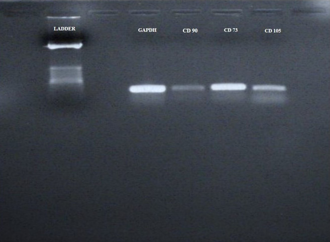
WJMSCS were characterized by conventional PCR gel Electrophoresis Method Expression of CD 90, CD105, CD73 was analyzed. GAPDH gene was used as internal standard. All these markers (CD 90, CD105, CD73) were detected positive for the WJMSCs.
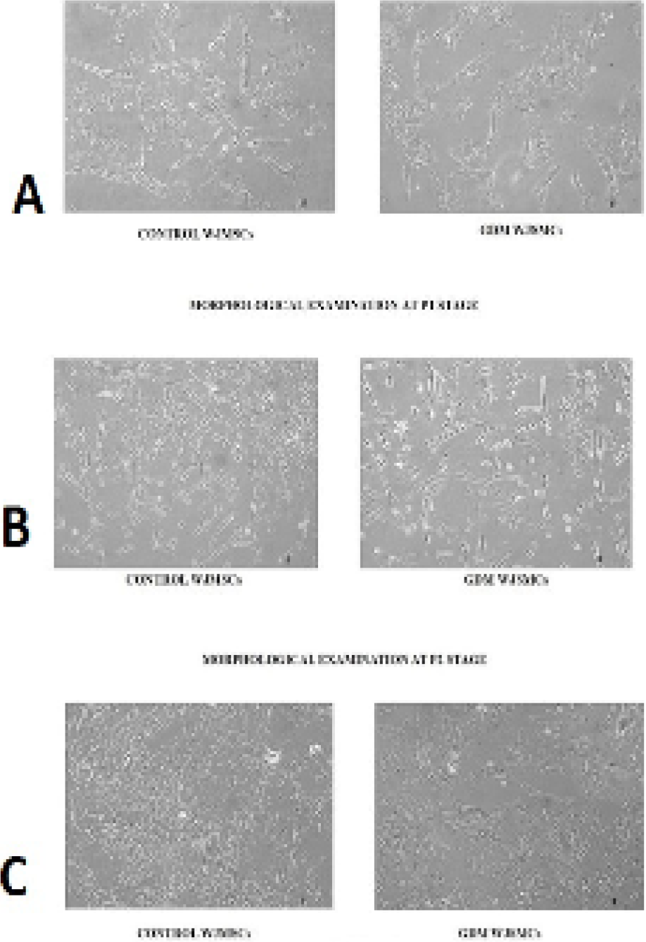
Morphological examination of control and GDM WJMSCs at P0, P1, P2 (passage 0, 1, 2) stage and magnification power, under inverted microscope (Floid Cell Imaging System). WJMSCs of both groups were analyzed under inverted microscope at different passaging stages (P0, P1, P2) at 10x magnification power. At P0 stage WJMSCs showed large spindle shaped morphology compared to control WJMSCs (A). At P1 stage GDM WJMSCs appeared as flattened as well as spindle shaped as compared to control (B). While at P2 there was no difference seen in morphology of WJMSCs in both groups showed normal spindle shaped morphology (C).
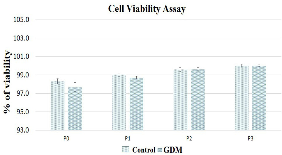
Trypan blue exclusion assay was used to evaluate the viability of GDM- WJMSCs as compared to NWMSCs. Cells of both groups were stained with trypan blue and in both groups viable cells were seen. The percentage of viability significantly increased with each passing of cells in both groups. Whereas no statically significant difference was observed between two groups.
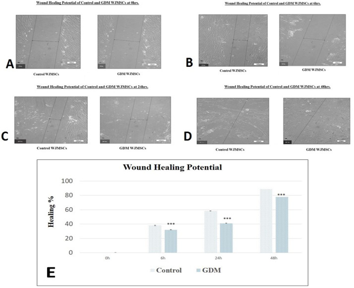
Wound healing potential of control and GDM WJMSCs at different interval of time (0h, 6h, 24h, 48h). The healing area of the scratch was calculated at different hours (6th, 24th, 45th h) in both groups. Significant difference in the healing potential was observed in GDM WJMSCs compared to control WJMSCs. The healing area of the scratch was calculated at different hours (6th, 24th, 45th h) in both groups. Significant difference in the healing potential was observed in GDM WJMSCs compared to control WJMSCs (E).
References
- Amrithaj, A.I., Kodali, A., Nguyen, L., Teo, A.K.K., Chang, C.W., Karnani, N., Ng, K.L., Gluckman, P.D., Chong, Y.S. and Stünkel, W., 2017. Gestational diabetes alters functions in offspring’s umbilical cord cells with implications for cardiovascular health. Endocrinology, 158: 2102-2112. https://doi.org/10.1210/en.2016-1889[CrossRef]
- An, B., Kim, E., Song, H., Ha, K.S., Han, E.T., Park, W.S., Ahn, T.G., Yang, S.R., Na, S. and Hong, S.H., 2017. Gestational diabetes affects the growth and functions of perivascular stem cells. Mol. Cells, 40: 434. https://doi.org/10.14348/molcells.2017.0053[CrossRef]
- Azzopardi, J.I. and Renald, B., 2018. Umbilical cord stem cells. Stem Cell Discov., 8: 1. https://doi.org/10.1155/2018/8329174[CrossRef]
- Buchanan, T.A., Xiang, A.H. and Kathleen, A., 2012. Gestational diabetes mellitus: Risks and management during and after pregnancy. Nat. Rev. Endocrinol., 8: 639. https://doi.org/10.1038/nrendo.2012.96[CrossRef]
- Chan, F.K., Moriwaki, K. and De Rosa, M.J., 2013. Detection of necrosis by release of lactate dehydrogenase activity. Methods mol. Biol., 979: 65–70. https://doi.org/10.1007/978-1-62703-290-2_7[CrossRef]
- Fong, C.Y., Chak, L.L., Biswas, A., Tan, J.H., Gauthaman, K., Chan, W.K. and Bongso, A., 2011. Human Wharton’s jelly stem cells have unique transcriptome profiles compared to human embryonic stem cells and other mesenchymal stem cells. Stem Cell Rev., 7: 1–16. https://doi.org/10.1007/s12015-010-9166-x[CrossRef]
- Forraz, N. and McGuckin, C.P., 2011. The umbilical cord: A rich and ethical stem cell source to advance regenerative medicine. Cell Prolif., 44: 60-69. https://doi.org/10.1111/j.1365-2184.2010.00729.x[CrossRef]
- Geissler, S., Textor, M., Kuhnisch, J., Konnig, D., Klein, O., Ode, A., Pfitzner, T., Adjaye, J., Kasper, G. and Duda, G.N., 2012. Functional comparison of chonological and in vitro aging: Differential role of the cytoskeleton and mitochondria in mesenchymal stromal cells. PLoS One, 7: e52700. https://doi.org/10.1371/journal.pone.0052700[CrossRef]
- Hadarits, O., Zoka, A., Barna, G., Al-Aissa, Z., Rosta, K., Rigo, J., Jr., Kautzky-Willer, A., Somogyi, A. and Firneisz, G., 2016. Increased proportion of hematopoietic stem and progenitor cell population in cord blood of neonates born to mothers with gestational diabetes mellitus. Stem Cells Dev., 25: 13-17. https://doi.org/10.1089/scd.2015.0203[CrossRef]
- Imam, K., 2012. Gestational diabetes mellitus. Adv. exp. Med. Biol., 771: 24–34. https://doi.org/10.1007/978-1-4614-5441-0_4[CrossRef]
- Kampmann, U., Shlush, E., Gat, I., Filice, M., Barretto, T., Jarvi, K., Lo, K., Gauthier-Fisher, A.S. and Librach, C.L., 2015. Gestational diabetes: A clinical update. World J. Diabetes, 6: 1065. https://doi.org/10.4239/wjd.v6.i8.1065[CrossRef]
- Kim, J., Piao, Y., Pak, Y.K., Chung, D., Han, Y.M., Hong, J.S., Jun, E.J., Shim, J.Y., Choi, J. and Kim, C.J., 2015. Umbilical cord mesenchymal stromal cells affected by gestational diabetes mellitus display premature aging and mitochondrial dysfunction. Stem Cells Dev., 24: 575-586. https://doi.org/10.1089/scd.2014.0349[CrossRef]
- Klinkhammer, B.M., Kramann, R., Mallau, M., Makowska, A., van Roeyen, C.R., Rong, S., Buecher, E.B., Boor, P., Kovacova, K., Zok, S., 2014. Mesenchymal stem cells from rats with chonic kidney disease exhibit premature senescence and loss of regenerative potential. PLoS One, 9: e92115. https://doi.org/10.1371/journal.pone.0092115[CrossRef]
- Kornicka, Katarzyna, Jenny, H. and Krzysztof, M., 2018. Dysfunction of mesenchymal stem cells isolated from metabolic syndrome and type 2 diabetic patients as result of oxidative stress and autophagy may limit their potential therapeutic use. Stem Cell Rev. Rep., 14: 337-345. https://doi.org/10.1007/s12015-018-9809-x[CrossRef]
- Manea, A., Manea, S.A., Todirita, A., Albulescu, I.C., Raicu, M., Sasson, S. and Simionescu, M., 2015. High-glucose-increased expression and activation of NADPH oxidase in human vascular smooth muscle cells is mediated by 4 hydroxynonenal activated PPARα and PPARβ/δ. Cell. Tissue Res., 361: 593-604. https://doi.org/10.1007/s00441-015-2120-0[CrossRef]
- Mantovani, C., Raimondo, S., Haneef, M.S., Geuna, S., Terenghi, G., Shawcross, S.G. and Wiberg, M., 2012. Morphological, molecular and functional differences of adult bone marrow and adipose derived stem cells isolated from rats of different ages. Exp. Cell Res., 318: 2034-2048. https://doi.org/10.1016/j.yexcr.2012.05.008[CrossRef]
- Montemurro, T., Andriolo, G., Montelatici, E., Weissmann, G., Crisan, M., Colnaghi, M.R., Rebulla, P., Mosca, F., Peault, B. and Lazzari, L., 2011. Differentiation and migration properties of human foetal umbilical cord perivascular cells: potential for lung repair. J. Cell. Mol. Med. 15: 796-808. https://doi.org/10.1111/j.1582-4934.2010.01047.x[CrossRef]
- Moon, H.E., Yoon, S.H., Hur, Y.S., Park, H.W., Ha, J.Y., Kim, K.H., Shim, J.H., Yoo, S.H., Son, J.H., Paek, S.L., 2013. Mitochondrial dysfunction of immortalized human adipose tissuederived mesenchymal stromal cells from patients with Parkinson’s disease. Exp. Neurobiol., 22: 283-300. https://doi.org/10.5607/en.2013.22.4.283[CrossRef]
- Phelps, J., Sanati-Nezhad, A., Ungrin, M., Duncan, N.A. and Sen, A., 2018. Bioprocessing of mesenchymal stem cells and their derivatives: Toward cell free therapeutics. Stem Cells Int., 2018: 9415367. [PMC free article] [PubMed] [Google Scholar]. https://doi.org/10.1155/2018/9415367[CrossRef]
- Rochette, L., Zeller, M., Cottin, Y. and Vergely, C., 2014. Diabetes, oxidative stress and therapeutic strategies. Biochim. biophys. Acta, 1840: 2709–2729. https://doi.org/10.1016/j.bbagen.2014.05.017[CrossRef]
- Tsang, W.P., Shu, Y., Kwok, P.L., Zhang, F., Lee, K.K., Tang, M.K., Li, G., Chan, K.M., Chan, W.Y. and Wan, C., 2013. CD146+ human umbilical cord perivascular cells maintain stemness under hypoxia and as a cell source for skeletal regeneration. PLoS One, 8: e76153. https://doi.org/10.1371/journal.pone.0076153[CrossRef]
- Van Rhijn-Brouwer, F.C.C., Kumar, M. and Verfaillie, C.M., 2018. Mesenchymal stromal cell characteristics and regenerative potential in cardiovascular disease: Implications for cellular therapy. Cell Transplant., 27: 765-785. https://doi.org/10.1177/0963689717738257[CrossRef]
- Vrachnis, N., Antonakopoulos, N., Iliodromiti, Z., Dafopoulos, K., Siristatidis, C., Pappa, K.I., Deligeoroglou, E. and Vitoratos, N., 2012. Impact of maternal diabetes on epigenetic modifications leading to diseases in the offspring. Exp. Diabetes Res., https://doi.org/10.1155/2012/538474[CrossRef]
- Wajid, N., Naseem, R., Anwar, S.S., Awan, S.J., Ali, M., Javed, S. and Ali, F., 2015. The effect of gestational diabetes on proliferation capacity and viability of human umbilical cord-derived stromal cells. Cell Tissue Bank., 16: 389-397. https://doi.org/10.1007/s10561-014-9483-4[CrossRef]
- Wang, R., Lv, L., Zhao, Y. and Yang, N., 2014. Okadaic acid inhibits cell multiplication and induces apoptosis in a549 cells, a human lung adenocarcinoma cell line. Int. J. clin. exp. Med., 7: 2025–203.
- Xu, S., Evans, H., Buckle, C., De Veirman, K., Hu, J., Xu, D., Menu, E., De Becker, A., Vande Broek, I., Leleu, X., 2012. Impaired osteogenic differentiation of mesenchymal stem cells derived from multiple myeloma patients is associated with a blockade in the deactivation of the Notch signaling pathway. Leukemia, 26: 2546- 2549. https://doi.org/10.1038/leu.2012.126[CrossRef]
- Yu, K.-R. and Kang, K.-S., 2013. Aging-related genes in mesenchymal stem cells: A mini-review. Gerontology, 59: 557-563. https://doi.org/10.1159/000353857[CrossRef]
Services
Article usage statistics
Loading…