
Cadmium is a heavy metal that is toxic and poisonous to the human body and could cause damage to different organs of the body, including testicles. Quercetin (Que), which is widely distributed in fruits and plants, belongs to the class of flavonoids and is thought to have health benefits and protect many tissues from heavy metals. This experiment aimed to explore the effect of Que on Cd-induced PERK-CHOP apoptosis pathway in rat testis. A total of 24 8-week-old Sprague–Dawley rats were divided into four groups: control group, CdCl2 group, CdCl2+Que group, and Que group. The control group was gavaged daily with distilled water and 0.9% sodium chloride solution, and the Cd and/or Que-treated groups were treated by daily intraperitoneal injection of CdCl2 solution and/or daily Que solution by gavage, respectively. Body weight, testis organ coefficient, and morphological changes of testis were measured. The related genes and protein expression levels of the PERK-CHOP pathway were also detected. The results showed that body weight and testicular organ coefficient decreased in cadmium-exposed rats compared with the control group, and pathological tissue sections showed atrophy of the internal convoluted vasculature structure, pathological conditions such as testicular interstitial cell hyperplasia, nuclear consolidation, and cell necrosis in cadmium-stained rats, and an increase in the number of apoptotic cells in the testis of TUNEL. In addition, cadmium exposure significantly increased the mRNA and protein expression levels of PERK, CHOP, ATF4, eIF2α, GRP78, and the pro-apoptotic factor Bax, while the expression of the apoptosis inhibiting factor Bcl-2 was significantly decreased, and compared with the CdCl2 group, the CdCl2 + Que group showed significantly decreased gene expression of PERK, CHOP, ATF4, eIF2α, GRP78, and Bax and Bcl-2 gene expression increased significantly, indicating that Que significantly attenuated Cd induced testicular toxicity. In conclusion, Que attenuates Cd toxicity and reduces apoptosis in rat testis by inhibiting the expression of mRNA and proteins involved in the PERK-CHOP pathway.
Research Article · Volume 56, Issue 6, 2 October 2024 · Open Access
Effect of Quercetin on Cadmium-Induced PERK-CHOP Apoptosis Pathway in Rat Testis
· · · · ·
Affiliations & Notes ⌄
- 1College of Animal Science and Technology, Henan University of Science and Technology, No. 263, Kaiyuan Avenue, 471023, Luoyang, PR China 2Jiangxi Agricultural Ecology and Resource Protection Station, No. 2 Dongerlu Donghu Dist, 330046, Nanchang, PR China * Corresponding author: wangjicang@126.com
Article Info ⌄
- Journal:
- Pakistan Journal of Zoology
- Published:
- 2 October 2024

From the article
- Compared with the Cd group, the Cd + Que group had significantly decreased body weight, and the relative organ coefficient of testis increased.
- In summary, cadmium can cause tissue damage in rat testis, leading to apoptosis and increased expression of related proteins and genes.
- Que inhibited cadmium toxicity, reduced the number of cadmium-induced apoptosis, and decreased apoptosis.
- Que attenuates cadmium toxicity in rat testis by inhibiting the expression of genes and proteins involved in the PERK-CHOP pathway.
Selected from the article's own text.
Abstract
Introduction
Cadmium (Cd) is a toxic heavy metal with a biological half-life of 10–35 years in humans and animals . In general, people are exposed to Cd by drinking contaminated water or by smoking or inhaling contaminated air. Daily intake of Cd through diet varies with pollution . For people working in mines and other environments, they could wear a good mask to reduce the inhalation of harmful substances . Cd could enter the body through respiration and cause damage to the liver, kidneys , and testicles, the testicles are very sensitive to cadmium toxicity . Low-dose Cd exposure can increase the mortality rate of prostate cancer and affect female reproduction and embryonic development .
Testis is a part of the reproductive system and the endocrine system; it is mainly used for spermatogenesis, synthesis, and secretion of male hormones to maintain the male physiological function of secondary sex characteristics. Spermatogenesis relies on microtubule-specific motor proteins, such as power egg 1, which act as engines to support the transport of germ cells and organelles through spermatogenic epithelium at different stages of the epithelial cycle . Testicular interstitial cells are mainly used to synthesize and secrete male hormones. Sertoli cells are mainly used to maintain spermatogenesis , participate in the formation of blood–testosterone barrier, and maintain androgen levels. Germ cells are mainly used to transmit genetic information from parents to off spring .
Apoptosis is recognized as a programmed cell death process that regulates cell growth and development . It plays a crucial role in immune homeostasis, and either insufficient or excessive apoptosis could cause disorders. Numerous in-vivo and in-vitro studies have demonstrated that Cd induces apoptosis in the testes, liver, and kidneys, which, in turn, causes damage to the body ; ; ; . The three potential mechanisms of Cd2+ toxicity in cells include the following: (1) excessive reactive oxygen species (ROS) generation directly attacks the mitochondrial membrane, reduces mitochondrial membrane potential, releases apoptotic factors, and then activates caspase to cause apoptosis; (2) the mitochondrial apoptosis pathway disrupts calcium (Ca) homeostasis and increases Ca concentration, leading to endoplasmic reticulum (ER) stress (ERS) and ER-mediated apoptosis. ERS apoptosis pathway ; and (3) activation of death receptor pathway induces apoptosis, namely, death-receptor-mediated apoptosis pathway of tumor cells .
Quercetin (Que) is a naturally active flavonoid that is not produced by the human body and is abundant in fruits and vegetables. The most common forms of Que are Que glycosides and Que sulfate . Que could be absorbed in the small intestine and stomach, but mainly in the small intestine. These bound Que reacts in the liver and are transported to muscle and brain through internal circulation. During the use of Que, it could produce a large number of metabolites due to its unique properties, the most representative and abundant metabolite being quercetin-3-O-β-D-glucuronide. When Que is distributed in tissues, a large number of functions and benefits are activated . Evidence proved that Que has various biological characteristics, including antioxidation, pro-oxidation, antivirus, antiallergic, and analgesic properties and antithrombosis, anti-ischemia, anti-apoptosis, antitumor, antibacterial, and antiviral activities . In a word, Que has multiple biological activities and no obvious toxicity. It is of great importance for the prevention and treatment of aging, cancer, and cardiovascular disease, so it has high research value.
In this study, the variations of apoptosis associated genes and protein expression levels on the testicular PERK-CHOP apoptosis pathway was examined on the basis of a rat Cd poisoning model. Then, the effects of Que on the expression of signaling molecules on the testicular PERK-CHOP apoptosis pathway in Cd-infected rats were investigated. The study provides a theoretical basis for the clinical treatment of Cd poisoning with Que.
Materials and Methods
Animals
A total of 24 8-week-old Sprague–Dawley (SD) rats were prepared and placed in the animal room, fed with standard chow, kept on adequate diet for 7 days, and then treated in accordance with the experimental requirements. Animals were purchased from Henan Provincial Animal Experiment Center.
Chemicals
Que (98.5%) (No. 117-39-5) and CdCl2 (99.99%)(No. 10108-64-2) were obtained from Aladdin Biochemical Technology Co., Ltd (Shanghai, China). PCR primers were obtained from Shanghai Sanko Biotechnology Co., Ltd.and TRIzol isolation kit (No. 15596-026) were obtained from Thermo Fisher Scientific (China) Co., Ltd.
Animals and treatments
Twenty-four male SD rats weighing approximately 110 g were fed in a 12-h light-dark cycle. The temperature was kept in the range of 25°C±2°C, and then the rats were free to eat under standard laboratory conditions. During the period, the rats could also drink freely. After 7 days of acclimatization, they were randomly divided into four groups: control, Cd, Cd+Que, and Que groups. The control group was gavaged daily with distilled water and 0.9% sodium chloride solution. The Cd group was intraperitoneally injected with 2 mg/kg b.w. of CdCl2. The Que group was gavaged with 100 mg/kg b.w. of Que, and the Cd+Que group received Que (100 mg/kg b.w.) by gavage and CdCl2 (2 mg/kg b.w.) intoxication for 28 days. Cd was administered by intraperitoneal injection, and Que was administered by gavage. The doses of Cd and Que could be found from previous literature .
After 4 weeks of treatment, the rats were fasted for 12h, then and euthanized and placed in anticoagulation and non-anticoagulation tubes, then, the rats were executed by breaking their necks and incised along the abdominal midline with surgical scissors to expose the abdominal cavity. Next, the testes were removed. The weight of the testes was registered, and the relative testicular weight (testicular weight/body weight × 100%) was immediately calculated in accordance with the listed formula. An appropriate amount of testicular tissue was collected, and 0.9% saline was added to prepare a 10% tissue homogenate before grinding the testicular samples at low temperature (4℃). The undamaged testes were then placed in 10% formaldehyde solution to make testicular sections for later use.
Histopathological analysis
Fresh testicular tissue was collected and immediately fixed in 10% formaldehyde solution for 24 h. Then, it was rinsed with purified water for 12 h, dehydrated in alcohol, embedded in paraffin, and cut into sections with a thickness of 5 μm. A drop of glycerol protein was taken onto a slide, and then several drops of cold water were dripped to make the sections suspended and placed in a 37° C oven. Afterwards, the slides were removed from the oven and examined under the light microscope.
TUNEL evaluation
Apoptosis was detected by terminal dUTP nick-end labeling (TUNEL) assay to evaluate the apoptosis rate of testicular cells (C1086, Beyotime Biotechnology Co., Ltd.). The TUNEL steps and analysis were carried out in strict accordance with the manufacturer’s instructions.
Quantitative real-time PCR analysis
The collected fresh testicular tissue was ground and stored in a refrigerator at −80 °C. RNA concentration was quantified at 260/280 nm by using an ultra-micro nucleic acid concentration meter in accordance with the manufacturer’s instructions. Samples that showed intact RNA bands were used for reverse transcription using a HiScript III RT SuperMix for qPCR (+gDNA wiper) Reverse-Transcription Kit (No. R323–01, Novizin BiotechnologyCo., Ltd., Nanjing, China). Fluorescence quantification was performed using cDNA templates and 2× ChamQ Universal SYBR qPCR Master Mix. The relative abundance of mRNA for each gene is always measured using 2−ΔΔCt method, where β-actin is chosen as the endogenous control. The primers for GRP78, PERK, CHOP, eIF2α, ATF4 were listed in Table I.
Western blot analysis
Testicular tissue blocks were collected, and the testicular tissue was lysed using RIPA lysis buffer (Beyotime, China) supplemented with ImM benzenesulfonyl fluoride (PMSF). The protease inhibitor mixture was added for some time before use. The whole process was ground at low temperature, and the total protein was extracted by vortex shaking and centrifugation (12000 r, 4 °C, 10 min). The BCA Protein Assay Kit (Beyotime, P0013, Beyotime Institute of Biotechnology, Shanghai, China) was used to determine the total protein concentration, and the whole process followed the steps of the manufacturer’s instructions. The obtained protein samples were subjected to SDS-PAGE gel, transferred to polyvinylidene fluoride membrane, and blocked in TBST containing 5% skim milk for 2 h. Then, they were washed with TBST five times (8 min each time) and incubated with the primary antibody at 4 °C overnight. At room temperature, the horseradish peroxidase-conjugated goat anti-rabbit/mouse IgG culture membrane was exposed and photographed in the darkroom with an ECL kit, and then the protein gray scale was quantified by a software.
Table I. Primer sequences.
| Gene | Primer sequences (5' to 3') | Product length |
| GRP78 | F: ATGGTGTGGGAGATCCTGTTTTC | 124 |
| R: CAAGACGCACAGGGATACGC | ||
| PERK | F: ACAAGGCTGTCACTCAGGTG | 121 |
| R: GCTAGGAGCCTTGGAGCAC | ||
| eIF2α | F: TTTCCGGGACAAGATGGCG | 87 |
| R: AAGTGTGGGGGTCCATTCAC | ||
| ATF4 | F: TGTTGGCGGGGGACTTAATG | 200 |
| R: AAAGGCATCCTCCTTGCCG | ||
| CHOP | F: CTGGGAAACAGCGCATGAAG | 121 |
| R: GTGGTCTCTACCTCCCTGGT |
Statistical analysis
All data were analyzed by SPSS 25.0 software for single variable factor analysis to obtain the mean, SEM, and P values of the corresponding protein. The value is expressed by mean + SEM. P > 0.05 indicated no significant difference, P < 0.05 indicated significant difference, and P < 0.01 indicated extremely significant difference.
Results
Body weight and relative testicular weight of rats
Table II shows that compared with the control group, the Cd group exhibited a significant decrease in body weight (P < 0.01). Meanwhile, the body weight of the Cd + Que group increased significantly compared with that of the Cd group (P < 0.01).
Table III clearly shows that the relative organ weight of the Cd group had a certain decreasing trend compared with that of the control group. Moreover, the relative organ weight of the Cd + Que group increased to a certain extent compared with that of the Cd group.
Table II. Effect of Cdcl2 and Quercetin on body weight of rats.
| Control | CdCl2 | CdCl2+ Que | Que | |
| 7 Day | 145.03±8.73 | 122.80±22.13 | 125.30±13.38 | 123.6±2.52 |
| 14 Day | 198.00±36.59 | 154.10±1.73** | 156.00±11.53 | 196±19.97 |
| 21 Day | 251.33±26.58 | 159.33±26.23** | 192.77±20.43## | 220±10.54 |
| 28 Day | 296.37±19.52 | 186.00±13.40** | 194.33±36.47 | 276.67±19.22 |
** indicates the cadmium group compared with the control group, and indicates a highly significant difference. ## indicates a highly significant difference compared to the cadmium treatment group, P < 0.01 indicates a highly significant difference; CdCl2, Cadmium; Que, quercetin, same below.
Table III. Effect of cadmium and quercetin on the relative organ coefficients of rat testes.
| Group | 28 days relative organ weight/g |
| Control | 1.00±0.07 |
| CdCl2 | 0.57±0.11 |
| CdCl2+ Que | 0.75±0.27 |
| Que | 1.01±0.07 |
Histological evaluation of rat testis
The pathological changes in the testicular tissue of rats are shown in Figure 1A. Figure 1A showed a regular distribution of cells in the testicular tissue, and the overall staining of the tissue showed a lighter state. Compared with A, B showed atrophy of the inner seminiferous tubule structure, shedding of multinucleated or megakaryocytes in the tubule, proliferation of connective tissue between tubules, proliferation of testicular Leydig cells. It also contained pigment granules, loose cells, disordered arrangement, irregular, cell vacuolization, nuclear pyknosis, and cell necrosis. Compared with B, a large part of the cells in C were closely distributed, arranged regularly, and stained lightly. Moreover, the number of cell necrosis was small, showing the same state as A. The images presented by D and A showed almost no significant difference.
Apoptosis changes in rat testis
Comparison between Figure 2A and B showed little or no apoptosis in the testicular tissue of the control group, whereas a large number of apoptosis occurred in the Cd-treated group. Comparison between Figure 2B and C demonstrated that the numbers of apoptosis in the Cd + Que group was significantly reduced compared with that in the Cd group. Comparison between Figure 2C and A found that the Que group was similar to the control group, and almost no apoptosis was observed.
mRNA expression levels of testicular PERK-CHOP apoptosis pathway
The mRNA expression levels of PERK-CHOP pathway-related genes in rat testicular tissues were detected by RT-PCR, as shown in Figure 3. Compared with the control group, the Cd group demonstrated a significant increase in GRP78, CHOP, ATF4, PERK, eIF2α, and Bax mRNA expression levels and a significant decrease in Bcl-2 mRNA expression (P < 0.01). Meanwhile, the mRNA expression levels of GRP78, CHOP, ATF4, PERK, eIF2α, and Bax in the Cd + Que group decreased significantly compared with those in the Cd group, and the mRNA expression level of Bcl-2 increased significantly (P < 0.01).
Protein expression levels of the apoptotic pathway in testicular PERK-CHOP cells
Figure 4 shows that compared with the control group, the Cd group had significantly increased PERK, GRP78, and ATF4 protein expression levels and decreased Bcl-2 protein expression. Compared with the Cd group, the Cd + Que group exhibited a significant decrease in PERK, GRP78, and ATF4 protein expression levels and a decrease in Bcl-2 protein expression.
Discussion
Cd exists in many vegetables and thus is easy to cause damage to the body. Studies have shown that Cd could easily accumulate in the testis, leading to apoptosis and damage to the reproductive performance of the body . However, the specific mechanism is still unknown. Que, as a natural and easy-to-obtain substance, has antioxidation, antitumor, antivirus, anti-inflammatory, and other effects . Some therapeutic effects on adverse symptoms are caused by Cd. Therefore, in the present work, Cd-exposed rats were used as experimental subjects to determine the effects of Que on protein content, gene expression, and apoptosis in specific parts of Cd-exposed rats. This study aimed to explore whether Que had protective effects on Cd-exposed rats and the bias and strength of these effects.
Cd poisoning could have further effects on the body. The damage to the animal body differs in accordance with the length of Cd exposure time and the size of the dose. Studies have shown that low doses of Cd could induce cell proliferation, and high doses of Cd could induce ROS production and apoptosis (Zhu et al., 2021). In the present work, a rat model was used as the research object, and the most intuitive data showed changes in rat body weight and testicular relative weight. Cd significantly inhibited the increase of body weight in rats, and this effect was related to the decrease in appetite and feed intake of animals exposed to Cd . A study showed that Cd could significantly reduce the body weight and relative organ coefficient of testis in mice . Moreover, a reduction in testicular weight may represent the production of free radicals, which could lead to oxidation; and then to DNA, protein, and lipid deterioration; and eventually, testicular atrophy. The experimental results of the present study are basically consistent with these results of previous studies. The body weight and organ coefficients of the Cd group showed a significant decrease compared with those of the control group. In particular, the body weight of the Cd group decreased significantly, and the relative organ coefficient of testis decreased. Compared with the Cd group, the Cd + Que group had significantly decreased body weight, and the relative organ coefficient of testis increased. This finding proved that Cd may reduce the appetite of rats and lead to weight loss. It could also damage its reproductive performance, leading to testicular atrophy. Que could reduce the inhibitory effect of Cd on the body weight and relative organ weight of rats.
Cd causes testicular damage, and the cell structure of germ cell morphology is disturbed . Cd exposure in mice caused proliferation of blood vessel wall cells and endothelial cells, resulting in abnormal testicular function . In the present study results, the transverse section of rat testis showed degenerative atrophic tubules, damage and necrosis of seminiferous tubules, and loose arrangement and necrosis of cells after Cd treatment. Moreover, the complete basement membrane and complete and orderly cells were clearly shown after Cd and Que treatment. This finding showed that Que inhibited the toxicity of Cd, thereby making the testicular tissue cells gradually arranged closely, protecting cell survival, and playing a protective role.
Apoptosis is a programmed death. A large number of studies have shown that Cd could cause multiple tissue damage and apoptosis in various types of cells ; . As known, apoptosis plays a crucial role in Cd-induced reproductive toxicity ; Zhu et al., 2021). Studies have shown that Cd treatment could lead to ERS, which, in turn, leads to apoptosis . The images obtained by TUNEL staining of rat testis showed that the number of testicular cell apoptosis in the Cd group increased significantly compared with that in the control group. The qRT-PCR experiment also showed that compared with the control group, the Cd group exhibited a significant increase in the expression of Bax (pro-apoptotic gene) and a significant decrease in the expression of Bcl-2 (anti-apoptotic gene). New data showed that the accumulation of abnormally misfolded proteins leads to ERS, and to restore homeostasis in ERS, the body sequentially activates the UPR and thus activates autophagy, in which lysosomes play a crucial role in degrading misfolded proteins . After ERS occurs, the body activates the corresponding signaling pathways to counteract the stress response. When the stress level is low, cells are not greatly affected; when the stress level is severe or even overloaded, the body activates the downstream apoptotic pathway signaling molecules, thus inducing apoptosis. Sustained ERS promotes the initiation of apoptotic pathways. Caspase-12, one of the pathways mediating apoptosis by ERS, is detached from the ERS membrane and cleaved into active fragments under ERS, leading to apoptosis by caspase-3 cleavage .
CHOP is a downstream transcription factor of PERK/eukaryotic translation initiation factor 2α (eIF2α)/activating transcription factor 4 (ATF4). It plays a crucial role in ERS-induced apoptosis by inhibiting the anti-apoptotic protein Bcl-2 and inducing the pro-apoptotic molecule Bim . Heavy metal Cd could damage ER homeostasis and induce UPR through ERS. Studies have shown that the classical markers of UPR and ERS (ATF4, GRP78, PERK, eIF2α, ERDJ4, Ero1LB, PBGD, IRE1, ATF6, and sXBP1) increased their corresponding protein levels after Cd treatment, consistent with the results of the present study .
According to Figure 3, the change trend of the five genes was roughly the same. The gene expression levels of the Cd group significantly increased compared with those of the control group. According to Figure 4, compared with the control group, the Cd group showed an increase in the protein expression levels of PERK, ATF4, and GRP78.ER UPR occurs to fight against protein misfolding and maintain cell homeostasis. In the PERK pathway during UPR, the binding of GRP78 and PERK is broken, which increases the content of free genes and proteins, that is, the detected GRP78 and PERK genes and proteins. Compared with the Cd group, the Cd + Que group had decreased protein expression and significantly decreased gene expression.
Conclusion
In summary, cadmium can cause tissue damage in rat testis, leading to apoptosis and increased expression of related proteins and genes. Que inhibited cadmium toxicity, reduced the number of cadmium-induced apoptosis, and decreased apoptosis. Que attenuates cadmium toxicity in rat testis by inhibiting the expression of genes and proteins involved in the PERK-CHOP pathway.
Funding
This study was supported by grants from National Nature Science Foundation of China (No. 32273083, 31972753).
Ethical statement and IRB approval
The protocol of this study was approved by the Institutional Animal Care and the Ethics Committee of Henan University of Science and Technology (approval number HAUST 18010).
Availability of data and materials
The datasets used and/or analysed during the current study are available from the corresponding author on reasonable request.
Statement of conflict of interest
The authors have declared no conflict of interest.
Figures
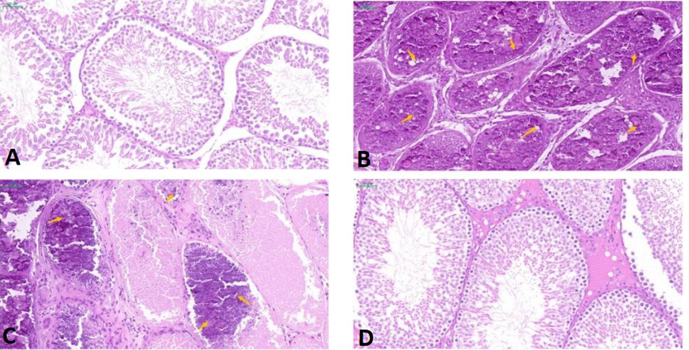
Histopathology of testicular sections from different groups (20×). Scale bar is 50 μm. (A) Control group; (B) CdCl2 group; (C) CdCl2 + Que group; (D) Que group. The orange arrow indicates the germinal epithelium and varicocele.
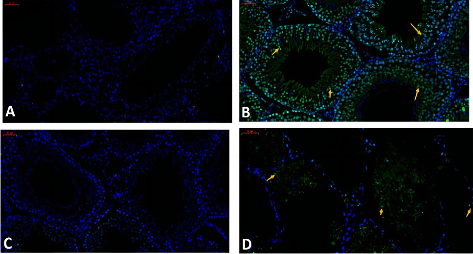
TUNEL assay for apoptosis in testicular tissue. Scale bar is 50 μm. The green ones are apoptotic cells (yellow arrows, 40×). (A) Control group; (B) CdCl2 group: (C) CdCl2 + Que group; (D) Que group.
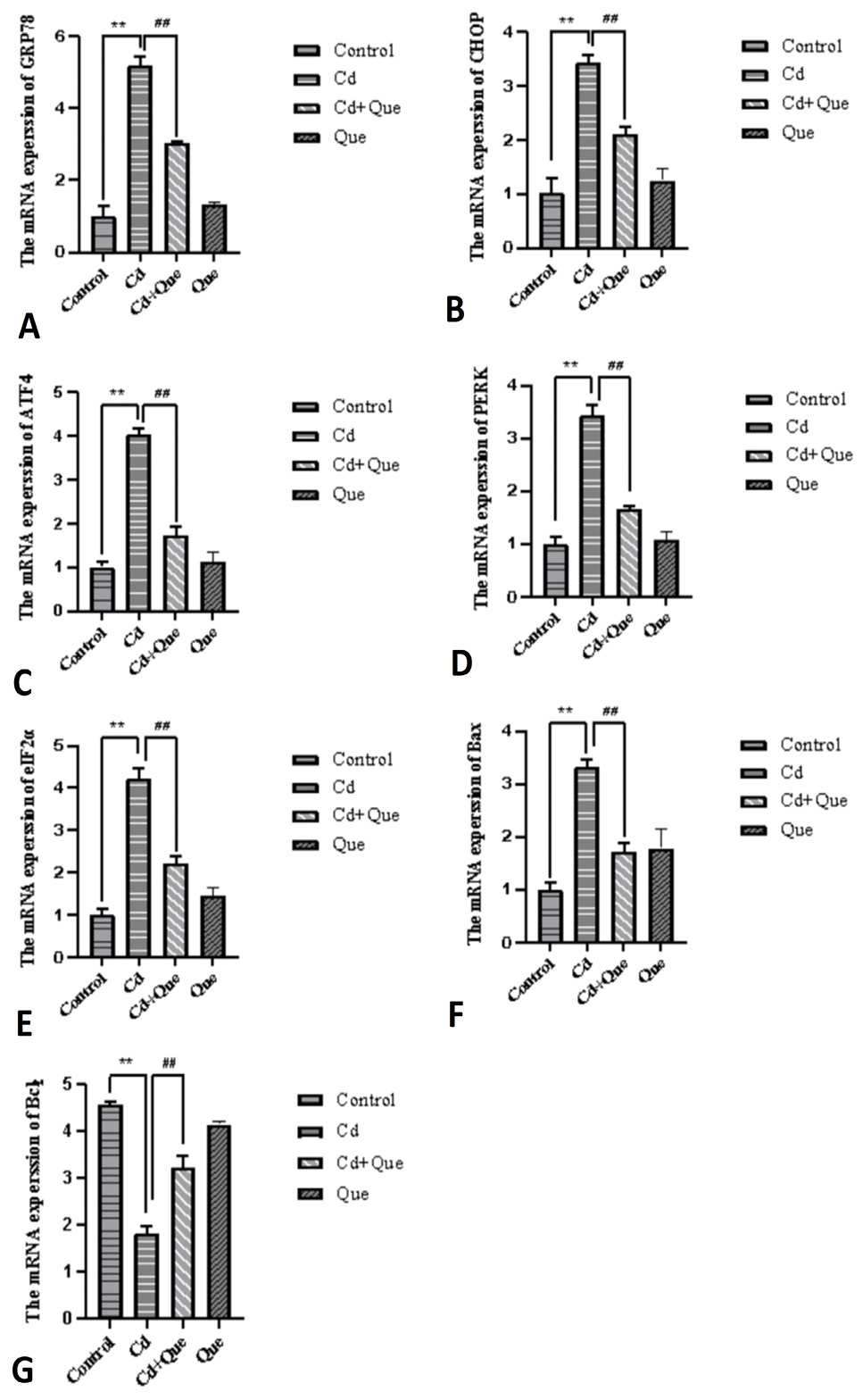
Effects of Que on mRNA expression levels of GRP78, CHOP, ATF4, PERK, elF2a, Bax, and Bcl-2. Bars indicate mean ± SEM (n = 3).
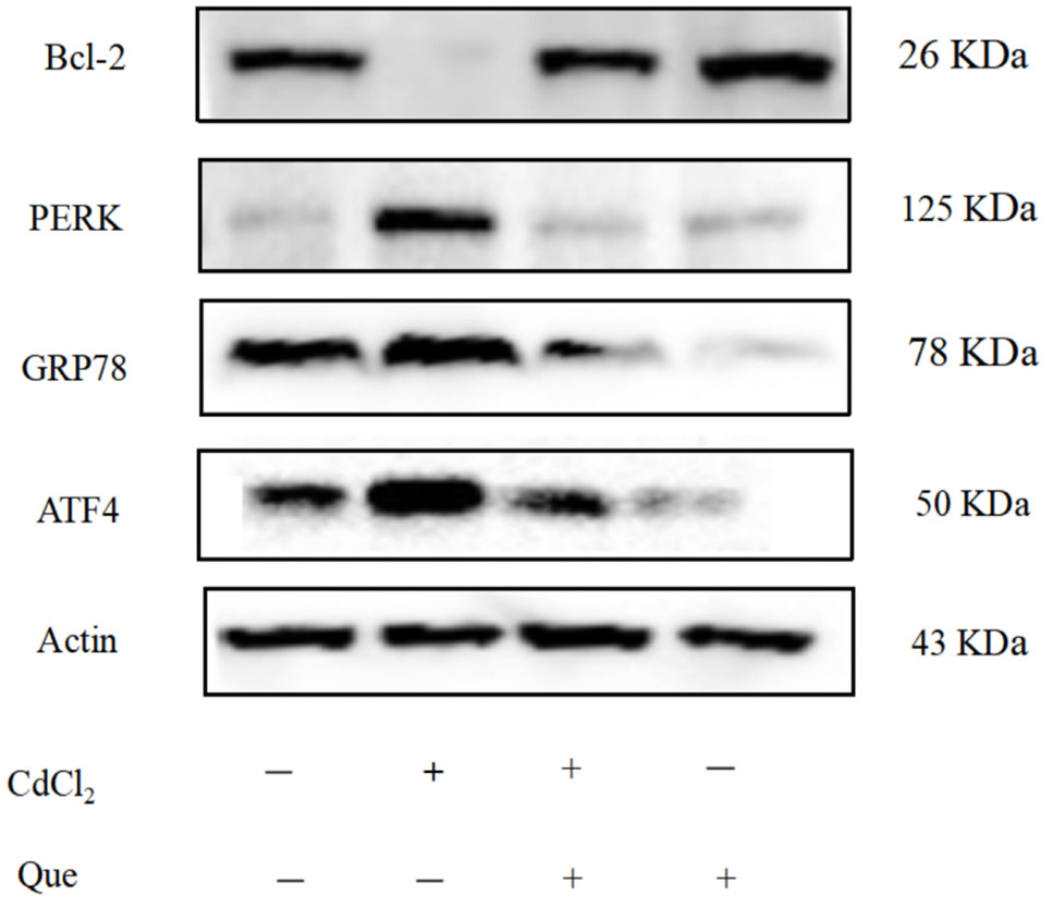
Western blot target band gray image + indicates added cadmium or quercetin, and - indicates no addition.
References
- Amanpour, P., Khodarahmi, P. and Salehipour, M., 2020. Protective effects of vitamin E on cadmium-induced apoptosis in rat testes. Naunyn Schmiedebergs Arch. Pharmacol., 393: 349–358. https://doi.org/10.1007/s00210-019-01736-w[CrossRef]
- Biagioli, M., Pifferi, S., Ragghianti, M., Bucci, S., Rizzuto, R. and Pinton, P., 2008. Endoplasmic reticulum stress and alteration in calcium homeostasis are involved in cadmium-induced apoptosis. Cell Calcium, 43: 184–195. https://doi.org/10.1016/j.ceca.2007.05.003[CrossRef]
- Brdarić, E., Soković Bajić, S., Đokić, J., Đurđić, S., Ruas-Madiedo, P., Stevanović, M., Tolinački, M., Dinić, M., Mutić, J., Golić, N. and Živković, M., 2021. Protective effect of an exopolysaccharide produced by Lactiplantibacillus plantarum BGAN8 against cadmium-induced toxicity in Caco-2 cells. Front. Microbiol., 12: 759378. https://doi.org/10.3389/fmicb.2021.759378[CrossRef]
- Cai, Y., Arikkath, J., Yang, L., Guo, M.L., Periyasamy, P. and Buch, S., 2016. Interplay of endoplasmic reticulum stress and autophagy in neurodegenerative disorders. Autophagy, 12: 225–244. https://doi.org/10.1080/15548627.2015.1121360[CrossRef]
- Cao, S.S., 2015. Endoplasmic reticulum stress and unfolded protein response in inflammatory bowel disease. Inflamm. Bowel. Dis., 21: 636–644. https://doi.org/10.1097/MIB.0000000000000238[CrossRef]
- Chen, C.Y., Zhang, S.L., Liu, Z.Y., Tian, Y. and Sun, Q., 2015. Cadmium toxicity induces ER stress and apoptosis via impairing energy homoeostasis in cardiomyocytes. Biosci. Rep., 35: e00214. https://doi.org/10.1042/BSR20140170[CrossRef]
- Cheung, M.R., Kang, J., Ouyang, D. and Yeung, V., 2014. Association between urinary cadmium and all cause, all cancer and prostate cancer specific mortalities for men: An analysis of national health and nutrition examination survey (NHANES III) data. Asian Pac. J. Cancer Prev., 15: 483–488. https://doi.org/10.7314/APJCP.2014.15.1.483[CrossRef]
- Dai, X., Nie, G., Cao, H., Xing, C., Hu, G. and Zhang, C., 2019. In vivo assessment of molybdenum and cadmium co-induced the mRNA levels of heat shock proteins, inflammatory cytokines and apoptosis in shaoxing duck (Anas platyrhyncha) testicles. Poult. Sci., 98: 5424–5431. https://doi.org/10.3382/ps/pez328[CrossRef]
- Deepika, N. and Maurya, P.K., 2022. Health benefits of quercetin in age-related diseases. Molecules, 27: 2498. https://doi.org/10.3390/molecules27082498[CrossRef]
- Fujiwara, Y., Lee, J.Y., Tokumoto, M. and Satoh, M., 2012. Cadmium renal toxicity via apoptotic pathways. Biol. Pharm. Bull., 35: 1892–1897. https://doi.org/10.1248/bpb.b212014[CrossRef]
- Gong, Z.G., Wang, X.Y., Wang, J.H., Fan, R.F. and Wang, L., 2019. Trehalose prevents cadmium-induced hepatotoxicity by blocking Nrf2 pathway, restoring autophagy and inhibiting apoptosis. J. Inorg. Biochem., 192: 62–71. https://doi.org/10.1016/j.jinorgbio.2018.12.008[CrossRef]
- Gong, Z.G., Zhao, Y., Wang, Z.Y., Fan, R.F., Liu, Z.P. and Wang, L., 2022. Epigenetic regulator BRD4 is involved in cadmium-induced acute kidney injury via contributing to lysosomal dysfunction, autophagy blockade and oxidative stress. J. Hazard. Mater., 423: 127110. https://doi.org/10.1016/j.jhazmat.2021.127110[CrossRef]
- Kedhari, S.M., Raina, R., Afroze, N., Bajbouj, K., Hamad, M., Haque, S. and Hussain, A., 2019. Quercetin modulates signaling pathways and induces apoptosis in cervical cancer cells. Biosci. Rep., 39: BSR20190720. https://doi.org/10.1042/BSR20190720[CrossRef]
- Kim, J., Song, H., Heo, H.R., Kim, J.W., Kim, H.R., Hong, Y., Yang, S.R., Han, S.S., Lee, S.J., Kim, W.J. and Hong, S.H., 2017. Cadmium-induced ER stress and inflammation are mediated through C/EBP–DDIT3 signaling in human bronchial epithelial cells. Exp. mol. Med., 49: e372. https://doi.org/10.1038/emm.2017.125[CrossRef]
- Kumar, K.K.M., Kumar, N.M., Patil, R.H., Nagesh, R., Hegde, S.M., Kavya, K., Babu, R.L., Ramesh, G.T. and Sharma, S.C., 2016. Cadmium induces oxidative stress and apoptosis in lung epithelial cells. Toxicol. Mech. Methods, 26: 658–666. https://doi.org/10.1080/15376516.2016.1223240[CrossRef]
- Li, Y., Yao, J., Han, C., Yang, J., Chaudhry, M.T., Wang, S., Liu, H. and Yin, Y., 2016. Quercetin, inflammation and immunity. Nutrients, 8: 167. https://doi.org/10.3390/nu8030167[CrossRef]
- Liu, W., Zhao, H., Wang, Y., Jiang, C., Xia, P., Gu, J., Liu, X., Bian, J., Yuan, Y. and Liu, Z., 2014. Calcium-calmodulin signaling elicits mitochondrial dysfunction and the release of cytochrome c during cadmium-induced apoptosis in primary osteoblasts. Toxicol. Lett., 224: 1–6. https://doi.org/10.1016/j.toxlet.2013.10.009[CrossRef]
- Marettová, E., Maretta, M. and Legáth, J., 2015. Toxic effects of cadmium on testis of birds and mammals: A review. Anim. Reprod. Sci., 155: 1–10. https://doi.org/10.1016/j.anireprosci.2015.01.007[CrossRef]
- Markiewicz-Górka, I., Pawlas, K., Jaremków, A., Januszewska, L., Pawłowski, P. and Pawlas, N., 2019. Alleviating effect of α-lipoic acid and magnesium on cadmium-induced inflammatory processes, oxidative stress and bone metabolism disorders in Wistar rats. Int. J. environ. Res. Publ. Hlth., 16: 4483. https://doi.org/10.3390/ijerph16224483[CrossRef]
- Martel, J., Ojcius, D.M., Ko, Y.F. and Young, J.D., 2020. Phytochemicals as prebiotics and biological stress inducers. Trends Biochem. Sci., 45: 462–471. https://doi.org/10.1016/j.tibs.2020.02.008[CrossRef]
- Mezynska, M. and Brzóska, M.M., 2018. Environmental exposure to cadmium a risk for health of the general population in industrialized countries and preventive strategies. Environ. Sci. Pollut. Res., 25: 3211–3232. https://doi.org/10.1007/s11356-017-0827-z[CrossRef]
- Nabavi, S.F., Russo, G.L., Daglia, M. and Nabavi, S.M., 2015. Role of quercetin as an alternative for obesity treatment: You are what you eat! Fd. Chem., 179: 305–310. https://doi.org/10.1016/j.foodchem.2015.02.006[CrossRef]
- Rebourcet, D., Darbey, A., Monteiro, A., Soffientini, U., Tsai, Y.T., Handel, I., Pitetti, J.L., Nef, S., Smith, L.B. and O’Shaughnessy, P.J., 2017. Sertoli cell number defines and predicts germ and leydig cell population sizes in the adult mouse testis. Endocrinology, 158: 2955–2969. https://doi.org/10.1210/en.2017-00196[CrossRef]
- Ren, M.T., Gu, M.L., Zhou, X.X., Yu, M.S., Pan, H.H., Ji, F. and Ding, C.Y., 2019. Sirtuin 1 alleviates endoplasmic reticulum stress-mediated apoptosis of intestinal epithelial cells in ulcerative colitis. World J. Gastroenterol., 25: 5800–5813. https://doi.org/10.3748/wjg.v25.i38.5800[CrossRef]
- Shi, L., Cao, H., Luo, J., Liu, P., Wang, T., Hu, G. and Zhang, C., 2017. Effects of molybdenum and cadmium on the oxidative damage and kidney apoptosis in duck. Ecotoxicol. environ. Saf., 145: 24–31. https://doi.org/10.1016/j.ecoenv.2017.07.006[CrossRef]
- Venditti, M., Ben Rhouma, M., Romano, M.Z., Messaoudi, I., Reiter, R.J. and Minucci, S., 2021. Evidence of melatonin ameliorative effects on the blood-testis barrier and sperm quality alterations induced by cadmium in the rat testis. Ecotoxicol. environ. Saf., 226: 112878. https://doi.org/10.1016/j.ecoenv.2021.112878[CrossRef]
- Wang, J., Ding, L., Wang, K., Huang, R., Yu, W., Yan, B., Wang, H., Zhang, C., Yang, Z. and Liu, Z., 2022. Role of endoplasmic reticulum stress in cadmium-induced hepatocyte apoptosis and the protective effect of quercetin. Ecotoxicol. environ. Saf., 241: 113772. https://doi.org/10.1016/j.ecoenv.2022.113772[CrossRef]
- Wang, J., Zhu, H., Wang, K., Yang, Z. and Liu, Z., 2020. Protective effect of quercetin on rat testes against cadmium toxicity by alleviating oxidative stress and autophagy. Environ. Sci. Pollut. Res. Int., 27: 25278–25286. https://doi.org/10.1007/s11356-020-08947-2[CrossRef]
- Xu, D., Hu, M.J., Wang, Y.Q. and Cui, Y.L., 2019. Antioxidant activities of quercetin and its complexes for medicinal application. Molecules, 24: 1123. https://doi.org/10.3390/molecules24061123[CrossRef]
- Xu, S., Pi, H., Chen, Y., Zhang, N., Guo, P., Lu, Y., He, M., Xie, J., Zhong, M., Zhang, Y., Yu, Z. and Zhou, Z., 2013. Cadmium induced Drp1-dependent mitochondrial fragmentation by disturbing calcium homeostasis in its hepatotoxicity. Cell Death Dis., 4: e540. https://doi.org/10.1038/cddis.2013.7[CrossRef]
- Yang, S.H., Chen, S.T., Liang, C., Shi, Y.H. and Chen, Q.S., 2022. Effects of cadmium exposure on leydig cells and blood vessels in mouse testis. Int. J. environ. Res. Publ. Hlth., 19: 2416. https://doi.org/10.3390/ijerph19042416[CrossRef]
- Zhang, R., Yi, R., Bi, Y., Xing, L., Bao, J. and Li, J., 2017. The effect of selenium on the Cd-induced apoptosis via no-mediated mitochondrial apoptosis pathway in chicken liver. Biol. Trace Elem. Res., 178: 310–319. https://doi.org/10.1007/s12011-016-0925-7[CrossRef]
- Zhao, J., Lu, P., Wan, C., Huang, Y., Cui, M., Yang, X., Hu, Y., Zheng, Y., Dong, J., Wang, M., Zhang, S., Liu, Z., Bian, S., Wang, X., Wang, R., Ren, S., Wang, D., Yao, Z., Chang, G., Tang, F. and Zhao, X.Y., 2021. Cell-fate transition and determination analysis of mouse male germ cells throughout development. Nat. Commun., 12: 6839. https://doi.org/10.1038/s41467-021-27172-0[CrossRef]
- Zhu, M., Miao, S., Zhou, W., Elnesr, S.s., Dong, X. and Zou, X., 2021. MAPK, AKT/FoxO3a and mTOR pathways are involved in cadmium regulating the cell cycle, proliferation and apoptosis of chicken follicular granulosa cells. Ecotoxicol. Environ. Saf., 214. https://doi.org/10.1016/j.ecoenv.2021.112091[CrossRef]
- Zhuang, J., Nie, G., Yang, F., Dai, X., Cao, H., Xing, C., Hu, G. and Zhang, C., 2019. Cadmium induces cytotoxicity through oxidative stress-mediated apoptosis pathway in duck renal tubular epithelial cells. Toxicology In Vitro, 61: 104625. https://doi.org/10.1016/j.tiv.2019.104625[CrossRef]
- Zou, H., Sun, J., Wu, B., Yuan, Y., Gu, J., Bian, J., Liu, X. and Liu, Z., 2020. Effects of cadmium and/or lead on autophagy and liver injury in rats. Biol. Trace Elem. Res., 198: 206–215. https://doi.org/10.1007/s12011-020-02045-7[CrossRef]
Services
Article usage statistics
Loading…