
Bilateral ovaries of 20 female SD rats were surgically resected. Four weeks after surgery, osteoporotic rat models were copied and randomly divided into a calcitriol group and a model group, with 10 rats in each group. The ovaries of another 10 rats were detached but not removed as a sham-operated group. After 12 weeks of continuous administration in each group of rats, the results showed that calcitriol increased the BMD values, reduced the levels of bone formation indicators ALP, BALP, UcOC and PINP, increased the levels of bone resorption indicators TRAP5a, TRAP5b and NTX, reduced CTX levels, and increased mRNA expression levels of Bglap and Runx2 in osteoporotic rats. In conclusion, calcitriol may become an effective drug therapy for treating osteoporosis, and its mechanism of action may be related to improving bone metabolism, promoting bone formation, and increasing bone mineral density.
Short Communication · Volume 54, Issue 4, 25 April 2022 · Open Access
Effect of Calcitriol on Bone Mineral Density and Bone Metabolism in Osteoporotic Rats
· · ·
Affiliations & Notes ⌄
- 1Joint surgery, Changyi People’s Hospital of Shandong Province, 261300, China. 2Second Department of Orthopaedics, The Third Hospital of Zhangzhou, No.5 Yingbin Road, Longwen District, Fujian Province 363001, China. * Corresponding author: vgi3vc@163.com
Article Info ⌄
- Journal:
- Pakistan Journal of Zoology
- Published:
- 25 April 2022

Abstract
Materials and methods
Thirty healthy adult SPF female rats with a body weight of 200-230 (210.57 ± 6.24) g were selected. They were raised underneath the indoor temperature of 22-25 °C and the moisture of 55%-65% and exposed to alternating cycles of 12 h of light and darkness with free access to water and food.
The bilateral ovaries of 20 female SD rats were surgically resected. Four weeks after surgery, osteoporotic rat models were randomly divided into a calcitriol group (1 μg/kg calcitriol) and a model group (equivalent distilled water), with 10 in each group. The ovaries of another 10 rats were detached but not removed as a sham-operated group. In the first 3 days after surgery, 40,000 u penicillin was injected intramuscularly into each rat to prevent infection. The calcitriol group was intragastrically administered with 1 μg/kg calcitriol and the volume of perfusion was 10 mL/kg. The model group and the sham-operated group were given the same amount of distilled water. Medicine was intragastrically administered once a day continuously for 12 weeks.
After 12 weeks of administration in each group, the right femur of the rats was taken, and the muscles and soft tissues were removed. BMD in the metaphysis of the right femur of the experimental rats in each group was measured by X-ray bone densitometer.
Twelve weeks after administration, laparotomy was performed in each group; blood was collected from the abdominal aorta and centrifuged at 3000 r/min for 15 min to collect serum. Enzyme-linked immunosorbent assay was used to measure serum bone metabolism. Bone metabolism indicators included bone formation such as ALP, BALP, UcOC and amino-terminal propeptide (PINP) of type I procollagen; and bone resorption indicators such as tartrate-resistant acid phosphatase (TRAP5a and TRAP5b), the N-terminal telopeptide (NTX) of type I collagen, and the C-terminal telopeptide (CTX) of type I collagen.
After 12 weeks of administration in each group, the right femur of the rat was taken, with the length of about 1 cm. After the tissue was homogenized, the mRNA was extracted with Trizol RNA extract, and the final precipitate was dissolved with DEPC-treated water before the volume was adjusted to 20 μL. A reverse transcription kit was used for RNA transformation to obtain cD-NA samples. FastStart SYBR Green (TakaRa Bio Inc kit) was added together with 0.4 μL Master (ROX) and 2 μL cDNA, 0.8 μL each primers GAPDH, Bglap and Runx2 for Real-Time PCR reaction. The reaction conditions were pre-denaturation for 1 min; then 35 cycles were performed at 95°C for 20s, 56°C for 20s, and 72°C for 30s; finally, elongation was performed at 75°C for 5 min. The relative mRNA expressions of Bglap and Runx2 were calculated using GAPDH as an internal reference.
All values were shown as means±SD. Three or more independent groups were compared by one-way ANOVA and LSD test with the SPSS 25.0 (Chicago, IL, USA) software. Differences were considered statistically significant with a probability value of less than 0.05.
Results and discussion
Compared with sham-operated group, BMD value in model group was reduced (p<0.01, Fig. 1), indicating that the animal model of osteoporosis was successfully replicated. Compared with model group, BMD value in calcitriol group increased (p < 0.05, Fig. 1), but was slightly lower than that in the sham-operated group.The results showed that calcitriol increased BMD values in osteoporotic rats, which was basically consistent with the research report by Del Pino-Montes et al. (2004).
Increased osteoblast activity enhances the secretion of ALP and BALP . UcOC is also a specific indicator for evaluating bone formation and turnover , while PINP is a specific and sensitive indicator of bone formation. There were significant differences in the levels of ALP, BALP, UcOC and PINP among the three groups (P <0.05, Table I). Compared with the sham-operated group, the levels of ALP, BALP, UcOC and PINP in the model group were significantly increased (P<0.01, Table I). Compared with the model group, the levels of ALP, BALP, UcOC and PINP in the calcitriol group were reduced (P <0.05, Table I); however, their levels in both groups were slightly higher than those in the sham-operated group. This study revealed that calcitriol reduced the levels of bone formation indicators ALP, BALP, UcOC and PINP.
Table I. Comparison of serum bone formation indicators in each group.
| Parameter | Sham-operated group (n=10) | Model group (n=10) | Calcitriol group (n=10) |
| ALP(U/L) | 10.61±0.52 | 13.04±0.71** | 10.92±0.35# |
| BALP(U/L) | 7.33±0.58 | 8.15±0.26** | 7.66±0.83# |
| UcOC(pg /mL) | 757.42±10.93 | 982.72±18.44** | 791.05±15.67# |
| PINP (μg /L) | 16.09±0.64 | 18.13±0.97** | 16. 88±0.52# |
ALP, alkaline phosphatase; BALP, bone alkaline phosphatase; UcOC, undercarboxylated osteocalcin; PINP, amino-terminal propeptide.
**, P < 0.01 vs. sham-operated group; #, P< 0.05 vs. model group.
TRAP includes two subtypes of TRAP5a and TRAP5b. There were significant differences in the levels of TRAP5a, TRAP5b, NTX and CTX among the three groups (P <0.05, Table II). Compared with the sham-operated group, the levels of TRAP5a, TRAP5b and NTX in the model group decreased (P<0.01, Table II), while CTX level increased (P<0.01, Table II). Compared with the model group, the levels of TRAP5a, TRAP5b and NTX increased in calcitriol group (P<0.05, Table II); however, their levels in both groups were slightly lower than those in sham-operated group; CTX level in the calcitriol group was lower than that in the model group (P <0. 05, Table II), but slightly higher than that in the sham-operated group. It suggested that calcitriol could increase the levels of TRAP5a, TRAP5b and NTX, reduce the increase of CTX caused by osteoporosis, and might exert the effect of reducing bone resorption. This study also found that calcitriol increased NTX levels.
Table II. Comparison of serum bone resorption indicators in each group.
| Parameter | Sham-operated group (n=10) | Model group (n=10) | Calcitriol group (n=10) |
| TRAP5a (U/L) | 10.73±0.46 | 9.04±0.18** | 10.03 ±0.25# |
| TRAP5b (U/L) | 6.03±0.38 | 4.56±0.43** | 5.81±0.12# |
| NTX (nmol /L) | 18.24±0.67 | 17.17±0.35** | 18.05±0.78# |
| CTX (nmol /L) | 64.82 ±3.14 | 70.95±4.08** | 65.36±2.23# |
TRAP5a and TRAP5b, tartrate-resistant acid phosphatase; NTX, N-terminal telopeptide of type I collagen; CTX, C-terminal telopeptideof type I collagen.
**, P < 0.01 vs. sham-operated group; #, P< 0.05 vs. model group.
Bglap is a commonly used genetic indicator for studying osteogenic activity . Runx2 is a special transcription factor controlling osteoblasts. It can increase the expression of osteoblast genes such as bone sialoprotein and type I collagen and promote osteogenic differentiation of ADSCs and maturation of osteoblasts .
The relative mRNA expressions of Bglap and Runx2 in the three groups were significantly different (P <0.05, Figs. 2A and 2B). Compared with the sham-operated group the relative mRNA expressions of Bglap and Runx2 in the model group decreased significantly (P <0.01, Figs. 2A and 2B). Compared with the model group, the relative mRNA expressions of Bglap and Runx2 in the calcitriol group increased (P <0. 05, Figs. 2A and 2B), but slightly lower than those in the sham-operated group. This study revealed that calcitriol increased mRNA expression levels of Bglap and Runx2. It suggested that calcitriol could promote bone formation and repair body bones.
Conclusion
Calcitriol may become an effective drug therapy for treating osteoporosis, and its mechanism of action may be related to improving bone metabolism, promoting bone formation, and increasing BMD.
Statement of conflict of interest
The authors have declared no conflict of interest.
Figures
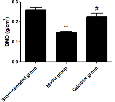
BMD values in metaphysis of right femur in each group. **; P< 0.01 vs. sham-operated group. #; P< 0.05 vs. model group.
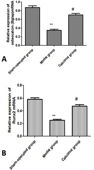
Relative osteocalcin (Bglap) (A) and Runx2 (B) gene expression in each group(n=10). **; P < 0.01 vs. sham-operated group. #; P< 0.05 vs. model group.
References
- Ambroszkiewicz, J., Gajewska, J., Rogowska, E., Szamotulska, K., Chelchowska, M., Rowicka, G. and Rychlowska-Pruszynska, M., 2015. Neoplasma, 62: 288-294. https://doi.org/10.4149/neo_2015_034[CrossRef]
- Aonuma, H., Miyakoshi, N., Hongo, M., Kasukawa, Y. and Shimada, Y., 2009. Tohoku J. exp. Med., 218: 201-205. https://doi.org/10.1620/tjem.218.201[CrossRef]
- Carmona, H., Valadez, H., Yun, Y., Sankar, J., Estala, L. and Gomez, F.A., 2015. Talanta, 132: 676-679. https://doi.org/10.1016/j.talanta.2014.10.025[CrossRef]
- Cui, C., Cui, J., Jin, F., Cui, Y., Li, R., Jiang, X., Tian, Y., Wang, K., Jiang, P. and Gao, J., 2017. Cell Physiol. Biochem., 42: 1888-1896. https://doi.org/10.1159/000479571[CrossRef]
- Del, Pino-Montes, J., Benito, G.E., Fernández-Salazar, M.P., Coveñas, R., Calvo, J.J., Bouillon, R. and Quesada, J.M., 2004. Calcif. Tissue Int., 75: 526-532. https://doi.org/10.1007/s00223-004-0118-9[CrossRef]
- Komori, T., 2017. Adv. Exp. Med. Biol., 962: 83-93. https://doi.org/10.1007/978-981-10-3233-2_6[CrossRef]
- Kuźniewski, M., Fedak, D., Dumnicka, P., Kapusta, M., Stępień, E., Chowaniec, E., Krzanowska, K., Krzanowski, M., Chmiel, G., Solnica, B. and Sułowicz, W., 2016. Ren. Fail., 38: 451-457. https://doi.org/10.3109/0886022X.2016.1138830[CrossRef]
- Lev, D., Binson, I., Foldes, A.J., Watemberg, N. and Lerman-Sagie, T., 2003. Isr. Med. Assoc. J., 5: 419-421.
- Lyngstad-Brechan, M.A., Taubøll, E., Nakken, K.O., Gjerstad, L., Godang, K., Jemtland, R. and Bollerslev, J., 2008. Scand. J. Clin. Lab. Invest., 68: 759-766. https://doi.org/10.1080/00365510802233442[CrossRef]
- Ogata, H., Koiwa, F., Shishido, K., Takahashi, K., Ito, H., Kinugasa, E. and Taguchi, S., 2007. Ther. Apher. Dial., 11: 202-209. https://doi.org/10.1111/j.1744-9987.2007.00422.x[CrossRef]
- Prahasanti, C., Ulfah, N., Kusuma, I.I., Hayati, N., Ernawati, D.S., Krismariono, A. and Bramantoro, T., 2018. Contemp. Clin. Dent., 9: 574-576. https://doi.org/10.4103/ccd.ccd_609_18[CrossRef]
- Silva, T.S., Primo, B.T., Silva, Júnior, A.N., Machado, D.C., Viezzer, C. and Santos, L.A., 2011. Acta Cir. Bras., 26: 7-11. https://doi.org/10.1590/S0102-86502011000100003[CrossRef]
Cited by 1 article
In journals published by ResearchersLinks.
Services
Article usage statistics
Loading…