Research Article · Volume 14, Issue 4, 8 September 2026 · Open Access
Unveiling The Protective Effects of Sage Extract on Bone SEM, EDX Analysis and Associated Biomarkers in Osteoporotic Rats
·
Affiliations & Notes ⌄
1Department of Physiology, Biochemistry and Pharmacology, College of Veterinary Medicine, University of Baghdad, Baghdad, Iraq. *Correspondence | Huda Abdul-Zahra Naji, Department of Physiology, Biochemistry and Pharmacology, College of Veterinary Medicine, University of Baghdad, Baghdad, Iraq
Osteoporosis is a metabolic bone disease characterized by alterations in bone microarchitecture and mineral composition, and it is commonly managed by addressing the underlying causes of calcium deficiency. The present study was designed to investigate the role of alcoholic extract of Salvia officinalis (sage) extract in mitigating the detrimental effects of osteoporosis induced by ovariectomy and retinoic acid on bone-related biochemical markers in adult female rats. A total of forty adult female rats were randomly allocated into four equal experimental groups. One group served as the negative control group (-ve) untreated to laparotomy without removing ovaries, while the remaining thirty animals underwent bilateral ovariectomy (OVX) to establish an experimental model of osteoporosis. Among the ovariectomized groups, one group of ovariectomized (OVX) rats served as the positive control, the second group received retinoic acid (OVX+RA) for thirty days to exacerbate bone loss, and the third group was treated with retinoic acid for thirty days followed by administration of crude sage extract for forty days. At the end of the experimental period, serum samples were collected to assess osteocalcin and osteopontin, while femoral bone specimens were obtained to evaluate energy-dispersive X-ray analysis (EDX) and scanning electron microscopy (SEM). The findings demonstrated significant alterations in serum bone-related biomarkers, osteocalcin and osteopontin, in the OVX group, along with pronounced deterioration of trabecular bone architecture. Administration of retinoic acid to ovariectomized rats (OVX+RA) show of severe impairment of hydroxyapatite stability, reduced mineral deposition. In contrast, treatment with crude Salvia officinalis extract markedly improved bone microarchitecture and mineral integrity in ovariectomized rats exposed to retinoic acid as evidenced by partial restoration of trabecular thickness, reduced porosity, enhanced collagen fiber density, and improved trabecular surface smoothness. These ultrastructural improvements were closely associated with normalization of osteocalcin and osteopontin levels and enhanced mineral composition, indicating improved hydroxyapatite deposition and stabilization. In conclusion, the present study indicates that crude sage extract has protective and restorative effects against osteoporosis , suggesting that sage extract may represent a promising natural therapeutic approach for the prevention or adjunctive treatment of osteoporosis.
Keywords | Retinoic acid, Osteoporosis, Sage extract, Bone biomarkers, Bone EDX and SEM
Introduction
Osteoporosis, a systemic metabolic bone disorder characterized by decreased bone mineral density (BMD) and microarchitectural deterioration of bone tissue, poses a significant and increasing global health burden, with projections indicating a rise in related fractures as populations age . This condition leads to increased bone fragility, resulting in substantial morbidity, mortality, and healthcare costs for humans and animal (Tarantino et al ., 2022: . Although pharmacological treatments, compounds, and biologic agents are available, their long-term use is often limited by high costs and potential adverse effects : Kendler et al., 2023). This situation has generated considerable interest in exploring safe, effective, and accessible alternative therapies, with medicinal plants offering a promising source of bioactive compounds for preventive and adjunctive management.
Salvia officinalis (sage), a medicinal plant belonging to the Lamiaceae family, has been shown through phytochemical studies to be rich in phenolic acids (such as rosmarinic acid), flavonoids, diterpenes, and phytoestrogen-like compounds. Modern science has confirmed its strong antioxidant and anti-inflammatory properties, mainly due to its high content of phenolic compounds like rosmarinic acid . These bioactive components are known to affect bone metabolism through various mechanisms, including the modulation of oxidative stress pathways, regulation of inflammatory cytokines, and activation of osteogenic signaling cascades . It has also been traditionally used for multiple therapeutic purposes, such as hormonal regulation, inflammation control, and reducing oxidative stress . In addition, sage has protective effects on bone mass .
Oxidative stress plays a crucial role in the development of osteoporosis, especially in models induced by ovariectomy and retinoic acid , where excessive reactive oxygen species (ROS) impair osteoblast differentiation and increase osteoclast activity (Tobeiha et al., 2022; . Antioxidant-rich plant extracts, such as sage, others have been shown to restore redox balance by boosting total antioxidant capacity and reducing lipid peroxidation (LPO), thereby promoting bone formation and inhibiting bone resorption : ; . Furthermore, the phytoestrogenic compounds in Salvia officinalis may partly offset estrogen deficiency, aiding in improving bone biomarkers and normalizing markers of bone remodeling . However, a comprehensive mechanistic understanding of sage’s anti-osteoporotic effects remains lacking. A significant gap exists in directly linking the phytochemical profile of sage extracts with measurable, multi-level improvements in bone quality, including microstructural and elemental composition ; . Consequently, this study aims to investigate the anti-osteoporotic effects of Salvia officinalis using an integrated bioanalytical and bone examination approach.
Materials and Methods
Animal use statement
The experimental design adhered to the ethical guideline established by the institutional animal care abd use committee at the College of Veterinary Medicine, University of Baghdad, and received animal use protocol approval obtained (P.G. 551, date 10-3-2024) which complies with the international standards for the care and use of laboratory animals.
Experimental design
Forty adults female Wistar rats (10–12 weeks old) were been randomly divided into four equal experimental groups: Control negative: C- ve received dimethyl sulfoxide (DMSO), Control positive bilateral ovariectomized rats (OVX) and OVX+RA (Retinoic acid 75mg/kg B.W for 30 days, respectively) and the fourth group subjected to OVX+RA at the same dose ,and after that received sage extract for 40 days.
Preparation of alcoholic extraction of Salvia official’s leaves
The extract was prepared using 500 mL of 70% ethanol in a Soxhlet apparatus, according to the method described by Sultana et al. (2012). The extract was then administered orally for 40 days.
Ovariectomy procedure
Thirty female rats were bilaterally ovariectomized using general anesthesia (with ketamine 80mg/kg and xylazine 10 mg/kg), using aseptic surgical techniques, followed by a 7 days convalescence period allows the animal to recover ; .
Blood collection
At the end of the experimental periods (at 30 and 70 days), blood samples were drawn by cardiac puncture from anesthetized rats using intramuscular injections of Ketamine (100mg/kg B.W) and Xylazine (10mg/kg B.W) Then, blood samples were kept in non-heparinized gel tubes, and serum was isolated as described by Veilleux-Lemieux et al. (2013), for further analysis of rat osteocalcin (OCN) using the ELISA Kit instruction (NSL1304r) and rat osteopontin (OPN) kit instruction (NSL1307r).
Bone specimens’ collection
Femur Bone specimens were prepared for SEM examination and energy dispersive X-ray to assess. Samples were cleaned, fixed in 2.5% glutaraldehyde (0.1 M phosphate buffer, 4°C, 24 h), rinsed with PBS, dehydrated through graded ethanol, and dried using a critical point dryer. Specimens were gold-coated and examined under SEM (e.g., JEOL JSM-6360) to evaluate surface morphology, trabecular structure, and porosity ; .
Energy dispersive X-ray (EDX)
The image of SEM of femur bone of rats was used to conduct EDX analysis of (Ca, Mg, Si, Fe, N, C, P and Ca/P) .
Statistical analysis
The descriptive statistics analysis was expressed as mean ± SE. Data subjected to analysis using SPSS (Version 22) Two-Way Analysis of variance (ANOVA) and the Least Significant Differences (LSD) tests were used to assess significant mean differences at P< 0.05.
Results and Discussion
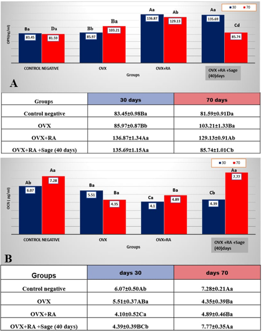
The mean values of serum osteopontin concentrations in the four experimental groups are illustrated (Figure 1A). A significant (p<0.05) increase in serum osteopontin concentration in OVX rats after 70 days, and in the OVX+RA group after 30 and 70 days, compared to the control group, was observed. Whereas OVX rats received RA for 30 days and were administered sage extract for 40 days, revealed a significant (P<0.05) decrease in OPN value (85.74±1.01) compared to OVX and OVX+RA groups, with mean values of (103.21±1.33) and (129.13±0.91), respectively. After 40 days of treatment with sage extract, osteopontin level became significantly lower than in the control group, documenting the effectiveness of S. officinalis in overwhelming the adverse effects of osteoporosis.
After 30 days and 70 days of the experiment, the results showed a significant decrease (P<0.05) in serum osteocalcin concentration in OVX and OVX+RA after 30days and 70 days compared with the control group, as shown in Figure 1B. Whereas rats administered Salvia officinalis extract after 40 days of OVX+RA (30 days) showed a significant (P<0.05) increase in serum OCN concentration compared with OVX and OVX+RA treatment groups. A non-significant difference in OCN concentrations between OVX+RA+sage extract and the control group was observed at the end of the experiment. The values were 7.77±0.35 and 7:28±0.21, respectively. Along with time, ovariectomized rats that received sage extract for 40 days showed a significant (P<0.05) increase in this parameter compared with 30 days of the experiment
Current results, showed that serum OPN, serum osteopontin (OPN) levels were significantly elevated in response to estrogen deficiency and retinoic acid–induced bone loss and then normalized by Salvia officinalis treatment.
These results were in line with Chang et al. (2010) and Morgan et al. (2021). The increase in OPN in the OVX and OVX+ retinoic acid groups likely reflect intensified bone catabolic remodeling and resorption. OPN is a matricellular protein that plays a crucial role in osteoclast adhesion and activation, thereby promoting bone resorption. Estrogen deficiency via ovariectomy has been shown to up-regulate OPN expression in osteocytes and bone-resorbing surfaces, which may be due to increased inflammatory cytokines and enhanced RANKL signaling in the absence of estrogen ; ; .
The reduction of OPN following sage treatment suggests that Salvia officinalis extract may suppress osteoclast activation and modulate bone remodeling (Salevic et al., 2022; . The rich phytochemical profile of sage especially its flavonoids and phenolic compounds could downregulate signaling pathways of OPN expression (Salevic et al., 2022; . Besides, phytoestrogenic activity of its constituents likely restores some estrogen-like regulation over bone cells, reducing the overexpression of OPN. This aligns with previous reports ; ; . Overall, current data support the idea that estrogen deficiency and retinoic acid promote bone resorption via up-regulation of OPN and down regulate OCL, while Salvia officinalis supplementation mitigates this effect by modulating the levels of bone biomarkers. Collectively, the OCN results corroborate our earlier biochemical and antioxidant findings estrogen deficiency and retinoic acid suppress osteoblast function and bone formation, whereas Salvia officinalis supplementation restores osteocalcin levels and promotes bone health through phytoestrogenic and antioxidant mechanisms. This supports the potential therapeutic use of sage in mitigating postmenopausal osteoporosis and oxidative bone deterioration. Significant change in serum OCN levels in ovariectomized rats and those that received retinoic acid compared to the control group, reflecting impaired osteoblastic activity and reduced bone formation due to estrogen deficiency. The administration of retinoic acid for 30 days further suppressed OCN levels, indicating a pronounced inhibition of bone matrix protein synthesis and accelerated bone resorption. Mechanistically, this reduction can be attributed to the combined effects of estrogen withdrawal and retinoic acid–induced oxidative stress, which impairs osteoblast differentiation, downregulates OCN gene expression, and enhances osteoclast-mediated bone turnover ; ; a).
Conversely, treatment with Salvia officinalis for 40 days after retinoic acid exposure restored OCN levels, slightly higher than the control value. This increase highlights the osteogenic potential of the sage extract This result was agreement with ; . The mechanism likely involves the bioactive phytochemicals identified in our GC-MS analysis, particularly quercetin derivatives, 3-O-glycosides, and other phenolic compounds, which exhibit phytoestrogenic activity. These compounds can bind to estrogen receptors, stimulate osteoblast differentiation, and upregulate OCN expression, while simultaneously scavenging reactive oxygen species to protect osteoblasts from oxidative damage ; Ghorbel -Koubaa et al., 2026). These findings align with recent studies ; . Current results confirm the dual osteogenic and antioxidant roles of these bioactive compounds ; .
Energy dispersive X-ray (EDX)
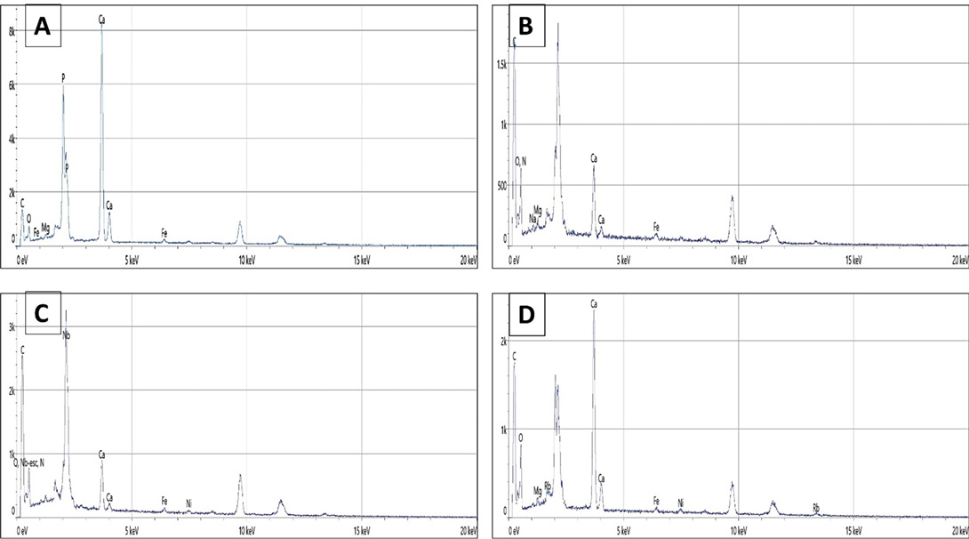
Composition across EDX spectroscopy revealed distinct alterations in bone mineral stoichiometry and organic/mineral balance among the four experimental groups, providing insights into hydroxyapatite (HA) crystal integrity and matrix remodeling under estrogen deficiency and therapeutic interventions.
Comparative mineral composition (at%) and (Wt%)
The results of atomic element percentage demonstrate general decline in both the OVX and OVX+RA groups for calcium, phosphorus, calcium/phosphorus ratio, and nitrogen compared to the control group (Tables 1 and 2). Carbon content was an exception to this trend. In contrast, a marked improvement was observed in the OVX+RA+sage extract group, which was treated with sage extract. This group showed recovery in levels of calcium, phosphorus, calcium/phosphorus ratio, and nitrogen, alongside an increase in carbon content. The results of weight % element show a decrease in the OVX and OVX+RA groups for calcium, magnesium, and silicon compared to the control group, except for iron, which increases. Meanwhile, a noticeable improvement is observed in the OVX+RA+sage extract group. The group treated with sage extract for calcium, magnesium, silicon, and nitrogen, with a decrease in iron as shown in Tables 1, 2 and Figure 2A, B, C, D).
Table 1: Atomic percentage of elements.
Groups/ Atomic % element
Control
OVX
OVX+RA
OVX +RA+ sage extract (40) days
Ca
9.7
5.7
1.9
5.8
P
4.1
2.1
0.6
2.8
Ca/P
2.37
2.71
3.17
2.07
N
13.9
12.6
19.1
8.7
C
25.9
53.3
49.3
31.7
Table 2: Weight percentage of elements.
Groups/ weight % element
Control
OVX
OVX+RA
OVX+RA+ sage extract (40) days
Ca
26.6
14.4
5.1
14.6
Mg
2.7
1.2
0.4
1.2
Si
1.5
1.1
0.6
0.0
Fe
0.4
0.5
0.6
0.2
Control (-ve negative): subjected to laparotomy without removal of ovaries. OVX (+ve control) : subjected to bilateral ovariectomy only. OVX +RA: bilateral ovariectomy and then given retinoic acid (75 mg/kg) for 30 days. OVX +RA + Sage extract: bilateral ovariectomy with given retinoic acid (75 mg/kg) for 30 days, after that given Salvia officinalis crude extract for 40 days.
EDX analysis of femoral bone confirmed optimal mineralization in the control group, characterized by high Ca+P content and a Ca/P ratio within the physiological range of non-stoichiometric biological apatite. This composition reflects stable hydroxyapatite with ionic substitutions typical of healthy cortical bone b; . Ovariectomy (OVX) markedly reduced total mineral content and increased carbon levels, indicating a collagen-rich but poorly mineralized matrix, a hallmark of estrogen-deficiency–induced osteopenia. The relatively preserved Ca/P ratio suggests that mineral loss was primarily quantitative rather than a major alteration in apatite chemistry ; . All-trans retinoic acid (RA) severely exacerbated OVX-induced bone loss, producing the lowest Ca+P values and a pathological Ca/P ratio exceeding 3.0. This Ca-rich, phosphate-deficient pattern is consistent with RA-induced enhancement of RANKL-mediated osteoclastogenesis and accelerated mineral resorption ; .
Co-treatment with sage significantly restored mineralization, increasing Ca+P content and normalizing Ca/P stoichiometry toward physiological values. These findings align with evidence that Salvia/Rosmarinus officinalis polyphenols promote osteoblast differentiation while suppressing osteoclast formation in estrogen-deficient models . The concurrent reduction in carbon and nitrogen further suggests more complete hydroxyapatite deposition on the collagen matrix ; . Overall, from a crystal-chemistry perspective, the control and sage-treated groups-maintained Ca/P ratios characteristic of chemically stable, non-stoichiometric biological apatite, supporting the protective role of sage against RA-exacerbated OVX bone loss.
Scanning electron microscope (SEM) examination of femur bone specimens
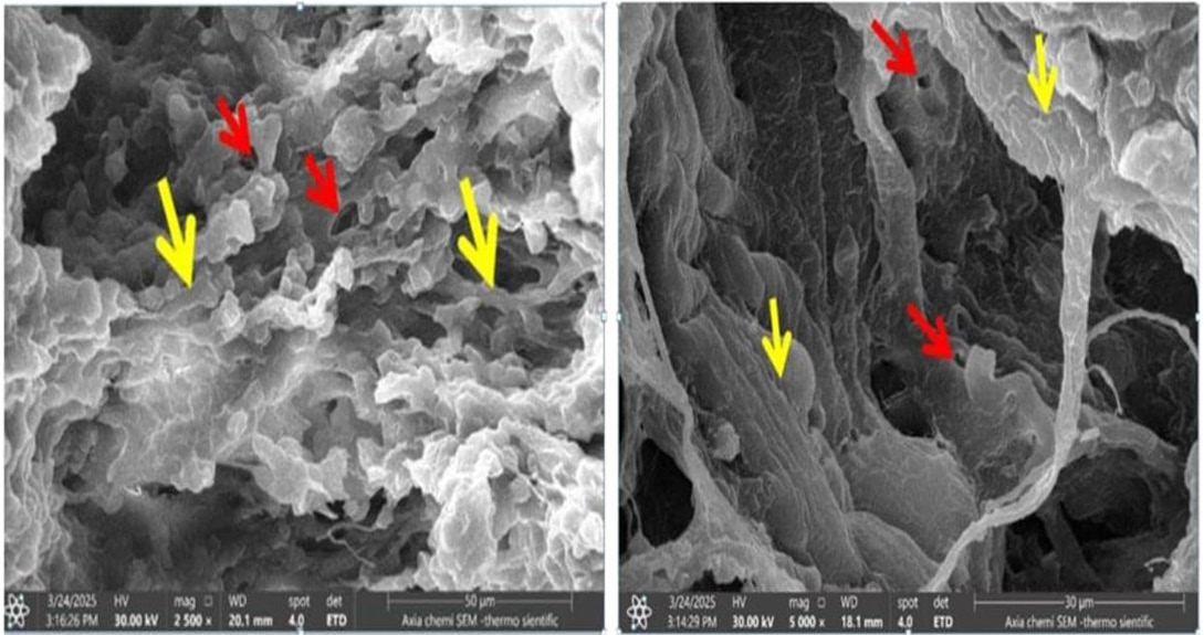
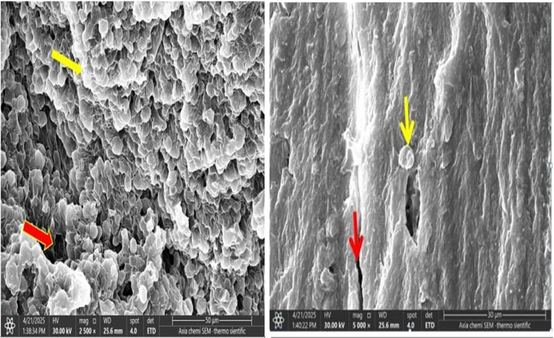
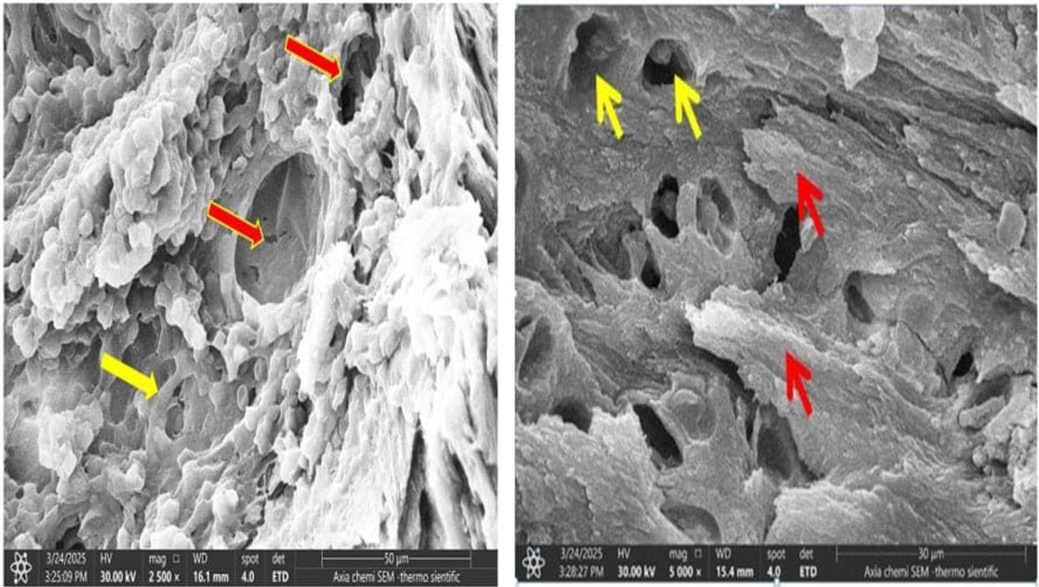
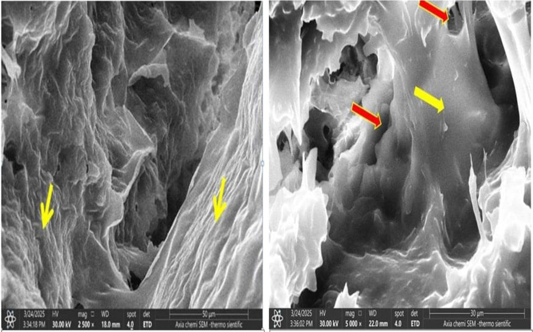
SEM analysis revealed marked microarchitectural differences among the experimental groups. The control group exhibited dense, continuous trabeculae with smooth surfaces and minimal porosity, indicating preserved collagen organization and optimal mineralization (Figure 3A, B) comparing to control. Ovariectomy caused pronounced trabecular thinning, fractures, enlarged pores, and loss of connectivity, consistent with estrogen-deficiency–induced osteoporosis (Figure 4A, B). These alterations were further aggravated by retinoic acid, which produced extensive trabecular perforation, widened marrow spaces, reduced collagen fibers, and severe cracking (Figure 5A, B). Meanwhile, sage extract treatment partially restored trabecular architecture, as evidenced by increased collagen density, reduced porosity, and improved trabecular thickness (Figure 6A, B).
SEM analysis demonstrated that the control group possessed dense, continuous trabeculae with smooth surfaces, minimal porosity, and well-organized collagen fibers, reflecting mechanically stable bone in which collagen provides an optimal scaffold for uniform hydroxyapatite (HA) deposition and balanced bone remodeling ; . In contrast, the OVX group exhibited severe trabecular deterioration, associated with estrogen-deficiency–induced osteoporosis. Estrogen loss enhances osteoclast-mediated resorption and disrupts collagen–HA interactions, resulting in mineral dissolution, micro-crack formation, and reduced bone strength ; ; . The OVX + retinoic acid group showed the most severe damage, with extensive perforations, cracks, markedly thinned trabeculae, and reduced collagen fibers, reflecting RA-induced stimulation of osteoclastogenesis, inhibition of osteoblast function, impaired collagen synthesis, and defective HA crystal formation ; . Conversely, treatment with sage extract partially restored trabecular architecture. These effects are attributed to the antioxidant, anti-resorptive, and phytoestrogen-like properties of sage polyphenols, which enhance osteoblast activity, suppress osteoclasts, and promote stable HA integrity (Ibrahim and aheed, 2022; El-Fiqi et al., 2020; Miaoa et al., 2024).
CONCLUSION
Collectively, the study findings achieved by EDX and SEM scanning could conclude that the deleterious changes in the microarchitectures of bone due to excessive exposure to the retinoic acid in estrogen-lacked rats could be reversed by administration of sage extract. These findings contribute to a deeper understanding of the interaction between estrogen deficiency and natural antioxidants in bone remodeling and may serve as a foundation for future translational and clinical researches.
ACKNOWLEDGMEMT
Not applicable.
NOVELTY STATEMENT
The current study presents deep insight and anew strategy to avoid the effects of osteoporosis and to ameliorate the hormonal profile, bone-related biomarkers and bone microarchitectures by using sage extract, which has an estrogenic activity.
AUTHOR’S CONTRIBUTION
BA and HN designed, conceptualization, reviewed the draft, and the last version of this manuscript. HN did all the experiments and wrote the draft, revised and wrote the last version of this manuscript.HN, and BA have reviewed and approved the submitted version of the manuscript.
Data availability
The data is available from the corresponding author upon reasonable request.
Ethical approval
In full compliance with the ethical guidelines for animal research, all animals used in this study were cared for and treated at the College of Veterinary Medicine, University of Baghdad. The ethical permission was obtained prior to experimentations (AUP# P.G./551, dated on 10-3-2024).
Generative AI and AI assisted technology statement
The authors declare that no generative AI and AI assisted technology was used in the creation of this manuscript.
Conflict of interest
The authors have declared no conflict of interest.
Figures
Figure 1:.
A: Shows the serum osteopontin concentration of four experimental groups in adult female rats. Values expressed as mean±SE. n=10 female rats. Control (-ve): subjected to laparotomy without removal of ovaries. OVX (+ve control): subjected to bilateral ovariectomy only. OVX +RA: bilateral ovariectomy and then given retinoic acid (75 mg/kg) for 30 days. OVX+RA + Sage extract: bilateral ovariectomy with given retinoic acid (75 mg/kg) for 30 days, after that given Salvia officinalis crude extract for 40 days. B: Shows the serum OCN concentration of four experimental groups in adult female rats. Values expressed as mean±SE. n=10 female rats. Control (-ve): subjected to laparotomy without removal of ovaries. OVX (+ve control): subjected to bilateral ovariectomy only. OVX +RA: bilateral ovariectomy and then given retinoic acid (75 mg/kg) for 30 days. OVX +RA + Sage extract: bilateral ovariectomy with given retinoic acid (75 mg/kg) for 30 days, after that given Salvia officinalis crude extract for 40 days.
Figure 2:.
EDX analysis of the femur bone in female rats. (A) Negative control; (B) Ovariectomized (+ve control); (C) Ovariectomized + retinoic acid (OVX + RA); and (D) Ovariectomized + retinoic acid + sage extract (OVX + RA + Sage).
Figure 3:.
A: Photography of Bone (control negative) show density of collagen fibers (yellow arrows) with small pores intermingled through it (red arrow) 2500X. B: Photography of bone (control negative group) show the strong, tightly packed frameworks (yellow arrows) and low porosity of the bone’s architecture (red arrows). 5000X.
Figure 4:.
A: Photography of bone (control positive group) reveal that the marrow spaces are noticeably enlarged, and the pores appear larger and more widely spaced than in the control negative group (red arrow). Occasionally surfaces with rough texture and a large number of resorption lacunae (yellow arrows). 2500X. B: Photography of bone (control positive group) show the osteocytes (yellow arrow) and the trabeculae appeared roughly and fractured (red arrows), and widely separated. 5000X.
Figure 5:.
A: Photographs of the bone (group 3) show that the large pores with visible cracked in trabeculae (red arrow) and thin with small amount of collagen fibers (yellow arrow) 2500X. B: Photographs of the bone (group 3) show There were many absorbent lacunae, expanded pores, and tiny cracks on the surface (yellow arrows). Trabeculae looked disorganized in a number of places (red arrows).5000X.
Figure 6:.
A: Photographs of the bone (group 4) show that the additionally, lesser decay pits, a reduced porosity, and less friction outermost portion of the bone (yellow arrows) 2500X. B: Photographs of the bone (group 4) show that the thicken of trabeculae and smoothly of surface near to control negative group (yellow arrow), and decrease in numbers and diameter of pores (red arrows).5000X.
References
Ahmed HS (2017). Correlations between serum interleukins-2,-4 levels and some biochemical parameters in Iraqi patients with osteoporosis. J. Fac. Med. Baghdad, 59(3): 275-279.
Ajaghaku DL, Chinwuba P, Ajaghaku AA (2025). Neuroprotective and cognitive-enhancing effects of a phytoestrogen rich fraction from Ochna schweinfurthiana bark in a rat model of menopause and cholinergic dysfunction. Asian J. Res. Med. Pharma. Sci., 14(3): 310-323. https://doi.org/10.9734/ajrimps/2025/v14i3340[CrossRef]
Alabsawy SK, Alghetaa HFK (2025a). Resveratrol administration reverses the osteoporotic bone in independent-manner of Ahr-Esr1 axis in rats. J. Neonatal Surg., 14(15S): 1134-1154. https://doi.org/10.63682/jns.v14i15S.3722[CrossRef]
Alabsawy SK, Alghetaa HFK (2025b). A novel practice for experimental osteoporosis model in rats mediated by combining phosphoric acid administration with ovariectomy. Kufa J. Vet. Med. Sci., 16(1): 26-38. https://doi.org/10.36326/kjvs/2025/v16i118383[CrossRef]
Al-Ajalein AA, Ibrahim NI, Fauzi MB, Mokhtar SA, Naina Mohamed I, Shuid AN, Mohamed N (2024). Evaluating the anti-osteoporotic potential of Mediterranean medicinal plants: A review of current evidence. Pharmaceuticals, 17(10): 1341. https://doi.org/10.3390/ph17101341[CrossRef]
Alghetaa H, Mohammed A, Singh NP, Bloomquist RF, Chatzistamou I, Nagarkatti M, Nagarkatti P (2023). Estrobolome dysregulation is associated with altered immunometabolism in a mouse model of endometriosis. Front. Endocrinol., 14: 1261781. https://doi.org/10.3389/fendo.2023.1261781[CrossRef]
Al-Haideri DH, Al-Timmemi HA (2023). Differentiated stem cells derived from rabbit adipose tissue exhibited in vitro adipogenesis and osteogenesis. Iraqi J. Vet. Med., 47(2): 59-63. https://doi.org/10.30539/ijvm.v47i2.1509[CrossRef]
Al-Okaily BN (2024). Unveiling the mysteries of oxidative stress: An insightful review of recent studies. J. Anim. Health Prod., 1(2): 395-412. https://doi.org/10.17582/journal.jahp/2024/12.3.395.412[Full text][CrossRef]
Alqahtani HM, Haq I, Almofarej N (2025). Mapping the trends of peer-reviewed research on osseodensification. Saudi Dent. J., 37(1): 5. https://doi.org/10.1007/s44445-025-00004-3[CrossRef]
Burguera B, Hofbauer LC, Thomas T, Gori F, Evans GL, Khosla S, Turner RT (2001). Leptin reduces ovariectomy-induced bone loss in rats. Endocrinol., 142(8): 3546-3553. https://doi.org/10.1210/endo.142.8.8346[CrossRef]
Castagna A, Pujia C, Mazza E, Maurotti S, Ferro Y, Rizzo V, et al. (2026). Underlying mechanisms of osteoporosis in the context of multimorbidity: Clinical challenges and management strategies. Nutrients, 18(2): 262. https://doi.org/10.3390/nu18020262[CrossRef]
Chang IC, Chiang TI, Yeh KT, Lee H, Cheng YW (2010). Increased serum osteopontin is a risk factor for osteoporosis in menopausal women. Osteoporosis Int., 21(8): 1401-1409. https://doi.org/10.1007/s00198-009-1107-7[CrossRef]
Chetehouna M (2019). Investigating the stress and strain fields in porous synthetic bone graft substitute materials with varied porosity levels. Doctoral dissertation, Queen Mary University of London. https://qmro.qmul.ac.uk/xmlui/handle/123456789/64267
El-Fiqi A, Kim JH, Kim HW (2020). Novel bone-mimetic nanohydroxyapatite/collagen porous scaffolds biomimetically mineralized from surface silanized mesoporous nanobioglass/collagen hybrid scaffold: Physicochemical, mechanical and in vivo evaluations. Mater. Sci. Eng. C, 110: 110660. https://doi.org/10.1016/j.msec.2020.110660[CrossRef]
Elghareeb MM, Elshopakey GE, Elkhooly TA, Salama B, Samy A, Bazer FW, Rezk S (2022). Estradiol and zinc-doped nano hydroxyapatite as therapeutic agents in the prevention of osteoporosis; oxidative stress status, inflammation, bone turnover, bone mineral density, and histological alterations in ovariectomized rats. Front. Physiol., 13: 989487. https://doi.org/10.3389/fphys.2022.989487[CrossRef]
El-Habibi MF (2023). Automated detection of cardiovascular diseases using deep learning and electrocardiogram (ECG) images: A convolutional neural network approach. Complexity, 7(8): 10-20. https://www.researchgate.net/publication/374227588.
Elkomy MM, Elsaid FG (2015). Anti-osteoporotic effect of medical herbs and calcium supplementation on ovariectomized rats. J. Basic Appl. Zool., 72: 81-88. https://doi.org/10.1016/j.jobaz.2015.04.007[CrossRef]
Erhardt W, Hebestedt A, Aschenbrenner G, Pichotka B, Blümel G (1984). A comparative study with various anesthetics in mice (pentobarbitone, ketamine-xylazine, carfentanyl-etomidate). Res. Exper. Med., 184(3): 159-169. https://doi.org/10.1007/BF01852390[CrossRef]
Ghorbel-Koubaa F, Jdidi H, Chaâbane M, Aoiadni N, El-Feki A (2026). Medicago sativa alleviates behavioral impairments and oxidative stress in the cerebrum and cerebellum of ovariectomized mice. Reprod. Fert. Dev., 38(2): RD25113. https://doi.org/10.1071/RD25113[CrossRef]
Gourion-Arsiquaud S, Faibish D, Boskey AL (2022). Early bone turnover changes following estrogen depletion: Implications for osteocalcin expression. Bone Rep., 16: 101131.
Hamdi RA (2013). Evaluation of serum osteocalcin level in Iraqi postmenopausal women with primary osteoporosis. J. Fac. Med. Baghdad, 55(2): 166-169. https://doi.org/10.32007/jfacmedbagdad.552649[CrossRef]
Hameed ZA, Radhi NJM (2023). The impact of osteoporosis on teeth decay in relation to salivary vitamin D among menopause in Baghdad city, Iraq. Al-Kindy Coll. Med. J., 19(2): 163-167. https://doi.org/10.47723/kcmj.v19i2.911[CrossRef]
Hsu SH, Chen LR, Chen KH (2024). Primary osteoporosis induced by androgen and estrogen deficiency: The molecular and cellular perspective on pathophysiological mechanisms and treatments. Int. J. Mol. Sci., 25(22): 12139. https://doi.org/10.3390/ijms252212139[CrossRef]
Ibrahim ZA, Waheed IN (2022). Histopathological effects of phytoestrogen (Genistein) on thymus gland of adult and post-natal female albino mice. Sci. J. Univ. Zakho, 10(2): 29-34. https://doi.org/10.25271/sjuoz.2022.10.2.889[CrossRef]
Jameel MG, Alhakeem ZM, Al-Osami MH (2022). Prevalence of AGER gene polymorphism in post menopause Iraqi sample with osteoporosis and osteopenia in type 2DM. Iraqi J. Pharma. Sci., 31(2): 202-210. https://doi.org/10.31351/vol31iss2pp202-210[CrossRef]
Jin Y, Du J, Wang Z, Ge S, Ma B (2025). Macrophage-targeted self-assembled nanosystem for periodontitis treatment. J. Dental Res., pp. 00220345251355267. https://doi.org/10.1177/00220345251355267[CrossRef]
Kayalar E, Goger F, Tas Deynek G, Tok OE, Kucuk S (2022). New bone-generative effect of Salvia officinalis L. in the expanded midpalatal suture: An in vivo and in vitro study. J. Orofacial Orthoped. Fortsch. Kieferorthopädie, 83(Suppl 1): 85-95. https://doi.org/10.1007/s00056-021-00366-3[CrossRef]
Kendler KS, Ohlsson H, Sundquist J, Sundquist K (2025). Profiles of genetic risks for psychotic disorders. JAMA Psych., 82(9): 926-933. https://doi.org/10.1001/jamapsychiatry.2025.1289[CrossRef]
Khudiar KK, Hussein GJ (2017). Effect of alcoholic extract of Salvia officinalis leaves on some physiological parameters aspects in acrylamide-treated rats. Adv. Anim. Vet. Sci., 5(1): 47-55. https://doi.org/10.14737/journal.aavs/2017/5.1.47.55[Full text][CrossRef]
Labuschagne L (2020). The use of histological examination methods to distinguish between the burnt remains of human and non-human bones.
Li J, Liu H, Lim J, Xing H, Chen Y, Yang S, Fu X (2025). Molecular and biological markers for assessing endometrial receptivity in infertile women: A narrative review. J. Int. Med. Res., 53(4): 03000605251328893. https://doi.org/10.1177/03000605251328893[CrossRef]
Li T, Chen X, Ma H (2022a). Ovariectomized rat as a model for postmenopausal osteoporosis: A review of methods and outcomes. Exper. Therapeut. Med., 24(3): 251.
Li W, Zhao X, Sun Y (2023). Estrogen deficiency and retinoid exposure synergistically suppress bone formation markers in rodent models. Front. Endocrinol., 14: 1132420. https://doi.org/10.3389/fendo.2023.1256406[CrossRef]
Li X, Zuo X, Lu L, Xu R, Wang Y, Chang S, Li G (2024). Vertebral marrow fat fraction is associated with circulating RANKL in postmenopausal females. Front. Endocrinol., 15: 1442046. https://doi.org/10.3389/fendo.2024.1442046[CrossRef]
Li Y, Li L, Li X, Luo B, Ye Q, Wang H, Wang P (2022). A mechanistic review of Chinese medicine polyphenols on bone formation and resorption. Front. Pharmacol., 13: 1017538. https://doi.org/10.3389/fphar.2022.1017538[CrossRef]
Lin X, Xu F, Zhang KW, Qiu WX, Zhang H, Hao Q, Qian AR (2022b). Acacetin prevents bone loss by disrupting osteoclast formation and promoting type H vessel formation in ovariectomy-induced osteoporosis. Front. Cell Dev. Biol., 10: 796227. https://doi.org/10.3389/fcell.2022.796227[CrossRef]
Micheletti C (2023). Multimodal and multiscale characterization of bone and bone interfaces in health and disease.
Mohanty S, Sahu A, Mukherjee T, Kispotta S, Mal P, Gupta M, Prabhakar PK (2025). Molecular mechanisms and treatment strategies for estrogen deficiency-related and glucocorticoid-induced osteoporosis: A comprehensive review. Inflammopharmacology, 33(5): 2409-2445. https://doi.org/10.1007/s10787-025-01749-3[CrossRef]
Morgan EN, Alsharidah AS, Mousa AM, Edrees HM (2021). Irisin has a protective role against osteoporosis in ovariectomized rats. BioMed. Res. Int., 2021(1): 5570229. https://doi.org/10.1155/2021/5570229[CrossRef]
Nguyen KD (2022). Effects of nacre supplementation on bone loss (induced or natural) in rodent models. Doctoral dissertation, Université Jean Monnet-Saint-Etienne.
Palanisamy S (2025). The impact of estrogen on periodontal tissue integrity and inflammation: A mini review. Front. Dental Med., 6: 1455755. https://doi.org/10.3389/fdmed.2025.1455755[CrossRef]
Pereira AR, Costa M, Costa GG, Carvalho AS, Costa G (2024). Anorexia nervosa and osteoporosis: A possible complication to remember. Cureus, 16(1): e52670. https://doi.org/10.7759/cureus.52670[CrossRef]
Perrone P, De Rosa C, D’Angelo S (2025). Polyphenols and bone health: A comprehensive review of their role in osteoporosis prevention and treatment. Molecules, 30(21): 4154. https://doi.org/10.3390/molecules30214154[CrossRef]
Radulescu DE, Vasile OR, Andronescu E, Ficai A (2023). Latest research of doped hydroxyapatite for bone tissue engineering. Int. J. Mol. Sci., 24(17): 13157. https://doi.org/10.3390/ijms241713157[CrossRef]
Safar M (2022). Telmisartan: An angiotensin receptor blocker regulates osteoclastogenesis via inhibition of the ERK triggering in osteoporotic male rats.
Salević A, Stojanović D, Lević S, Pantić M, Đorđević V, Pešić R, Nedović V (2022). The structuring of sage (Salvia officinalis L.) extract-incorporating edible zein-based materials with antioxidant and antibacterial functionality by solvent casting versus electrospinning. Foods, 11(3): 390. https://doi.org/10.3390/foods11030390[CrossRef]
Shamsulddin HH, Salih LA, Eleiwe SA (2020). Relationship between osteopontin biochemical parameters and BMD status in Iraqi postmenopausal women with osteoporosis. Iraqi J. Sci., 2494-2503. https://doi.org/10.24996/ijs.2020.61.10.6[CrossRef]
Silva JR, Balbas MCDM, Corrêa CÁ, Zanela M, Okamoto R, Pereira RDS, Hochuli-Vieira E (2022). The role of bone grafts in preventing medication-related osteonecrosis of the jaw: Histomorphometric, immunohistochemical, and clinical evaluation in animal model. Craniomax. Trauma Reconst., 15(4): 304-311. https://doi.org/10.1177/19433875211048367[CrossRef]
Su Z, Yao B, Liu G, Fang J (2024). Polyphenols as potential preventers of osteoporosis: A comprehensive review on antioxidant and anti-inflammatory effects, molecular mechanisms, and signal pathways in bone metabolism. J. Nutr. Biochem., 123: 109488. https://doi.org/10.1016/j.jnutbio.2023.109488[CrossRef]
Subhi DAA, Al-Okaily BN (2024). Effectiveness of resveratrol supplement on some biochemical criteria and bone in lead-exposed rats. Iraqi J. Vet. Sci. https://doi.org/10.47278/journal.ijvs/2024.126[CrossRef]
Sultana S, Talegaonkar S, Ali R, Mittal G, Ahmad FJ, Bhatnagar A (2012). Inhalation of alendronate nanoparticles as dry powder inhaler for the treatment of osteoporosis. J. Microencapsul., 29(5): 445–454. https://doi.org/10.3109/02652048.2012.655428[CrossRef]
Svydenko L, Vergun O, Ivanišová E, Korablova O, Šramková KF (2022). Polyphenol compounds and antioxidant activity of Salvia officinalis L. and Salvia sclarea L. Agrobiodiv. Improv. Nutr. Health Life Qual., 6(2). https://doi.org/10.15414/ainhlq.2022.0015[CrossRef]
Tambuzzi S, Gentile G, Cattaneo C (2022). How to perform SEM/EDX analysis on bone. Procedural aspects and main anthropological applications. Anthropol. Ethnol. Open Access J., 5(2): 1-5. https://doi.org/10.23880/AEOAJ-16000178[CrossRef]
Tarantino P, Jin Q, Tayob N, Jeselsohn RM, Schnitt SJ, Vincuilla J, Tolaney SM (2022). Prognostic and biologic significance of ERBB2-low expression in early-stage breast cancer. JAMA Oncol., 8(8): 1177-1183. https://doi.org/10.1001/jamaoncol.2022.2286[CrossRef]
Tobeiha M, Amin N, Shahaboddin ME, Tabatabaizadeh M, Jafarnejad S (2025). The effect of oral consumption of sesame oil on anthropometric, metabolic and oxidative stress markers of patients with type 2 diabetes: A double-blind, randomized controlled trial. Int. J. Diabetes Dev. Count., 45(2): 389-399. https://doi.org/10.1007/s13410-024-01350-4[CrossRef]
Turki KM, Al-Osami MH, Mohammed NS (2013). Evaluation of serum osteocalcin and osteopontin levels as bone biochemical markers in postmenopausal women. Al-Kindy Coll. Med. J., 9(2): 40-44. https://jkmc.uobaghdad.edu.iq/index.php/MEDICAL/article/view/526
Veilleux-Lemieux D, Castel A, Carrier D, Beaudry F (2013). Stability of blood biomarkers in rats: Comparison of serum and plasma. J. Pharmacol. Toxicol. Methods, 68(3): 424-429.
Wang X, Liang T, Zhu Y, Qiu J, Qiu X, Lian C, Huang D (2019). Melatonin prevents bone destruction in mice with retinoic acid-induced osteoporosis. Mol. Med., 25(1): 43. https://doi.org/10.1186/s10020-019-0107-0[CrossRef]
Wei L, Yue Q, Yang D, Miao S, Lu M, Zhang Z, Zhang Y (2026). The retinoic acid-related orphan receptors (RORs) signaling axis in skeletal homeostasis: Mechanistic insights and chronotherapeutic prospects. Ann. Med., 58(1): 2615534. https://doi.org/10.1080/07853890.2026.2615534[CrossRef]
Ye H, Wang K, Li K (2023). Anti-osteoporosis effect of cynaropicrin against ovariectomy-induced osteoporosis in rats. Pharmacogn. Magaz., 19(2): 448-460. https://doi.org/10.1177/09731296221137415[CrossRef]
Zhang G, Jia X, Li Z, Wang Q, Gu H, Liu Y, Mao H (2024). Comprehensively characterizing heterogeneous and transversely isotropic properties of femur cortical bones. J. Mech. Behav. Biomed. Mater., 151: 106387. https://doi.org/10.1016/j.jmbbm.2024.106387[CrossRef]
Zhao L, Feng X (2021). Retinoids and estrogen deficiency activate RANKL/NF-κB pathway to increase osteopontin expression. Bone Res., 9: 38.
Zhao Q, Li Y, Zhang J, Lei X, Tang J, Chen J, Li Y (2025). Bienzyme-engineered fibrous membranes: A mitochondrial-targeted strategy to reverse bone loss in osteoporotic models. Adv. Fiber Mater., 7(6): 1803-1829. https://doi.org/10.1007/s42765-025-00580-3[CrossRef]
Zych M, Wolan R, Włodarczyk A, Londzin P, Borymska W, Kaczmarczyk-Żebrowska I, Folwarczna J (2026). Effects of rosmarinic acid and sinapic acid on the skeletal system in ovariectomized rats. Nutrients, 18(2): 301. https://doi.org/10.3390/nu18020301[CrossRef]