
Amid the numerous health concerns faced by the world, osteoporosis a silent epidemic is a major health concern, since it affects millions of people all around the world. Recognizing the limited treatment options for osteoporotic patients, along with their long-term use safety concerns, our research group focused on investigating efficacious, inexpensive and safe alternative therapies for osteoporosis. Investigating the osteogenic potential of CQ, our research group previously reported osteogenic stimulatory effects of CQ (hexane fraction). The present study explores anti-resorptive effects of CQ-H on bones. We utilized RAW264.7 murine macrophage cell line as osteoclast precursors, and resveratrol (RSV) as a standard for its anti-osteoclast activity. Upon RANK-L induction of RAW264.7 cells to differentiate into osteoclasts, we found 10ng/ml CQ-H as most efficacious concentration for inhibiting osteoclastogenesis with 57.8% inhibition of TRAP activity. Whereas, in comparison 20ng/ml RSV showed 49.9% inhibition of TRAP activity. Expression of osteoclast marker genes such as NFATc1, ACP-5, CTSK, MMP-9 and CTR corroborated our results. Distinct downregulation of marker genes was observed in CQ-H and RSV treated differentiated cells compared to the positive control. Moreover, the downregulation was enhanced in CQ-H treated osteoclasts compared to RSV treatment. This study presents in vitro findings on the osteoclast inhibition activity of CQ and provide evidence of anti-osteoporotic therapeutic property of CQ. Further studies on isolation and functional characterization of active compounds of signaling pathways that mediate osteoclastogenesis will contribute further insights on the anti-osteoporotic therapeutic activity of CQ.
Research Article · Volume 55, Issue 6, 9 October 2023 · Open Access
Cissus quadrangularis (Hexane Fraction) Inhibits RANK-L Induced Osteoclast Differentiation of Murine Macrophage RAW264.7 Cell Line
· · · · ·
Affiliations & Notes ⌄
- 1School of Biological Sciences, University of the Punjab, Quaid-i-Azam Campus, Lahore, Pakistan 2Department of Biochemistry, Kinnaird College for Women University, Jail Road, Lahore, Pakistan 3Department of Biochemistry, University of Vermont College of Medicine, Burlington, Vermont, USA * Corresponding author: arshakoori.sbs@pu.edu.pk, arshaksbs@yahoo.com
Article Info ⌄
- Journal:
- Pakistan Journal of Zoology
- Published:
- 9 October 2023

From the article
- Upon RANK-L induction of RAW264.7 cells to differentiate into osteoclasts, 10ng/ml CQ-H was found to be the most efficacious concentration for inhibiting osteoclastogenesis with 57.8% inhibition of TRAP activity, compared to 20ng/ml of resveratrol (RSV) which showed 49.9% inhibition of TRAP activity.
- Expression of osteoclast marker genes such as NFATc1, ACP-5, CTSK, MMP-9 and CTR was significantly downregulated in CQ-H and RSV treated differentiated cells compared to the positive control.
Selected from the article's own text.
Abstract
INTRODUCTION
Bone remodeling is a continuous, complex, and physiologically coordinated process primarily involving properly functioning osteoblasts cells that form bone and osteoclasts cells that resorb bone in order to maintain structural integrity of the bones, maintain healthy bone mass, and regulate mineral homeostasis ; . Several factors and signaling pathways are involved in the process of bone remodeling, however, disruptions in the process can occur as a result of hormonal fluctuations, imbalance of growth or inflammatory factors, age, cancer, or menopause. Such disruptions in the normal functionality of the bone remodeling process can result in reduced bone mass and weakening of bones, a condition designated Osteoporosis .
Osteoporosis is a major health challenge that affects millions of people worldwide with incidences of fractures at every 3 seconds . Most treatment options for osteoporosis focus on reducing the rate of bone resorption and limited therapies focus on restoring bone formation. Treatment options for osteoporosis include hormone therapies such as SERMs, estrogen replacement, calcitonin, and PTH peptides, RANK-L antibodies, bisphosphonates, Cathepsin K inhibitors and nutritional supplements ; ; Sozen et al., 2017). However, despite the limited effectiveness of these therapies, all have side effects that include vaginal bleeding, breast tenderness, irregular menstruation and cancer ; . Consequently, there is a requirement to investigate alternative treatments for osteoporosis that are both efficacious and have limited side effects.
Cissus quadrangularis (CQ; common name: Haddjod) is a straggling shrub belonging to Vitaceae plant family. Native to hot tropical climate such as Pakistan, India, China, Thailand, Malaya, Java, Bangladesh, Africa, Philippines, and Sri-Lanka, the herb is well known in ayurvedic literature for its pharmacological properties especially healing bone fractures, as implied by the common name ; ; . Studies have reported presence of over eighty bioactive phytochemical compounds such as resveratrol, quercetin, carotenes, tannins, vitamins, minerals, ascorbic acid, steroids, flavonoids and stilbenoids, all of which collectively attribute to medicinal properties of CQ ; ; , 1991; , 2012, 2019; ; ; ; ; ; . Resveratrol (RSV), a polyphenolic compound, and its derivatives have been reported as a predominant compound in CQ ; ; , and has been reported for its osteogenic , 2012; Tseng et al.., 2011; and osteoclast inhibition ; properties. Studies on mice models have also indicated anti-osteoporotic property of RSV ; ; ; ; . Therefore, RSV was used as a standard for osteoclast inhibitory activity in present study.
Evidence of an anti-osteoporotic property for CQ has been reported in numerous in-vivo studies ; , 2010; and clinical studies ; ; , 2013). However, to best of our knowledge, the in-vitro osteoclast inhibition activity of CQ has not been reported. The present study is the first attempt from our research group to investigate the in-vitro osteoclast inhibitor activity of CQ in order to establish its potential therapeutic anti-osteoporotic property. We used the murine macrophage cell line RAW264.7 as pre-osteoclast precursor cells which, upon induction with RANK-ligand, fuses to form large, multinucleated cells that stain positive for tartrate resistant acid phosphatase (TRAP) and express osteoclast marker genes that include NFATc1, cathepsin K, metalloproteinase 9, and calcitonin receptor. Our results indicate that CQ is more efficacious compared to RSV to inhibit osteoclast differentiation, thereby providing evidence for its anti-osteoporotic therapeutic potential.
MATERIALS AND METHODS
Chemicals and reagents
The biological reagents for in vitro experiments utilized in present study were acquired from Thermofisher Scientific (Carlsbad, CQ, USA). The chemical reagents were acquired from Sigma Aldrich (Merck) KGaA, Darmstadt, Germany). Other consumables were acquired from SPL Life Sciences Co. Ltd (Korea) and Nunc (Nunc Cell Culture, Thermofisher Scientific, CA, USA), unless stated otherwise. All experiments were conducted with technical and biological replicates.
Raw264.7 murine macrophage cell culture
The mouse macrophage cell line RAW264.7 (ATCC® TIB-71) is capable of fusing to form TRAP positive multinucleated cells that exhibit osteoclast-like characteristics. The cell line was procured from American Type Culture Collection (ATCC; Manassas, Virginia. USA). The cells were maintained in complete growth medium i.e., alpha MEM medium (without ascorbic acid; Hyclone Laboratories, Inc), 1% penicillin/ streptomycin (cat# 15140122), 2mM L-glutamine (cat# 21051024), and 10% fetal bovine serum (FBS; cat# SH30071.03); under standard culture conditions i.e., 37oC, 5% CO2, humidified conditions. Raw 264.7 cells grew in the form of colonies. Upon 60–70% confluence, the cells were sub-cultured and used for further experimentation. Cells of passage number 6–16 were used for all experiments. For cell viability/ metabolic activity/ proliferation and growth curve analysis, cells were seeded at 3000/cm2 density. For differentiation, 12000/cm2 seeding density was used.
Preparation of standard and CQ-H solutions
The fractionation of ethanolic extract of CQ herb and preparation of CQ-H stock and working solutions was performed as previously described by our research group , 2020). The non-cytotoxic working concentrations of CQ-H fraction (0.1, 1.0, 10 ng/ml) explored for their osteogenic potential in our previous study were further explored for their effects on the growth, viability, and osteoclast differentiation of RAW264.7 cells.
Resveratrol (Sigma-Aldrich cat# R5010) was utilized as a standard for comparison of anti-osteoporotic activity of CQ-H. The stock solution of 40mg/ml was prepared in DMSO, which was further diluted to 4, 0.4, 0.04, 0.004, and 0.0004 mg/ml working stock solutions. The working concentrations of RSV (20, 2, 0.2, 0.02, 0.002, 0.0002 µg/ml) were prepared by adding 0.5µl/ml of the working stock solutions into the complete growth medium. The concentration of DMSO was maintained at 0.05%.
Cell viability, proliferation, and growth kinetics
To examine the effect of CQ-H and RSV on the viability, proliferation, and growth parameters of RAW264.7 cells, the cells were seeded in triplicates at the density of 3000 cells/cm2 in 24-well plates (cat# 142475) in independent experiments. In order to analyze the cell viability, neutral red assay was performed as described by Repetto et al. (2008). The assay is based on the principal that the cells lysosomes uptake the vital dye i.e., neutral red (cat# N4638), the amount of dye up-taken by the cells is directly proportional to the number of viable cells. The control cells were treated with complete growth medium supplemented with 0.05% DMSO, whereas, treated cells were fed with complete medium supplemented with 0.1, 1, and 10 ng/ml CQ-H and 20, 2.0, 0.2, 0.02, 0.002, 0.0002µg/ml RSV. Absorbance was taken using BioTek Elx808 reader at 570nm.
To study the effect of CQ-H and RSV on the growth kinetics i.e., doubling time, specific growth rate of RAW264.7 cells, the cells were treated with CQ-H (0.1, 1.0, 10 ng/ml) and RSV (2, 20, and 200 ng/ml) 24h after seeding. Cells were scrapped and counted on day 1, 3, 5, and 7 of treatment. Control cells were treated with 0.05% DMSO supplemented medium. Medium was changed every 48h. Logarithmic growth curve of cells number against duration of treatment showing lag, log, stationery and decline phase of cells was plotted on Microsoft Excel 2019. Doubling time and specific growth rate were calculated as explained by Butler (2004).
5-Bromo-2-deoxyuridine (BrdU) incorporation assay (BrdU cell proliferation kit Roche Pakistan, cat# 11647229001) and 3-4, 5-dimethylthiazol-2-yl-2,5-diphenyl tetrazolium bromide (MTT; cat# M6494) assay were performed to analyze cell proliferation and metabolic activity, respectively following manufacturer’s instructions. After 24h seeding, the cells were treated with CQ-H (0.1, 1.0, 10 ng/ml) and RSV (2, 20, and 200 ng/ml) up to 72h with media change every 48h. Control cells were treated with complete medium with 0.05% DMSO. The absorbance of cells was read at 450 and 570nm, respectively, using BioTek Elx808 ELISA reader.
Osteoclast differentiation
Raw 264.7 cells were cultured in 12-well cell culture plates at density of 12000/cm2 in complete growth medium. The cells were induced to differentiate into osteoclasts by adding 10ng/ml RANK ligand (cat# R0525) supplemented medium after 24 h. Complete growth medium with 0.05% DMSO was used as control. Positive control cells were fed with 10ng/ml RANK-L and 0.05% DMSO supplemented medium. Induction medium containing non-cytotoxic doses of CQ-H (0.1, 1, 10 ng/ml) and RSV (2, 20, and 200 ng/ml) was fed to the treated cultures for 72h. Medium was changed after 48h. The cells were fixed for TRAP staining and collected for TRAP activity estimation at the end of differentiation. Trizol was added at different time intervals until the end of differentiation for isolation of RNA and quantification of gene expression of osteoclast marker genes.
TRAP staining and activity quantification
Differentiated RAW264.7 cells were fixed in 10% ice cold NBF at 4oC for 20 min. The fixed cells were washed thrice with distilled water. 0.013g naphthol AS MX-PO4 (cat # N4875) dissolved in 100µl N, N-DMF was added to a solution prepared by mixing 0.025g fast red TS salt and 0.025g sodium tartrate dibasic (cat # S4979) dissolved in equal volume of 0.2 M tris HCl (pH 6) and distilled water. The contents were filtered and added to the cells for 45 min at room temperature. Images of stained cells after proper washing were taken using Eclipse TS-100 inverted microscope at 4x and 10x magnification.
For estimation of TRAP activity, the differentiated RAW264.7 cells were lysed using 200µl of 0.1% triton X-100 (cat # T8787). 200µl TRAP solution (1mM ascorbic acid, 0.1 M sodium acetate pH 5.8, 0.15 M KCl, 10mM disodium tartrate and 10mM para-nitorphenyl phosphate) was added to the cell lysate followed by 30 min incubation at room temperature. 0.3M NaOH was used to terminate the reaction. Absorbance was taken at 405nm using BioTek-808 ELISA reader in a microplate. The activity of TRAP was plotted with reference to positive control after normalizing with negative control.
Relative expression of osteoclast marker genes
Trizol was added to the differentiating cells at the time of induction (t=0) and after 24, 48, and 72h of osteoclast induction. Total RNA was extracted from the samples following manufacturer’s instructions. After treatment with DNase I (Ambion, ThermoFisher Scientific – Cat# AM2222) and purification using PureLinkTM RNA mini kit (Cat # 12183018A), RNA was quantified by Pico 200 Microliter Spectrophotometer (Picodrop™ by VWR International, PA, USA). First strand cDNA synthesis kit (Cat # K1622) was used to synthesize cDNA (2.5ng/µl) and qPCR was performed using Maxima SYBR Green/Rox qPCR master mix – 2x – (Cat # K0222) following manufacturer’s instructions and using 500nm of optimized forward and reverse primer sequences (Table I). The qPCR profile was; initial denaturation = 95oC for 3min, 40 cycles each of denaturation at 95oC for 30s, annealing/ extension at 60oC for 30s to amplify cDNA on PikoReal Real-Time PCR system (ThermoFisher Scientific); final extension at 72oC for 5 min. Melt curve was generated at 60–90oC with 0.2oC increment. Hydroxymethylbilane synthase (HMBS) was selected as housekeeping gene and was used to normalize the expression of osteoclast marker genes such as AP5, NFATc1, Cathepsin K (CTSK), Calcitonin receptor (CTR), and MMP-9. Relative expression of osteoclast marker genes was determined by delta-delta Ct method.
Gas chromatography/ Mass spectrometry (GC-MS)
GC-MS analysis was performed to identify the chemical constituents in CQ-H fraction. For sample preparation, 10mg/ml solution of CQ-H was prepared in n-hexane and was filtered through 0.22µm syringe filter (MiniSart-16534-K).
GC-MS analysis was performed using GC (Clarus 500 Perkin Elmer Gas Chromatogram system) consisting of AOC-20i auto-sampler and MS (Clarus 500 Perkin Elmer Mass Spectrometer system) interfaced with silica capillary column (30mm x 0.25mm I.D. x 1µm df, composed of 100% dimethylpolysiloxane). GC-MS was run with inlet temperature of 250oC, oven temperature set at 110oC to 270oC with 4oC ramp, and ion source temperature set at 280oC. Sample (0.5µl) with 10:1 split ratio was injected using micro-syringe with 99.999% helium as carrier gas with flow rate of 1ml/min. Mass spectra were collected using electron impact mode at 70eV, scan interval of 0.5 seconds and fragment size ranging from 45 to 450Da. The profile run was of 41 min.
The identification of the compound(s) was carried out based on the retention time and the mass spectrum of unknown compound(s) with a library of known compounds (NIST library). The spectrum of each unknown compound was matched with that of known compound in the library and compounds predicted based on the mass spectra that matched the best with the unknown compound.
Statistical analyses
The graphs for each experiment were plotted using Microsoft Excel 2019. For each experiment, mean ± SD was calculated for technical and biological replicates. One-way analysis of variance (ANOVA) along with Dunnett’s test (for multiple comparison) with statistical significance of p≤0.05 and confidence interval of 95% was performed where required using GraphPad Prism (version 7.03) software.
Table I. Details of primers used for quantitative determination of relative expression of osteoclast marker genes by real time PCR.
| Primer ID | Gene | Sequence 5’ – 3’ |
| NFAT | Nuclear factor-activated T cell c1 | F= GGAGCGGAGAAACTTTGCG R= GTGACACTAGGGGACACATAACT |
| ACP5 | Acid phosphatase 5, Tartrate resistant | F= CACTCCCACCCTGAGATTTGT R= CATCGTCTGCACGGTTCTG |
| CTSK | Cathepsin K | F= GAAGAAGACTCACCAGAAGCAG R= TCCAGGTTATGGGCAGAGATT |
| CLR | Calcitonin rReceptor | F= CATGCAGGTATGTCACAGGC R= CTCATCTTCGGTTTGCGGAG |
| MMP9 | Matrix metalloproteinase 9 | F= ATTCCCCAAATCCTGCCTCA R= CCTCTTCCTTGGGCTTCTGA |
| HMBS | Hydroxymethylbilane synthase | F= AAGGGCTTTTCTGAGGCACC R= AGTTGCCCATCTTTCATCACTG |
RESULTS
Analyses of cell viability and growth parameters
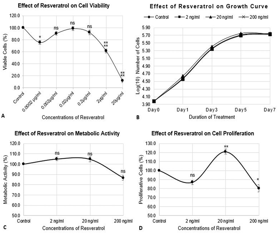
Previously, we reported non-cytotoxic concentrations of CQ-H (0.1, 1, 10, and 100ng/ml) for MC3T3-E1 cells . The same concentrations of CQ-H were evaluated for their effect on the viability and growth of RAW264.7 cells. Figure 1A shows the viability of cells after treatment with the aforementioned concentrations of CQ-H determined by neutral red assay. Treatment with 0.1ng/ml CQ-H exhibited 17% reduction (p≤0.05) in cell viability whereas, treatment with 1, 10, and 100 ng/ml CQ-H showed >90% cell viability. RSV was used as a standard to explore anti-osteoclast activity of CQ-H in this study. To determine the optimum concentrations of RSV, cell viability was assessed in response to treatment with 0.0002, 0.002, 0.02, 0.2, 2, and 20µg/ml RSV. Figure 2A shows the viability (%) of RSV treated cells. We observed >90% cell viability (p≥0.05) for cells treated with 2, 20, and 200 ng/ml whereas, other concentrations were non-cytotoxic for the cells. The concentrations exhibiting >90% cell viabilities were selected for further experimentation.
Growth curve of the cells treated with non-cytotoxic concentrations of CQ-H (1, 10, and 100 ng/ml) and RSV (2, 20, and 200 ng/ml) was generated by plotting logarithmic values of number of cells against duration of treatment. The growth curves of CQ-H and RSV treated cells are shown in Figure 1B and 2B, respectively. The images distinctly show different phases of growth cycle such as lag phase, log phase, stationary phase, and decline phase. Moreover, the growth curves of CQ-H and RSV treated cells is comparable to the control indicating no detrimental effect to the cells. Doubling time of control cells in both cases was 18.2h with specific growth rate of 0.017. CQ-H treatment to the cells reduced the doubling time to 17.8 (-2.2%),17.4 (-4.4%), and 17.4h (-2.2%) with specific growth rates of 0.017, 0.018, and 0.017, respectively for 1, 10 and 100 ng/ml concentrations of CQ-H, indicating slight increase in number of CQ-H treated cells, however, statistical analyses revealed the difference was not significant (p≥0.05). Similarly, doubling time for RSV treated cells was calculated to be 18.5, 17.8, and 18.4h with specific growth rate of 0.017 for all three concentrations, which is comparable to the control cells.
Figure 1C shows the metabolic activity of the cells treated with CQ-H. We observed that compared to control cells, metabolic activity of 1 and 10 ng/ml CQ-H treated cells was significant (p≤0.05) elevated i.e., 11.4% and 11.6%, respectively. The cells treated with 100ng/ml CQ-H indicated 7% increased metabolic activity, however the difference was not statistically significant compared to the control cells. Treatment with RSV showed 5%, 4%
and -13.3% (p≥0.05) metabolic activities compared to control cells for 2, 20, and 200 ng/ml RSV treated cells (Fig. 2C). Cell proliferation was measured as the amount of BrdU incorporated into DNA during the cell cycle. The colorimetric BrdU assay ELISA kit was utilized to quantify cell proliferation in response to CQ-H and RSV treatment. We observed 4.4, 2.4 and 7.2% reduction in cell proliferation compared to control cells in 1, 10 and 100 ng/ml CQ-H treated cells (Fig. 1D). The differences were not statistically significant. Figure 2D shows proliferation of RSV treated cells. We observed 21% (p≤0.05) elevated cell proliferation in cells treated with 20 ng/ml RSV. However, treatment with 2 and 200 ng/ml RSV showed -19.7% (p≤0.05) and 13.3% (p≥0.05) reduced cell proliferation compared to control cells.
Assessment of the results for the viability, proliferation, and growth kinetics of RAW264.7 cells in response to of CQ-H and RSV treatment indicated that the tested concentrations were non-cytotoxic and non-detrimental for the cells. Therefore, we selected these concentrations to examine their effect on the differentiation of RAW264.7 cells into osteoclasts.
RANK-l induced differentiation of raw264.7 cells into osteoclasts
To explore the efficacy of CQ-H to inhibit differentiation of RAW264,7 cells into osteoclasts, RAW264.7 cells were induced to differentiate into multinucleated osteoclasts by RANK Ligand. RSV treatment was employed as a standard to assess the osteoclast inhibition activity of CQ-H. RAW264.7 cells are murine macrophage cells that upon induction with RANK-L begin to fuse and form large multinucleated cells exhibiting osteoclast-like characteristics. Differentiation was induced after 24h of seeding, with differentiation medium containing CQ-H (1, 10, and 100ng/ml) and RSV (2, 20, and 200 ng/ml) along with appropriate controls.
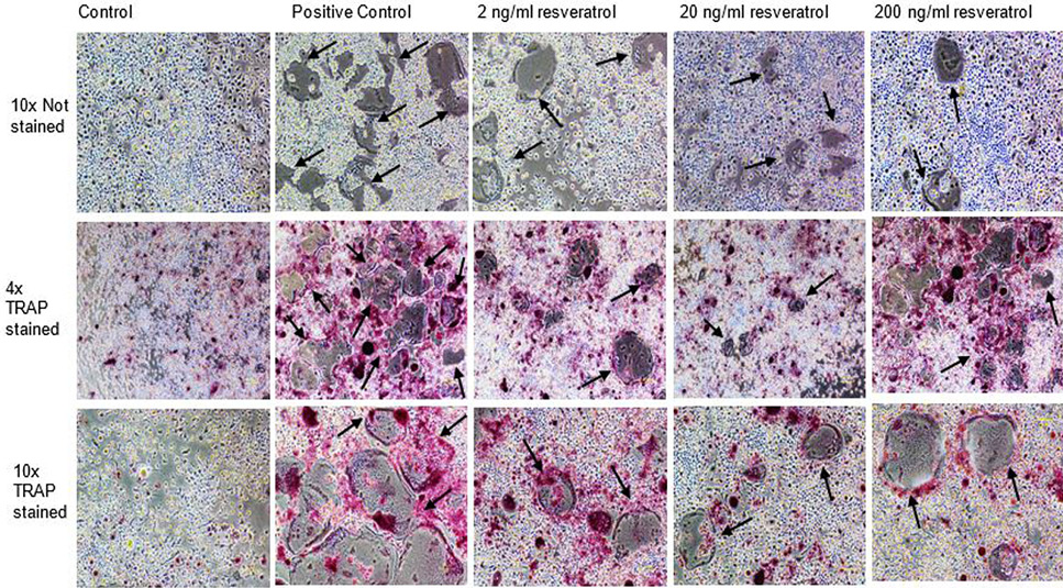
We observed formation of multinucleated cells in induced cell cultures after 48h, and by 72h of induction, mature large multinucleated cells were observed in the culture plates. Figure 3 shows differentiating CQ-H treated cells along with the controls after 24, 48 and 72h of differentiation induction. The figure distinctly shows a smaller number of multinucleated cells in CQ-H treated cultures compared to positive control cells. Similar observation was made for the cells treated with RSV (Fig. 4).
TRAP staining and activity estimation
Tartrate resistant acid phosphatase (TRAP) is an enzyme that is produced by mature osteoclasts in bone microenvironment and plays an important role in osteoclast differentiation, proliferation, and bone resorption. TRAP is therefore, an important marker of osteoclast formation and function. Histochemical staining of TRAP is an important indicator for osteoclast differentiation and TRAP activity is an important indicator of bone resorption ability of differentiated osteoblasts.
Figures 5 and 6 show TRAP stained CQ-H and RSV treated cultures of differentiated RAW264.7 cells after 72h of differentiation induction. The images clearly indicate a smaller number of differentiated stained cells in CQ-H and RSV treated cultures compared to the positive control cell cultures, whereas, the control cells fed with complete growth medium only does not show any differentiated or stained cells.
Estimation of TRAP activity reflects statistically significant osteoclast inhibitory activities of CQ-H and RSV. Figure 7A shows TRAP activity of CQ-H treated cells. We observed 57.8% (p≤0.05) reduced TRAP activity in cultures treated with 10ng/ml CQ-H, whereas, 46.5% and 45.8% (p≤0.05) reduced TRAP activities were observed in cultures treated with 1 and 100 ng/ml CQ-H. Treatment with RSV showed 38%, 49.9% and 46.6% (p≤0.05) reduced TRAP activities for 2, 20, and 200 ng/ml RSV treated cultures. Comparison of osteoclast inhibitory activity of CQ-H with that of RSV (standard) indicated that CQ-H is more efficacious anti-osteoclast agent.
Relative expression profiles of osteoclast marker genes
Further determination of the osteoclast inhibitory effect of CQ-H and RSV was carried out at the molecular level by analyses of expression profiles of osteoclast marker genes in the differentiating cultures. The osteoclast specific genes included transcription factors such as nuclear factor activated T cell c1 (NFATc1), enzymes such as cathepsin K (CTSK), acid phosphatase, tartrate resistant (ACP-5), matrix metalloproteinase 9 (MMP-9), and osteoclast specific proteins such as calcitonin receptors (CTR). We isolated RNA from differentiating cells at different time intervals, followed by cDNA synthesis and determination of Ct values for osteoclast specific genes by qPCR. Relative expression (fold) in terms of 2ΔΔCt was calculated as described by delta-delta Ct method ; .
NFATc1 is the key transcriptional regulator of osteoclast differentiation, hence it is expressed in the early phases of differentiation. It regulates the expression of several downstream osteoclast specific genes including ACP-5, CTSK, and CTR . In correlation with the above findings, we observed delayed and reduced expression of NFATc1 in CQ-H (Fig. 8) and RSV (Fig. 9) treated cultures. The transcription factor was not expressed in control cultures indicating their undifferentiated state. In positive control cultures, expression followed the normal expression profile with maximum expression at 24h which gradually decreased with time. Compared to positive controls at 24h interval, the CQ-H treated cultures showed 87.7, 74.2, and 75.9% reduced expression for 1, 10, and 100 ng/ml CQ-H treatment respectively, whereas, RSV treated cultures showed 39.6, 59.4, and 52.3% reduced NFATc1 expression for 2, 20, and 200ng/ml RSV treatment, respectively. In comparison to the standard, CQ-H treatment enhanced osteoclast differentiation inhibitory properties.
Cathepsin K (CTSK) is secreted by mature osteoclasts and is involved in bone resorption and matrix degradation . It is one of the late-phase osteoclast marker genes , with enhanced expression as the cells differentiate into mature osteoclasts as reflected by expression in positive control cultures. Compared to the positive control at 72h, we observed 53.3%, 75.6%, and 73.11% reduced expression in 1, 10, and 100ng/ml CQ-H treated cultures, respectively (Fig. 8). The RSV treated cells showed 38.8%, 55.09%, and 49.9% downregulation in CTSK expression in 2, 20, and 200 ng/ml RSV treated differentiating cells respectively (Fig. 9). Similar to the previously described results for differentiation, TRAP estimation and expression profile of NFATc1, CQ-H treated cultures showed enhanced inhibitory activity for CTSK compared to RSV, thereby indicating its efficacy to inhibit bone resorption by osteoclasts.
TRAP expression by the ACP-5 gene is also an enzyme secreted by mature osteoclasts that plays a role in resorption of bone matrix by dephosphorylation of phosphorylated proteins (osteopontin and IBSP) ; ; ; and is one of the late-phase osteoclast marker genes. Like CTSK, the expression of ACP-5 is enhanced as the cells differentiate into mature osteoclasts. For both CQ-H and RSV, the positive control cultures showed normal expression profiles for ACP-5 with expression gradually increasing over the period of 72h. Compared to the positive control, the CQ-H treated cells depicted 72%, 81.6%, and 71.3% reduced expression of ACP-5 for 1, 10, and 100 ng/ml CQ-H treatment respectively (Fig. 8). Correlating with the previous findings in this study, CQ-H treatment was more inhibitory to ACP-5 expression compared to RSV which showed 37.4%, 51.3% and 32.4% reduced expression for 2, 20 and 200 ng/ml RSV treatment (Fig. 9).
Matrix metalloproteinase 9 (MMP-9) is a collagenase secreted by mature osteoclasts that plays a role in matrix degradation ; ; and therefore, is one of the late-phase osteoclast marker genes. We observed that positive control cultures followed the normal expression profile, with enhanced expression of MMP-9 as cells differentiated into osteoclasts. However, in comparison to the positive control cells, we observed reduced expression of MMP-9 in CQ-H and RSV treated differentiating cells. Analogous to the previous observations, CQ-H exhibited elevated inhibitory effects on the expression of MMP-9 compared to RSV, indicating CQ-H is a more potent anti-osteoclast agent. We observed 83.9%, 91.4%, and 81.6% reduced MMP-9 expression in 1, 10, and 100 ng/ml treated cells (Fig. 8) compared to 50.3%, 70.6%, and 55.4% reduced expression in 2, 20 and 200 ng/ml RSV treated cells (Fig. 9).
Calcitonin is a hormone synthesized by the thyroid gland that plays a role in phosphate and calcium homeostasis in the bone resorption process and inhibits osteoclast activity . In the bone microenvironment, calcitonin receptors (CTR) are found only on the surfaces of osteoclasts ; 1999; ; , therefore it is considered an osteoclast specific marker. We observed the expression of CTR in the positive control as well as CQ-H and RSV treated differentiating cells. Corresponding to the reduced number of multinucleated osteoclasts in response to CQ-H and RSV treatment, we observed 47.7%, 65.1%, and 54.1% reduced expression of CTR in cells treated with 1, 10, and 100 ng/ml CQ-H respectively (Fig. 8), whereas, the cells treated with 2, 20 and 200 ng/ml RSV depicted 13.2%, 44.9% and 16.2% reduced expression of CTR in differentiating cells, respectively (Fig. 9).
The data presented in the present study together with the data reported in our previous study on the osteogenic effects of CQ-H, establishes the potential of CQ-H as an anti-osteoporotic agent. To fully elucidate the anti-osteoporotic potential of CQ-H, further in vitro and in vivo studies are required to isolate the bioactive compounds of CQ-H responsible for its anti-osteoporotic properties.
GC-MS analysis of CQ-H
Eighteen compounds were predicted in CQ-H i.e. (1) Undecane 2, 2-di methyl, (2) 2-methylpentyl propionate, (3) Oxirane, 2, 3-bis(1-methylethyl)-, trans-, (4) Butane, 2, 2, 3-trimethyl, (5) Butyl pentadecanoate, (6) Butanoic acid, 2-Methyl ester, (7) Borinic acid, diethyl-, (8) Neopentyl glycol (9) Heptane 1,1-Oxybis (10) 4,4-Trimethyl-1-pentanol (11) 1-Hexanol, 3, 5, 5-Trimethyl, (12) Oxalic acid, ethyl neopentyl ester, (13) 2, 4-Dipropyl-5-ethyl-1, 3-dioxane (14) 6, 10-Dimethyl-4-undecanol (15) 1, 2-benzenedicarboxylic acid, dinonyl ester (16) Butyl 4, 8, 12-trimethyl-tridecanoate, (17) 2-propenoic acid, butyl ester, (18) 1-Monolinoleylglycerol Trimethylsilyl ether. Table II shows list of all these compounds with their retention time (min) and reported biological activities.
DISCUSSION
Therapeutic options largely available for osteoporosis target bone resorption. The anti-absorptive drugs commercially available include bisphosphates, estrogen modulators, calcium, and calcitonin, while other therapies such as antibodies and inhibitors are also under development . However, for effective treatment of the disease, therapeutic drugs targeting bone anabolic processes should be developed. Although hormones such as parathyroid hormone (PTH) are being used, such treatments are costly and have long-term safety concerns . There is a need to investigate such therapies that are not only efficacious and inexpensive but are also safe for long term use. Alternative medicine employing bioactive compounds isolated from plants are now being explored for their therapeutic potential.
We previously reported the osteogenic potential of CQ (hexane fraction) . To further characterize the anti-osteoporotic activity of CQ-H, we studied its effect on osteoclastogenesis of the RAW264.7 cell line. The non-cytotoxic concentrations of CQ-H
Table II. Compounds predicted in CQ-H by GC-MS analysis with their retention time and their reported activities.
| No. | Retention time | Predicted compounds | Biological activity |
| 1 | 14.37/ 15.02/ 18.55 | Undecane 2,2-di methyl | Not detected |
| 2 | 14.51 | 2-methylpentyl propionate | Not detected |
| 3 | 15.47 | Oxirane, 2,3-bis(1-methylethyl)-, trans- | Not detected |
| 4 | 16.08 | Butane, 2,2,3-trimethyl- | Not detected |
| 5 | 18.02 | Butyl Pentadecanoate | Not detected |
| 6 | 18.22 | Butanoic acid, 2-Methyl ester | Not detected |
| 7 | 19.44 | Boronic acid, diethyl- | Antimicrobical and anti-inflammatory ; ; |
| 8 | 19.89/ 20.25 | Neopentyl glycol | Not detected |
| 9 | 21.16 | Heptane 1,1-Oxybis | Not detected |
| 10 | 22.29 | 4,4-Trimethyl-1-pentanol | Not detected |
| 11 | 24.80 | 1-Hexanol, 3,5,5-Trimethyl | Not detected |
| 12 | 25.14/ 26.28/27.29 | Oxalic acid, ethyl neopentyl ester | Not detected |
| 13 | 25.56 | 2,4-Dipropyl-5-ethyl-1,3-dioxane- | Not detected |
| 14 | 28.91 | 6,10-Dimethyl-4-undecanol | Not detected |
| 15 | 28.95 | 1,2-benzenedicarboxylic acid, dinonyl ester | Not detected |
| 16 | 29.18 | Butyl 4,8,12-trimethyl-tridecanoate | Not detected |
| 17 | 31.78 | 2-propinoic acid, butyl ester | Not detected |
| 18 | 32.38 | 1-MonolinoleylglycerolTrimethylsilyl ether | Antioxidant, anti-inflammatory, antimicrobial, diuretic, anti-arthritic, antiasthma ( |
(0.1, 1, and 10 ng/ml) determined by our research group on MC3T3-E1 cells were analyzed for their effect on the viability, proliferation and growth of RAW264.7 cells. We found that the cell line grew with the doubling time of 18.2h, while the doubling time of CQ-H treated cells was comparable to the control cells. The same concentrations of CQ-H had no detrimental effect on the viability, metabolic activity and proliferation of the cells.
RSV was employed as standard to compare the anti-osteoclastogenesis activity with that of CQ-H. The optimum concentrations found for RSV treatment of RAW264.7 cells were 2, 20, and 200ng/ml with no detrimental effects on viability, metabolic activity, proliferation, and growth parameters of the cells. He and colleagues reported similar findings with 0.3, 1, and 3µM as non-cytotoxic concentrations of RSV .
While osteogenic and anti-osteoclast activities of RSV via estrogen dependent and modulation of the SIRT-1/ FOXO3 pathway and ROS inhibition respectively have been reported , 2012; . there is no published evidence for in vitro anti-osteoclast activity of CQ. However, evidence on stimulatory effects of CQ for osteogenesis are reported ; ; ; ; , 2010; ; .
To analyze the effect of CQ-H and RSV on osteoclastogenesis, RANK-L induced RAW264.7 cells were treated with non-cytotoxic concentrations of CQ-H and RSV. The cells formed large multinucleated osteoclasts after 3 days of induction. In comparison with positive control, there were fewer large multinucleated cells in CQ-H and RSV treated cultures. TRAP is an important biochemical marker of osteoclast differentiation that is involved in bone resorption activity of osteoclasts therefore; TRAP staining was employed to confirm osteoclastogenesis. The induced cells in positive control, CQ-H and RSV treated cultures were positively stained while undifferentiated cells (control) were not stained.
TRAP activity was estimated to quantify the anti-osteoclast activities of CQ-H and RSV. Supporting the observations, we made in Figures 4-7, our results on quantification of TRAP activity showed 57.8% downregulation in TRAP activity in the 10ng/ml CQ-H treated differentiated cells whereas 49.9% downregulation was reported in 20ng/ml RSV treated cells. Table III summarizes the comparative results of CQ-H and RSV treatment on the viabilities, metabolic and proliferative activities of the cells as well as on their effect on osteoclastogenesis.
In the bone microenvironment, bone structure integrity is maintained by activity of mesenchymal stem cells forming osteoblasts and hematopoietic stem cells forming osteoclasts. Osteoclasts are bone resorbing cells derived from macrophage lineage and like osteoblastogenesis, several osteoclast specific genes are involved in formation of osteoclasts from HSCs . The differentiation process is associated with timely expression of transcription factors and marker genes. Expression profiles of osteoclast marker genes such as NFATc1, ACP-5, CTSK, MMP-9 and CTR was analyzed in CQ-H and RSV treated differentiated cells.
Expression analysis of osteoclast specific genes showed downregulation of all genes in both CQ-H and RSV treated cells indicating inhibition of osteoclastogenesis. There are no reports of anti-osteoclastogenic effects of CQ crude or solvent fractions, however, dose-dependent inhibition in TRAP activity and downregulation of ACP-5
Table III. Comparison of growth, viability, metabolic, proliferation parameters, and differentiation potential of CQ-H and resveratrol treated RAW264.7 cells. Osteoclast differentiation was induced by RANK-L and differentiation of RAW264.7 cells was estimated by determination of percentage TRAP activity of differentiated cells. The values are written as percentages ± SD compared to the appropriate controls (taken as 100%). The + sign indicates percentage increase and – sign indicates percentage decease in the viability/ metabolic activity/ proliferation/ differentiation of the RAW264.7 cells.
|
| Concentrations tested | Doubling time (h) | Cell viability (%) | Metabolic activity (%) | Cell proliferation (%) | TRAP activity (%) |
| CQ-H | Control 0.1 ng/ml 1 ng/ml 10 ng/ml | 18.2± 0.05 17.8± 0.01 17.4 ± 0.06 17.8 ± 0.05 | --- -8 ± 2.2 -8 ± 2.4 -7.1 ± 1.8 | --- +11.6 ± 3.1 +11.4 ± 2.0 +7.2 ± 1.9 | --- -4.4 ± 3.0 -2.4 ± 1.5 -7.2 ± 1.9 | --- -46.5 ± 1.8 -57.8 ± 2.5 -45.8 ± 2.8 |
| Resveratrol | Control 2 ng/ml 20 ng/ml 200 ng/ml | 18.2 ± 0.65 18.5 ± 0.71 17.8 ± 0.47 18.4 ± 0.64 | --- -9.5 ± 3.0 -1.4 ± 3.4 -7.9 ± 3.7 | --- +5 ± 1.7 +4.7 ± 2.4 -13.3 ± 2.5 | --- -13.1 ± 2.5 +21 ± 2.6 -19.7 ± 4.0 | --- -38 ± 1.5 -49.9 ± 2.1 -46.6 ± 1.9 |
and CTSK expression in RSV treated RAW264.7 cells has been reported by He et al. (2010) and Shakibaei et a;. (2012).
We predicted presence of eighteen compounds in CQ-H by GC-MS analysis (Table II). Although for many of the predicted compounds, no reported biological activity was found, however, derivatives of compounds such as oxirane, 2, 3-bis(1-methylethyl)-, trans, for example, L-3-trans-(Propylcarbamoyl) oxirane-2-carbonyl]-L-isoleucyl-L-proline methyl ester (CA-074Me) is a cathepsin inhibitor and inhibits osteoclast differentiation by altering NFATc1 and c-FOS pathways . Similarly, PT-100, a boronic acid based dipeptidyl peptidase (DPP) IV activity and/or structure homologues (DASH; a serine protease) inhibitor is also reported to inhibit osteoclast formation (Busek et al., 2004). Moreover, compounds such as butyric acid and boric acid are the backbone of some of the predicted compounds, and has been reported to promote osteoblast differentiation and inhibit osteoclast formation ; . Further purification of CQ-H to isolate and analyze bioactivities of individual compounds will provide more insight to identify the active compound(s) responsible for its bone resorption property.
The results of the present study indicated that compared to RSV treatment, CQ-H is not only an efficacious osteogenic agent but also has anti-osteoporotic potential exhibiting therapeutic potential as an anti-osteoporotic agent.
CONCLUSION
The present study explores anti-resorptive effects of CQ-H on bones. Upon RANK-L induction of RAW264.7 cells to differentiate into osteoclasts, 10ng/ml CQ-H was found to be the most efficacious concentration for inhibiting osteoclastogenesis with 57.8% inhibition of TRAP activity, compared to 20ng/ml of resveratrol (RSV) which showed 49.9% inhibition of TRAP activity. Expression of osteoclast marker genes such as NFATc1, ACP-5, CTSK, MMP-9 and CTR was significantly downregulated in CQ-H and RSV treated differentiated cells compared to the positive control. This study provides an evidence of anti-osteoporotic therapeutic property of CQ.
Acknowledgements
This work is financially supported by the research grant of Pakistan Academy of Sciences Ref. # 5-9/PAS/22 dated 27.7.2016 and NIH grant R01 AR039588 (Stein/Lian). Gary Stein is the AJ Perelman Professor, University of Vermont.
Ethics approval and consent to participate
Not Applicable.
Consent for publication
Not Applicable
Authors’ declaration
The authors hereby declare that the work presented in this article is original and that any liability for claims relating to the content of this article will be borne by them.
Statement of conflict of interest
The authors have declared no conflict of interest.
Figures
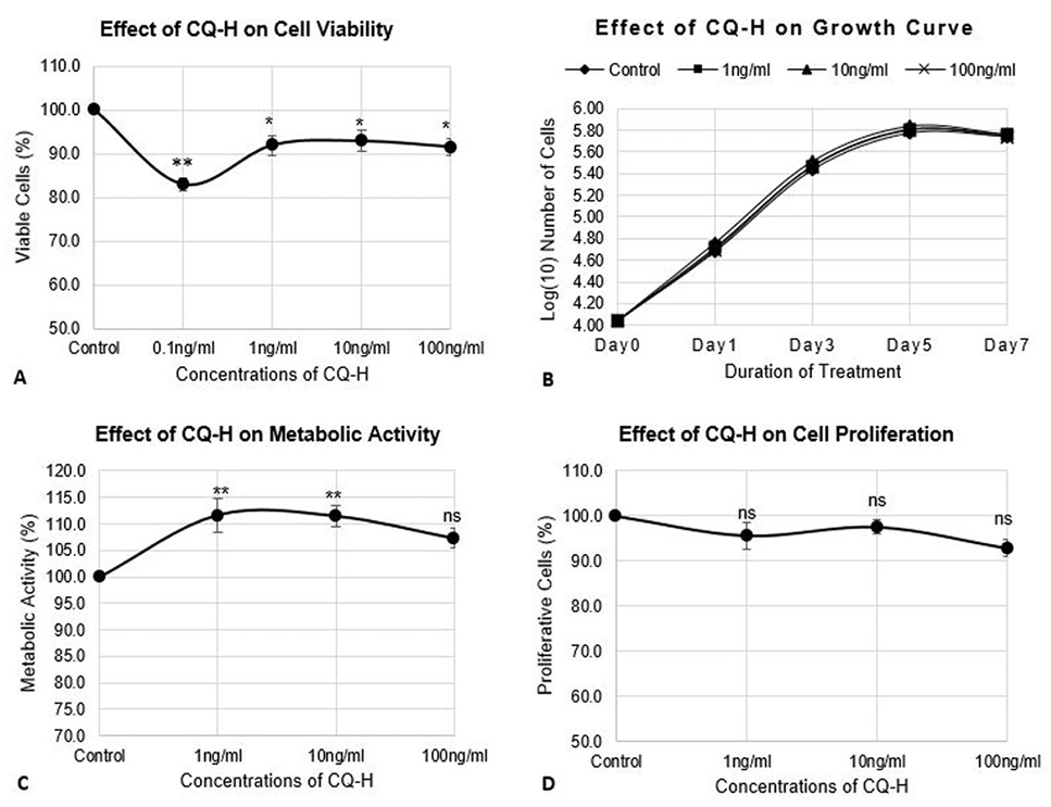
Effect of CQ-H on cell viability, growth parameters, and proliferation of RAW264.7 cell line. The cells were seeded at density of 3000 cells/cm2 in 24-well plates. The control cells were fed with 0.05% DMSO supplemented complete medium whereas, complete medium containing 1, 10, and 100 ng/ml CQ-H was fed to the treated cultures. (A) Neutral red assay was performed for analysis of cell viability. The cells were treated with CQ-H for 72h. Assay was performed at 24h and 72h. Averages of values were taken. (B) Growth curve was generated by treated cells with CQ-H for 7 days and counting them on alternate days (day 1, 3, 5, and 7). Logarithmic values of cell number were plotted to reveal lag, log, stationery and decline phase of cell growth. (C) MTT assay and (D) BrdU incorporation assay was performed to analyze effect of CQ-H on the metabolic activity and cell proliferation respectively. The cells were seeded for up to 72h and assays were performed at 24 and 72h. Averages of values were taken. Data are represented as Mean ± SD of technical and biological replicates for each experiment. For statistical analyses, ANOVA with Dunnett’s test for multiple comparison was performed using GraphPad Prism (v 7.03). (P≤0.05; ns = not significant, * P≤0.05, **P≤0.01, ***P≤0.001, ****P≤0.0001).
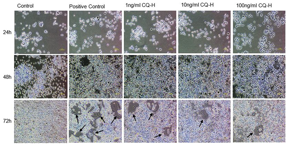
Effect of non-cytotoxic, non-detrimental concentrations of CQ-H (1, 10, and 100 ng/ml) on the differentiation of RAW264.7 cell line. The cells were cultured at 12000/cm2 density in 12-well culture plates. The control cells were fed with 0.05% DMSO supplemented medium. The positive control cells were fed with osteoclast induction medium (complete growth medium supplemented with 10ng/ml RANK-L and 0.05% DMSO). The CQ-H treated cells were fed with osteoclast induction medium containing 1, 10 and 100ng/ml CQ-H. After 48h of osteoclast induction, the cells fed with induction medium began to fuse with each other. After 72h of induction, mature, multinucleated cells were formed in the culture plate. The number of multinucleated cells was larger compared to CQ-H treated cultures. The black arrows show the large multinucleated cells. There were no large multinucleated cells in the control cultures. The images were taken at 4x magnification with Nikon Eclipse TS-100 inverted microscope.
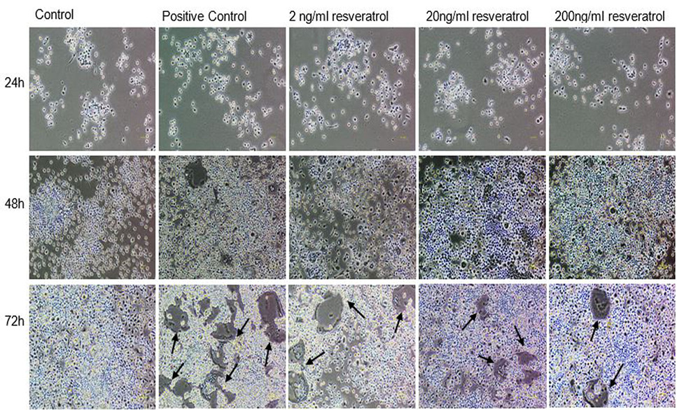
Effect of non-cytotoxic, non-detrimental concentrations of Resveratrol (2, 20, and 200 ng/ml) on the differentiation of RAW264.7 cell line. The cells were cultured at 12000/cm2 density in 12-well culture plates. The control cells were fed with 0.05% DMSO supplemented medium. The positive control cells were fed with osteoclast induction medium (complete growth medium supplemented with 10ng/ml RANK-L and 0.05% DMSO). The Resveratrol treated cells were fed with osteoclast induction medium containing 2, 20, and 200 ng/ml resveratrol. After 48h of osteoclast induction, the cells fed with induction medium began to fuse with each other. After 72h of induction, mature, multinucleated cells were formed in the culture plate. The number of multinucleated cells was larger compared to resveratrol treated cultures. The black arrows show the large multinucleated cells. There were no large multinucleated cells in the control cultures. The images were taken at 4x magnification with Nikon Eclipse TS-100 inverted microscope.
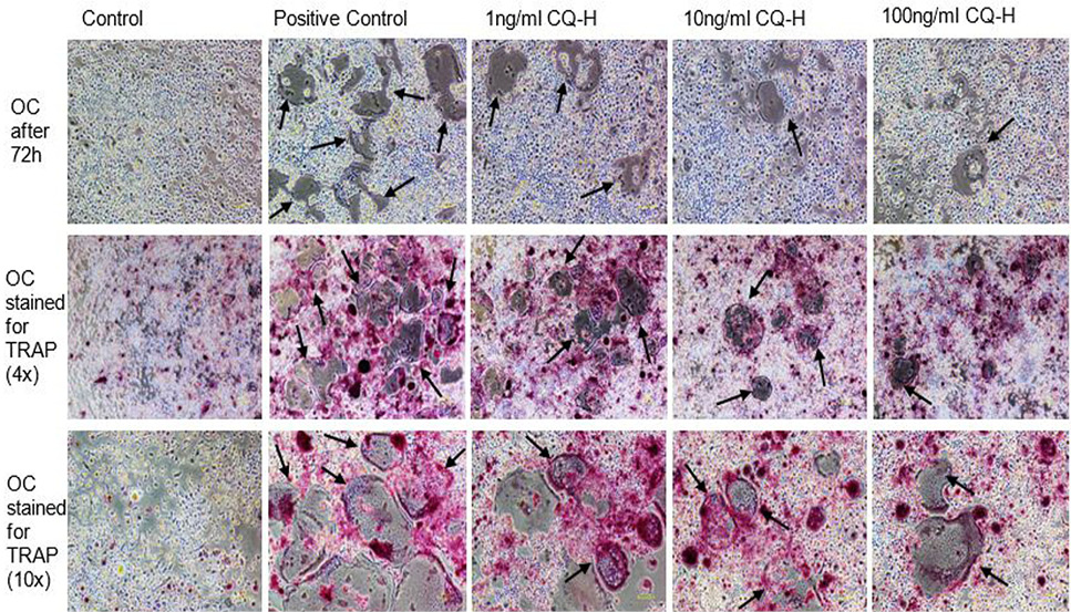
TRAP stained multinucleated cells formed after 72h of osteoclast induction of RAW264.7 cells. The images in first row show the unstained RAW264.7 cultures after 72h of induction and CQ-H treatment at 4x magnification. The second row shows the cells stained for TRAP at 4x magnification. The pink colored cells indicate presence of TRAP positive osteoclasts. The third row shows TRAP stained cells at 10x magnification. The figures indicate that the positive control cultures have more stained multinucleated cells compared to CQ-H treated cells. The black arrows show the multinucleated cells. Images were taken from Nikon Eclipse TS-100 inverted microscope.
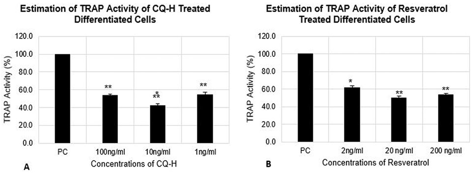
Estimation of TRAP activity of differentiated RAW264.7 cells. The cells were seeded at 12000 cells/cm2 density and differentiated for 72h. The control cells were fed with 0.05% DMSO supplemented medium. The positive control cells were fed with osteoclast induction medium (complete growth medium supplemented with 10ng/ml RANK-L and 0.05% DMSO). The CQ-H treated cells were fed with osteoclast induction medium containing 1, 10 and 100ng/ml CQ-H and Resveratrol treated cells were fed with osteoclast induction medium containing 0.002, 0.02, and 0.2 µg/ml resveratrol. TRAP activity was estimated after 72h of osteoclast differentiation induction. Data are represented as Mean ± SD of technical and biological replicates for each experiment. For statistical analyses, ANOVA with Dunnett’s test for multiple comparison was performed using GraphPad Prism (v 7.03). (P≤0.05; ns = not significant, * P≤0.05, **P≤0.01, ***P≤0.001, ****P≤0.0001).
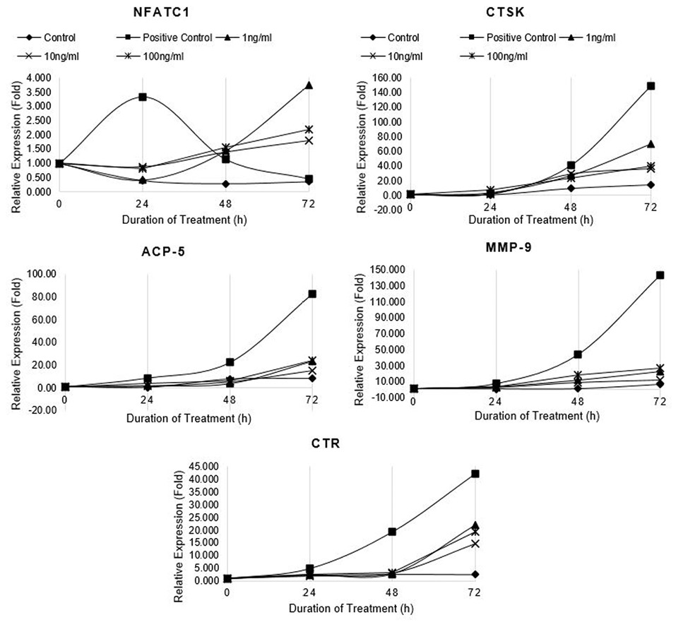
Relative expression of osteoclast marker genes (NFATc1, CTSK, ACP5, MMP-9 and CTR) in differentiating RAW264.7 cells treated with non-cytotoxic concentrations of CQ-H. Control cells were fed with 0.05% DMSO supplemented medium. The control culture was treated with 0.05% DMSO in normal complete medium. The positive control cells were fed with osteoclast induction medium (complete growth medium supplemented with 10ng/ml RANK-L and 0.05% DMSO). The CQ-H treated cells were fed with osteoclast induction medium containing 1, 10 and 100ng/ml CQ-H. The cells were allowed to differentiate for 72h with medium change after 48h. Total RNA was isolated at 0, 24, 48, and 72h of induction and cDNA was synthesized. Hydroxymethylbilane synthase (HMBS) was selected as housekeeping gene. Expression of genes was quantified by real-time. Relative expression (fold) was calculated by Pfaffl method.
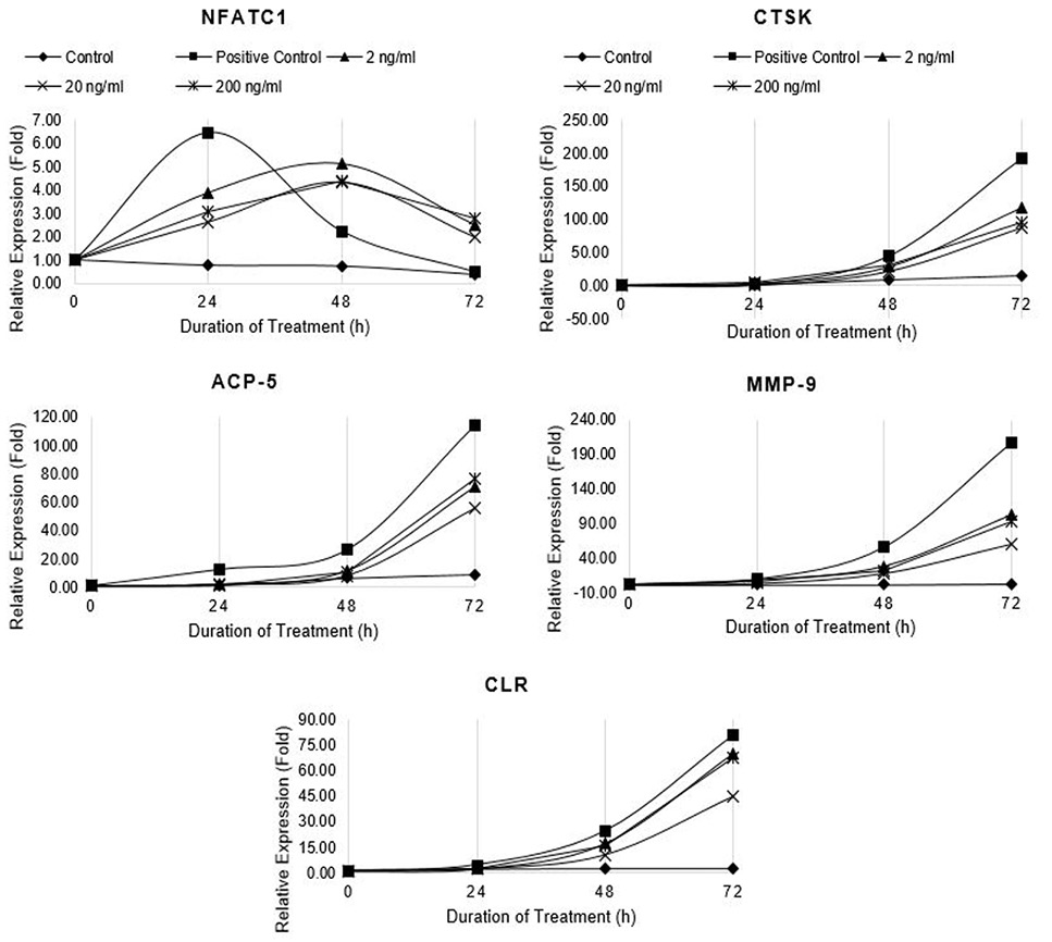
Relative expression of osteoclast marker genes (NFATc1, CTSK, ACP5, MMP-9 and CTR) in differentiating RAW264.7 cells treated with non-cytotoxic concentrations of Resveratrol. Control cells were fed with 0.05% DMSO supplemented medium. The control culture was treated with 0.05% DMSO in normal complete medium. The positive control cells were fed with osteoclast induction medium (complete growth medium supplemented with 10ng/ml RANK-L and 0.05% DMSO). The Resveratrol treated cells were fed with osteoclast induction medium containing 2, 20, and 200 ng/ml resveratrol. The cells were allowed to differentiate for 72h with medium change after 48h. Total RNA was isolated at 0, 24, 48, and 72h of induction and cDNA was synthesized. Hydroxymethylbilane synthase (HMBS) was selected as housekeeping gene. Expression of genes was quantified by real-time. Relative expression (fold) was calculated by Pfaffl method.
References
- Adesanya, S.A., Nia, R., Martin, M-T., Boukamcha, N., Montagnac, A., and Païs, M., 1999. Stilbene derivatives from Cissus quadrangularis. J. Nat. Prod., 62: 1694-1695. https://doi.org/10.1021/np9902744[CrossRef]
- Bafna, P.S., Patil, P.H., Maru, S.K. and Mutha, R.E., 2021. Cissus quadrangularis L: A comprehensive multidisciplinary review. J. Ethnopharmacol., 279: 114355. https://doi.org/10.1016/j.jep.2021.114355[CrossRef]
- Bailey, P., Cousins, G., Snow, G., and White, A., 1980. Boron-containing antibacterial agents: Effects on growth and morphology of bacteria under various culture conditions. Antimicrob. Agents Chemother., 17: 549-553. 0066-4804. https://doi.org/10.1128/AAC.17.4.549[CrossRef]
- Baker, S.J., Akama, T., Zhang , Y-K., Sauro, V., Pandit, C., Singh, R., Kully, M., Khan, J., Plattner, J.J., Benkovic, S.J., Lee, V., and Maples, K.R., 2006. Identification of a novel boron-containing antibacterial agent (AN0128) with anti-inflammatory activity, for the potential treatment of cutaneous diseases. Bioorg. Med. Chem. Lett., 16: 5963-5967. https://doi.org/10.1016/j.bmcl.2006.08.130[CrossRef]
- Benkovic, S.J., Baker, S.J., Alley, M.R.K., Woo, Y-H., Zhang, Y.K., Akama, T., Mao, W., Baboval, J., Rajagopalan, P.T.R., Wall, M., Kahng, L.S., Tavassoli, A., and Shapiro, L., 2005. Identification of borinic esters as inhibitors of bacterial cell growth and bacterial methyltransferases CcrM and MenH. J. med. Chem., 8: 7468-7476. https://doi.org/10.1021/jm050676a[CrossRef]
- Bhutani, K., Kapoor, R. and Atal, C., 1984. Two unsymmetric tetracyclic triterpenoids from Cissus quadrangularis. Phytochemistry, 23: 407-410. https://doi.org/10.1016/S0031-9422(00)80341-6[CrossRef]
- Blumer, M.J., Hausott, B., Schwarzer, C., Hayman, A.R., Stempel, J. and Fritsch, H., 2012. Role of tartrate-resistant acid phosphatase (TRAP) in long bone development. Mech. Dev., 129: 162-176. https://doi.org/10.1016/j.mod.2012.04.003[CrossRef]
- Boissy, P., Andersen, T.L., Abdallah, B.M., Kassem, M., Plesner, T. and Delaissé, J-M., 2005. Resveratrol inhibits myeloma cell growth, prevents osteoclast formation, and promotes osteoblast differentiation. Cancer Res., 65: 9943-9952. https://doi.org/10.1158/0008-5472.CAN-05-0651[CrossRef]
- Bušek, P., Malı́k, R., and Šedo, A., 2004. Dipeptidyl peptidase IV activity and/or structure homologues (DASH) and their substrates in cancer. Int. J. Biochem. Cell Biol., 36: 408-421. https://doi.org/10.1016/S1357-2725(03)00262-0[CrossRef]
- Butler. M., 2004. Animal cell culture and technology. Taylor and Francis; 2004. ISBN: 1135325383. https://doi.org/10.4324/9780203427835[CrossRef]
- Chanda, S., Baravalia, Y. and Nagani, K., 2013. Spectral analysis of methanol extract of Cissus quadrangularis L. stem and its fractions. J. Pharmacog. Phytochem., 2: 149-157.
- Chen, C., Dong, B., Wang, Y., Zhang, Q., Wang, B., Feng, S., and Zhu, Y., 2020. The role of Bacillus acidophilus in osteoporosis and its roles in proliferation and differentiation. J. Clin. Lab. Anal., 34: e234710887-8013. https://doi.org/10.1002/jcla.23471[CrossRef]
- Christensen, J., and Shastri, V.P., 2015. Matrix-metalloproteinase-9 is cleaved and activated by cathepsin K. BMC Res. Notes, 8: 322. https://doi.org/10.1186/s13104-015-1284-8[CrossRef]
- Dai, R., Wu, Z., Chu, H.Y., Lu, J., Lyu, A., Liu, J., and Zhang, G., 2020. Cathepsin K: the action in and beyond bone. Front. Cell Develop. Biol., 8: 433. https://doi.org/10.3389/fcell.2020.00433[CrossRef]
- Eijken, H.J.M., 2007. Human osteoblast differentiation and bone formation: growth factors, hormones and regulatory networks. 2007 ISBN: OCLC:210282353. Erasmus University Rotterdam. Retrieved from http://hdl.handle.net/1765/10597
- Eswaran, R., Anandan, A., Sangeetha, G. and Anand, S.P., 2012. Analysis of chemical composition of Cissus quadrangularis Linn by GC-MS. Asian. J. Pharm. clin. Res., 5: 139-140.
- Gabbay, K.H., Bohren, K.M., Morello, R., Bertin, T., Liu, J., and Vogel, P., 2010. Ascorbate synthesis pathway: Dual role of ascorbate in bone homeostasis. J. biol. Chem., 285: 19510-19520. https://doi.org/10.1074/jbc.M110.110247[CrossRef]
- Guerra, J.M., Hanes, M.A., Rasa, C., Loganathan, N., Innis-Whitehouse, W., Gutierrez, E., Nair, S., and Banu, J., 2019. Modulation of bone turnover by Cissus quadrangularis after ovariectomy in rats. J. Bone Miner. Metab., 37: 780-795. https://doi.org/10.1007/s00774-018-0983-3[CrossRef]
- Gupta, M., Arshad, M., Shivnath, N., Khan, M.S., and Gupta. R., 2018. µCT analysis reveals that Cissus quadrangularis L. stem and Trigonella foenum-graecum L. seed extracts prevent diabetic osteopathy. Indian J. Trad. Knowl. 17: 460-467.
- Gupta, M.M. and Verma, R.K., 1990. Unsymmetric tetracyclic triterpenoid from Cissus quadrangularis. Phytochemistry, 29: 336-337. https://doi.org/10.1016/0031-9422(90)89067-J[CrossRef]
- Gupta, M.M. and Verma, R.K., 1991. Lipid constituents of Cissus quadrangularis. Phytochemistry, 30: 875-878. https://doi.org/10.1016/0031-9422(91)85270-A[CrossRef]
- Habold, C., Momken, I., Ouadi, A., Bekaert, V. and Brasse, D., 2011. Effect of prior treatment with resveratrol on density and structure of rat long bones under tail-suspension. J. Bone Miner. Metab., 29: 15-22. https://doi.org/10.1007/s00774-010-0187-y[CrossRef]
- Hadjidakis, D.J. and Androulakis, I.I., 2006. Bone remodeling. Annls N. Y. Acad. Sci., 1092: 385-396 https://doi.org/10.1196/annals.1365.035.[CrossRef]
- Hayman, A.R., Bune, A.J., Bradley, J.R., Rashbass, J., and Cox, T., 2000. Osteoclastic tartrate-resistant acid phosphatase (Acp 5): its localization to dendritic cells and diverse murine tissues. J. Histochem. Cytochem., 48: 219-228. https://doi.org/10.1177/002215540004800207[CrossRef]
- He, X., Andersson, G. Lindgren, U. and Li, Y., 2010. Resveratrol prevents RANKL-induced osteoclast differentiation of murine osteoclast progenitor RAW 264.7 cells through inhibition of ROS production. Biochem. biophys. Res. Commun., 401: 356-362. https://doi.org/10.1016/j.bbrc.2010.09.053[CrossRef]
- Hsiao, C-Y., Chen, T-H., Chu, T-H., Ting, Y-N., Tsai, P-J., and Shyu, J-F., 2020. Calcitonin induces bone formation by increasing expression of Wnt10b in osteoclasts in ovariectomy-induced osteoporotic rats. Front. Endocrinol., 11: https://doi.org/10.3389/fendo.2020.00613[CrossRef]
- Jain, A., Dixit, J., and Prakash, D., 2008. Modulatory effects of Cissus quadrangularis on periodontal regeneration by bovine-derived hydroxyapatite in intrabony defects: exploratory clinical trial. J. Int. Acad. Periodontol., 10: 59-65.
- Jiang, C., Ma, Q., Wang, S., Shen, Y., Qin, A., Fan, S. and Jie, Z., 2021. Oxymatrine attenuates osteoclastogenesis via modulation of ROS-mediated SREBP2 signaling and counteracts ovariectomy-induced osteoporosis. Front. Cell Develop. Biol., 9: 684007. https://doi.org/10.3389/fcell.2021.684007[CrossRef]
- Kong, Y.Y., and Penninger, J., 2000. Molecular control of bone remodeling and osteoporosis. Exp. Gerontol., 35: 947-956. https://doi.org/10.1016/S0531-5565(00)00178-9[CrossRef]
- Kumar, M., Rawat, P., Dixit, P., Mishra, D., Gautam, A.K., Pandey, R.. Singh, D., Chattopadhyay, N. and Maurya, R., 2010. Anti-osteoporotic constituents from Indian medicinal plants. Phytomedicine, 17: 993-999. https://doi.org/10.1016/j.phymed.2010.03.014[CrossRef]
- Kumar, P., Dev, K., Sharma, K., Sahai, M., and Maurya, R., 2019. New lignan glycosides from Cissus quadrangularis stems. Nat. Prod. Res., 33: 233-238. https://doi.org/10.1080/14786419.2018.1443099[CrossRef]
- Kumar, T.S., Anandan, A., and Jegadeesan, M., 2012. Identification of chemical compounds in Cissus quadrangularis L. Variant I of different samples using GC-MS analysis. Arch. appl. Sci. Res., 4: 1782-1787. http://www.scholarsresearchlibrary.com/
- Liu, Z., Li, W., Yu, B., Huang, J., Sun, J., Huo, J., and Liu, C.X., 2005. Effects of trans-resveratrol from Polygonum cuspidatum on bone loss using the ovariectomized rat model. J. med. Fd., 8: 14-19. https://doi.org/10.1089/jmf.2005.8.14[CrossRef]
- Livak, K.J. and Schmittgen, T.D., 2001. Analysis of relative gene expression data using real-time quantitative PCR and the 2−ΔΔCT method. Methods, 25: 402-408. https://doi.org/10.1006/meth.2001.1262[CrossRef]
- Lotinun, S., Kiviranta, R., Matsubara, T., Alzate, J.A., Neff, L., Lüth, A., Koskivirta, I., Kleuser, B., Vacher, J., Vuorio, E., Horne, M.C., and Baron, R., 2013. Osteoclast-specific cathepsin K deletion stimulates S1P-dependent bone formation. J. clin. Investig., 123: 666-681. https://doi.org/10.1172/JCI64840[CrossRef]
- Luchian, I., Goriuc, A., Sandu,D. and Covasa, M., 2022. The role of matrix metalloproteinases (MMP-8, MMP-9, MMP-13) in periodontal and peri-implant pathological processes. Int. J. mol. Sci., 23: 1806. https://doi.org/10.3390/ijms23031806[CrossRef]
- Mehta, M., Kaur, N., and Bhutani, K., 2001. Determination of marker constituents from Cissus quadrangularis Linn. and their quantitation by HPTLC and HPLC. Phytochem. Anal., 12: 91-95. https://doi.org/10.1002/pca.569[CrossRef]
- Mishra, G., Srivastava, S. and Nagori, B.P., 2010. Pharmacological and therapeutic activity of Cissus quadrangularis: An overview. Int. J. Pharm. Tech. Res., 2: 1298-1310.
- Mizutani, K., Ikeda, K., Kawai, Y., and Yamori, Y., 1998. Resveratrol stimulates the proliferation and differentiation of osteoblastic MC3T3-E1 cells. Biochem. biophys. Res. Commun., 253: 859-863. https://doi.org/10.1006/bbrc.1998.9870[CrossRef]
- Mizutani, K., Ikeda, K., Kawai, Y., and Yamori, Y., 2000. Resveratrol attenuates ovariectomy-induced hypertension and bone loss in stroke-prone spontaneously hypertensive rats. J. Nutr. Sci. Vitaminol., 46: 78-83. https://doi.org/10.3177/jnsv.46.78[CrossRef]
- Okada, Y., Naka, K., Kawamura, K., Matsumoto, T., Nakanishi, I., Fujimoto, N., Sato, H., and Seiki, M., 1995. Localization of matrix metalloproteinase 9 (92-kilodalton gelatinase/type IV collagenase= gelatinase B) in osteoclasts: Implications for bone resorption Lab. Investig. J. Tech. Meth. Pathol., 72: 311-322. PMID 7898050.
- Parthipan, B., Mgt, S., and Mohan, V.R., 2015. GC-MS analysis of phytocomponents in Pleiospermium alatum (Wall. ex Wight and Arn.) Swingle, (Rutaceae). J. Pharmacog. Phytochem., 4: 216-222. e ISSN: 2278-4136.
- Patel, N., Nizami, S., Song, L., Mikami, M., Hsu, A., Hickernell, T., Chandhanayingong, C., Rho, S., Compton, J.T., Caldwell, J.M., Kaiser, P.B., Bai, H., Lee, H.G., Fischer, C.R., and Lee, F.Y., 2015. CA-074Me compound inhibits osteoclastogenesis via suppression of the NFATc1 and c-FOS signaling pathways. J. Orthopaed. Res., 33: 1474-1486. https://doi.org/10.1002/jor.22795[CrossRef]
- Pathomwichaiwat, T., Ochareon, P., Soonthornchareonnon, N., Ali, Z., Khan, I.A., and Prathanturarug, S., 2015. Alkaline phosphatase activity guided isolation of active compounds and new dammarane-type triterpenes from Cissus quadrangularis hexane extract, J. Ethnopharmacol., 160: 52-60. https://doi.org/10.1016/j.jep.2014.11.026[CrossRef]
- Patil, V.S., Biradar, P.R, Attar, V. and Khanal, P., 2019. In silico docking analysis of active biomolecules from Cissus quadrangularis L. against PPAR-γ. Indian J. Pharmaceut. Educ. Res., 53: S332-337. https://doi.org/10.5530/ijper.53.3s.103[CrossRef]
- Pearson, K.J., Baur, J.A., Lewis, K.N., Peshkin, L., Price, N.L., Labinskyy, N., Swindell, W.R., Kamara, D., Minor, R.K., Perez, E., Jamieson, H.A., Zhang, Y., Dunn, S.R., Sharma, K., Pleshko, N., Woollett, L.A., Csiszar, A., Ikeno, Y., Couteur, D.L., Elliott, P.J., Becker, K.G., Navas, P., Ingram, D.K., Wolf, N.S., Ungvari, Z., Sinclair, D.A., and deCabo, R., 2008. Resveratrol delays age-related deterioration and mimics transcriptional aspects of dietary restriction without extending life span. Cell Metab., 8: 157-168. https://doi.org/10.1016/j.cmet.2008.06.011[CrossRef]
- Pondel, M., 2000. Calcitonin and calcitonin receptors: Bone and beyond. Int. J. exp. Pathol., 81: 405-422. https://doi.org/10.1046/j.1365-2613.2000.00176.x[CrossRef]
- Potu, B.K., Rao, M.S., Nampurath, G.K., Chamallamudi, M.R., Nayak, S.R., and Thomas, H., 2010. Anti-osteoporotic activity of the petroleum ether extract of Cissus quadrangularis Linn. in ovariectomized wistar rats. Chang Gung med. J., 33: 252-257. https://www.scopus.com/inward/record.uri?eid=2-s2.0-77954502168andpartnerID=40 andmd5= 46a5dfecbd9cbab587316f08905b850d PMID: 20584502
- Potu, B.K., Rao, M.S., Nampurath, G.K., Chamallamudi, M.R., Prasad, K., Nayak, S.R., Dharmavarapu, P.K., Kedage, V., and Bhat, K.M.R., 2009. Evidence-based assessment of antiosteoporotic activity of petroleum-ether extract of Cissus quadrangularis Linn. on ovariectomy-induced osteoporosis. UPS J. med. Sci., 114: 140-148. https://doi.org/10.1080/03009730902891784[CrossRef]
- Raggatt, L.J. and Partridge, N.C., 2010. Cellular and molecular mechanisms of bone remodeling, J. biol. Chem., 285: 25103-25108. https://doi.org/10.1074/jbc.R109.041087[CrossRef]
- Rao, G.V., Annamalai, T., Mukhopadhyay, T. and Madhavi, M., 2011. Chemical constituents and melanin promotion activity of Cissus quadrangularis Linn. Res. J. chem. Sci., 1: 25-29.
- Rao, X., Huang, X., Zhou, Z., and Lin X., 2013. An improvement of the 2ˆ (–delta delta CT) method for quantitative real-time polymerase chain reaction data analysis. Biostat. Bioinform. Biomath., 3: 71-85.
- Reid, I.R., 2008. Anti-resorptive therapies for osteoporosis. Sem. Cell Develop. Biol., 19: 473-478. https://doi.org/10.1016/j.semcdb.2008.08.002[CrossRef]
- Reithmeier, A., Panizza, E., Krumpel, M., Orre, L.M., Branca, R.M.M., Lehtiö, J., Ek-Rylander, B., and Andersson, G., 2017. Tartrate-resistant acid phosphatase (TRAP/ACP5) promotes metastasis-related properties via TGFβ2/TβR and CD44 in MDA-MB-231 breast cancer cells. BMC Cancer, 17: 650. https://doi.org/10.1186/s12885-017-3616-7[CrossRef]
- Repetto, G., del Peso, A., and Zurita, J.L., 2008. Neutral red uptake assay for the estimation of cell viability/cytotoxicity. Nat. Protoc., 3: 1125-1131. https://doi.org/10.1038/nprot.2008.75[CrossRef]
- Roodman, G.D., 1996. Advances in bone biology: The osteoclast. Endocrine Rev., 17: 308-332. https://doi.org/10.1210/er.17.4.308[CrossRef]
- Roodman, G.D., 1999. Cell biology of the osteoclast. J. exp. Hematol., 27: 1229-1241. https://doi.org/10.1016/S0301-472X(99)00061-2[CrossRef]
- Sen, M.K. and Dash, B.K., 2012. A review on phytochemical and pharmacological aspects of Cissus quadrangularis L. Int. J. Green Pharm., 6: 169-173. https://doi.org/10.4103/0973-8258.104924[CrossRef]
- Shakibaei, M., Buhrmann, C., and Mobasheri, A., 2011. Resveratrol-mediated SIRT-1 interactions with p300 modulate receptor activator of NF-κB ligand (RANKL) activation of NF-κB signaling and inhibit osteoclastogenesis in bone-derived cells. J. biol. Chem., 286: 11492-11505. https://doi.org/10.1074/jbc.M110.198713[CrossRef]
- Shakibaei, M., Shayan, P., Busch, F., Aldinger, C., Buhrmann, C., Lueders, C., and Mobasheri, A., 2012. Resveratrol mediated modulation of Sirt-1/Runx2 promotes osteogenic differentiation of mesenchymal stem cells: Potential role of Runx2 deacetylation. PLoS One, 7: e35712. https://doi.org/10.1371/journal.pone.0035712[CrossRef]
- Shalehin, N., Hosoya, A., Takebe, H., Hasan, M.R. and Irie, K., 2020. Boric acid inhibits alveolar bone loss in rat experimental periodontitis through diminished bone resorption and enhanced osteoblast formation. J. Dental Sci., 15: 437-444. https://doi.org/10.1016/j.jds.2019.09.009[CrossRef]
- Shirwaikar, A., Khan, S. and Malini, S., 2003. Antiosteoporotic effect of ethanol extract of Cissus quadrangularis Linn. on ovariectomized rat. J. Ethnopharmacol., 89: 245-250. https://doi.org/10.1016/j.jep.2003.08.004[CrossRef]
- Singh, G., Rawat, P. and Maurya, R., 2007. Constituents of Cissus quadrangularis. Nat. Prod. Res., 21: 522-528. https://doi.org/10.1080/14786410601130471[CrossRef]
- Singh, N., Singh, V., Singh, R. K., Pant, A.B., Pal, U.S., Malkunje, L.R. and Mehta, G., 2013. Osteogenic potential of Cissus qudrangularis assessed with osteopontin expression. Natl. J. Maxillofac. Surg., 4: 52-56. https://doi.org/10.4103/0975-5950.117884[CrossRef]
- Singh, V., Singh, N., Pal, U., Dhasmana, S., Mohammad, S. and Singh, N., 2011. Clinical evaluation of Cissus quadrangularis and Moringa oleifera and osteoseal as osteogenic agents in mandibular fracture. Natl. J. Maxillofac. Surg., 2: 132-136. https://doi.org/10.4103/0975-5950.94466[CrossRef]
- Song, L., 2017. Calcium and bone metabolism indices. Adv. clin. Chem., 82: 1-46. https://doi.org/10.1016/bs.acc.2017.06.005[CrossRef]
- Sözen, T., Özışık, L. and Başaran, N.C., 2017. An overview and management of osteoporosis. Eur. J. Rheumatol., 4: 46-56. https://doi.org/10.5152/eurjrheum.2016.048[CrossRef]
- Tabatabaei-Malazy, O., Salari, P., Khashayar, P. and Larijani, B., 2017. New horizons in treatment of osteoporosis. J. Pharmaceut. Sci., 25: 1-16. https://doi.org/10.1186/s40199-017-0167-z[CrossRef]
- Thakur, A., Jain, V., Hingorani, L., and Laddha, K., 2009. Phytochemical studies on Cissus quadrangularis Linn. Pharmacog. Res., 1: 213-215.
- Toor, R. H., Malik, S., Qamar, H., Batool, F., Tariq, M., Nasir, Z., Tassaduq, R., Lian, J.B., Stein, J.L., Stein, G.S. and Shakoori, A.R., 2019. Osteogenic potential of hexane and dichloromethane fraction of Cissus quadrangularis on murine preosteoblast cell line MC3T3-E1 (subclone 4). J. cell. Physiol., 234: 23082-23096. https://doi.org/10.1002/jcp.28869[CrossRef]
- Toor, R.H., Khan, Z.N., Tariq, M., Tassaduq, R., Gardner, Q.A., Zaman, W., Lian, J.B., Stein, J.L., Stein, G.S. and Shakoori, A.R., 2020. Bioactivity-guided isolation and identification of anti-adipogenic constituents from the n-butanol fraction of Cissus quadrangularis. Crit. Rev. Eukary. Gene Expr., 30: 519-541. https://doi.org/10.1615/CritRevEukaryotGeneExpr.2020036843[CrossRef]
- Tseng, P.C., Hou, S. M., Chen, R.J., Peng, H.W., Hsieh, C.F., Kuo, M.L. and Yen, M.L., 2011. Resveratrol promotes osteogenesis of human mesenchymal stem cells by upregulating RUNX2 gene expression via the SIRT1/FOXO3A axis. J. Bone Miner. Res., 26: 2552-2563. https://doi.org/10.1002/jbmr.460[CrossRef]
- Vijayakumar, B., 2005. Studies on phytochemical antimicrobial activities and pharmacognosy of Cissus quadrangularis Linn and Hypericum mysorense. Thesis, University of Madras.
- Xie, J., Guo, J., Kanwal, Z., Wu, M., Lv, X., Ibrahim, N.A., Li, P., Buabeid , M.A., Arafa, E.S. A., and Sun, Q., 2020. Calcitonin and bone physiology: in vitro, in vivo, and clinical investigations. Int. J. Endocrinol., Article ID 3236828. https://doi.org/10.1155/2020/3236828[CrossRef]
- Yang, L., Chen, Q., Wang, F., and Zhang, G., 2011. Antiosteoporotic compounds from seeds of Cuscuta chinensis. J. Ethnopharmacol., 135: 553-560. https://doi.org/10.1016/j.jep.2011.03.056[CrossRef]
Services
Article usage statistics
Loading…