
Sirt1/AMPK and Akt/mTOR axes regulate cellular energy homeostasis and metabolism. Sodium butyrate (NaB) is a product of healthy gut microbiota and a histone deacetylase inhibitor. Recent studies have shown that NaB activates the intracellular pathways involving key metabolic regulator molecules. However, the effect of NaB on Sirt1/AMPK and Akt/mTOR axes is not reported. We compared genetic expression of Sirt1, AMPK, Akt, and mTOR in adipose tissue from mice treated with NaB versus placebo. Four groups of mice (n=27) mice were administered oral NaB, intraperitoneal (i.p) NaB, oral placebo or i.p placebo for 16 days. RNA was isolated from the epididymal fat pads and relative genetic expression of AMPK, Sirt1, mTOR and Akt was measured using real time PCR. Oral NaB group showed decreased genetic expression of AMPK (0.04 fold, p<0.001), mTOR (0.30 fold, p=0.04), Akt (0.29 fold, p=0.67) and Sirt1 (0.83 fold, p=0.18). In the I/P group AMPK (2.26 fold, p=0.33), Akt (1.3 fold, p=0.59) and Sirt1 (1.69, p=0.45) were increased while mTOR was unchanged (0.99 fold, p=0.81). Our study for the first time reports on change in genetic expression of Sirt1/AMPK and Akt/mTOR axes in adipose tissue of mice given oral and intraperitoneal NaB treatment.
Research Article · Volume 50, Issue 2, 10 February 2018 · Open Access
Effect of Butyrate on Genetic Expression of Sirt1/AMPK and Akt/mTOR Axes in Murine Adipose Tissue
· · ·
Affiliations & Notes ⌄
- 1Department of Biomedical Sciences, King Edward Medical University, Lahore 54000 2School of Biological Sciences, University of the Punjab, Lahore 54590 3Services Institute of Medical Science, Lahore 54000 4University of Health Sciences, Lahore 54600 , * Corresponding author: drtaniashakoori@kemu.edu.pk
Article Info ⌄
- Journal:
- Pakistan Journal of Zoology
- Published:
- 10 February 2018

From the article
- Sirt1/AMPK axis expression was over all reduced with oral NaB treatment but increased when it was administered i.p (Fig. 1).
- Our results show an overall reduced genetic expression both the signaling axes in mice treated with oral butyrate along with significant weight loss.
- In sharp contrast to the oral group, the mice given intraperitoneal NaB, did not experience significant weight loss, the expression of mTOR remain unchanged and that of AMPK, Akt and Sirt1 increased.
- Our study for the first time demonstrates effect of sodium butyrate on genetic expression of Sirt1/AMPK and Akt/mTOR axes in adipose tissue of mice.
Selected from the article's own text.
Abstract
Introduction
Cellular energy metabolism and proliferation are interdependent physiological processes. A delicate balance between the two is key for cellular homeostasis. Cells have developed signaling axes to coordinate their growth with nutrient availability. AMPK/Sirt1 axis is the key nutrient sensing axis which is activated with energy stress and nutrient depletion. It activates downstream catabolic pathways aimed at replenishing energy status. Akt/mTOR axis, on the other hand, regulates cellular proliferation in addition to metabolism. It is primarily activated by external growth factors and insulin as well as energy and nutrient availability .
The Sitr1/AMPK axis includes two serine threonine kinases silent information regulator 1 (Sirt1) and AMP-activated protein kinase (AMPK). Both these molecules affect similar biological process and it has also been discovered that Sirt1 phosphorylates LKB1, the upstream kinase of AMPK and AMPK activated Sirt1 though Nampt AMPK is a serine threonine kinase which acts as a metabolic fuel gauge. It senses intracellular AMP/ATP ratio . Typically AMPK is activated when intracellular concentration of ADP, AMP, or calcium increase as occurs in starvation, physical exercise, and hypoxia. Once activated it enhances the catabolic pathways and inhibits the anabolic pathways of glucose, lipid and protein metabolism. It replenishes depleted ATP stores by promotes cellular glucose and fatty acid uptake and then simulates their catabolism to generate ATP. In addition it activates mitochondrial biogenesis as well as mitophagy. Sirt1, the other key metabolic regulator of the Sirt1/AMPK axis, is the best studied of seven orthologs of sirtuins Sirt1 becomes active in response to rise in intracellular NAD+ levels as they occur during exercise and caloric restriction. The exact mechanisms have yet to be discovered. Once activated however, it stimulates a cascade of downstream metabolic pathways similar to AMPK. This axis is predominantly aimed at replenish body’s energy stores .
The Akt/mTOR axes is mainly involved in cell growth and proliferation. It’s main players are protein kinase B (Akt) and the mechanistic target of rapamycin (mTOR). mTOR is made of two large protein complexes called mTOR complex 1 and mTOR complex 2 (mTORC1 and mTORC2). mTOR is a well-known signaling node that integrates environmental nutrients, growth factors, cellular energy status, and other cellular cues into a variety of anabolic processes, cytoskeleton dynamics, and autophagy . mTORC1 is the better known of the two mTOR complexes with diverse upstream signals. These include growth factors, energy status, stress, oxygen, as well as amino acids. Typically a growth factor or insulin attaches to its receptor tyrosine kinase leading to activation of phosphatidylinositide 3-kinase (PI3K) which results in phosphatidylinositol (3,4,5)-tris phosphate (PIP3) formation. PIP3 stimulkates Akt which in turn activates mTORC1 . The principal downstream effect of this pathway is increased protein synthesis at the levels of translation, through phosphorylation of S6 kinase 1 (S6K1) and eukaryotic translation initiation factor 4E-binding protein 1 (4E-BP1). It also promotes lipid synthesis and glycolysis. The main functions of mTORC2 are cytoskeletal organization and promotion of cell survival. It also activates Akt, thereby enhancing mTORC1 functions. Akt also affects glucose uptake and glycolysis independent of mTOR. Together this axis functions to provide the machinery for cellular proliferation along with metabolic changes required to promote this proliferation .
There is significant cross talk between these two metabolic axes. Most importantly 4E-BP1 and s6k are downstream targets of both AMPK and mTOR, AMPK is an upstream inhibitor of mTOR and Akt’s effects on energy metabolism tends to reduce AMP which indirectly inhibit AMPK .
NaB is a short chain fatty acid produced primarily by the microbiota of the gut . It mediates energy homeostasis of the body ; , improves insulin sensitivity and is hypothesized to control obesity . NaB alters genetic expression of various molecules by modification of histones, transcription factors and various non-euchromatic targets . The exact effect on genetic expression is however differential. In some studies upregulation of certain genes has been reported while in other cases downregulation has been shown ; ; . Although scarce, but there is some evidence that NaB may affect metabolism and cell growth in cells other than adipose tissue, by interacting with key metabolic regulators under investigation in this study ; ; .
Adipose tissue has a central role in regulating metabolism and its dysfunction underlies obesity and related disorders ; . At the same time obesity and deregulated adipose tissue metabolism is increasingly being linked to cancer. Despite its importance in pathophysiology of metabolic disease and cancer, there is paucity of data regarding effect of butyrate on adipose tissue ; ; . There is thus a need to study the key regulatory molecules in adipose tissue and the changes in genetic expression which may be brought about by NaB treatment. We, therefore, designed an animal randomized control trial using sodium butyrate to compare genetic expression of AMPK, SIRT1, mTOR and Akt in adipose tissue from mice treated with NaB versus placebo. Percentage weight loss, changes in serum lipids in these groups have already been reported in our previous publication .
Table I.- Randomized animal groups, treatments and doses.
| Treatment group # | Dose per day | No: of mice |
| Oral NaBa | 1g/kg body weight | 11 |
| i.pb NaB | 500 mg/kg body weight | 8 |
| Oral Placebo (PBSc) | 100 μL | 4 |
| i.p Placebo (PBS) | 100 μL | 4 |
a Sodium butyrate; b Intraperitoneal; c Phosphate saline buffer.
Materials and methods
The details of the animal study have been reported previously . Briefly twenty seven (n=27) Swiss Webster albino mice aged 15-18 weeks were housed in the animal facility of the School of Biological Sciences, University of the Punjab, Lahore, Pakistan. They were divided into four groups and administered oral (1 g/kg body weight) or i.p (500mg/kg body weight) NaB and oral or i.p phosphate saline buffer as placebo, respectively, for 16 days as shown in Table I. Body weights were recorded daily. The animals were sacrificed in fasting state after 16 days, epididymal fat pads were dissected out and flash frozen in liquid nitrogen immediately. The adipose tissue samples were stored at -80 °C till RNA extraction. RNA was extracted using RNeasy Lipid Tissue Mini Kit (Qiagen, K1621). RNA was quantified using Picodrop P200 UV/Vis Spectrophotometer. A260/280 and A260/230 ratios showed adequate yield and purity of RNA. Genomic DNA was removed from the RNA sample by DNase I, RNase-free (Thermo Scientific#EN0521). The purified RNA (1 μg) was used to reverse transcribe cDNA using Thermo Scientific Maxima H Minus First Strand cDNA Synthesis Kit (#K1652). 10 μl real time polymerase reaction ( qPCR) was prepared using Maxima SYBR Green/ROX qPCR master mix- 2X (Thermo Scientific, #K0222) and 20 times diluted cDNA. Target gene expression was normalized to β actin using following primers (β actin Forward 5-GACCTCTATGCCAACACA-3,Reverse 5-TCAGTAACAGTCCGCCTA-3 ; AMPK forward 5′-AGAGGGCCGCAATAAAAGAT-3′, reverse 5′-TGTTGTACAGGCAGCTGAGG-3) Akt Forward 5’-CTTCCGTCCACTCTTCTCTTTC-3’ reverse 5’-ATCCCCTCAACAACTTCTCAGT-3’ ; Sirt1 forward 5’-GCAGATTAGTAAGCGGCTTGAGG-3’ reverse 5’-AGCACATTCGGGCCTCTCCGTA-3’ ; mTOR forward 5’-GAGAACCAGCCCATAAGA-3’; reverse 5’-ACCAGCCAATGTAGCACT-3’). The reaction conditions for the qPCR were as follows: 95°C for 10 min, then 40 cycles of denaturation at 95°C for 15 seconds and annealing of primers at 60°C for 1 min, followed by extension at 72°C for 2 min. The reaction was set up in PikoReal™ Real-Time PCR System (ThermoScientific). Each treatment group was run with its own placebo group. All reactions were done in triplicate. The amplification efficiencies of all the primer pairs were similar. Data Analysis was carried out as follows. Fold change in genetic expression was calculate by Livak method . Mean delta Cq values were compared between treatment and placebo groups using 2 sided student’s t-test. Pearson’s correlation test was applied to detect any link between gene expression and degree of weight loss.
Results
Sirt1/AMPK axis expression was over all reduced with oral NaB treatment but increased when it was administered i.p (Fig. 1). In mice treated with oral NaB, genetic expression of AMPK was 96% reduced (p<0.001) whereas Sirt1 expression was reduced by 17% (p=0.18). In case of the mice administered i.p NaB, genetic expression of AMPK was increased 125% (p=0.41) while Sirt1 expression was raised 69% (p=0.45).
Akt/mTOR axis showed a similar trend decreased expression in oral NaB group while there was neglibigle change in i.p group. In oral NaB group genetic expression of mTOR was reduced by 70% (p=0.04) while that of Akt was reduced by 71% (p=0.67). In case of mice give i.p NaB, mTOR expression was reduced only 1% reduced (p=0.81) while that of Akt was increased 30% (p=0.59).
All the genes showed highly statistically positive correlation with each other in their expression (Table II).
As we previously reported NaB produced significant weight loss when it orally administered (4.57±3.49% compared to placebo 1.81±2.89% at p=0.04) but not when given i.p (4.08±2.65% versus 4.78±2.23% in placebo at p=0.85). There was no significant correlation between genetic expression and weight change in any group.
Table II.- Relative genetic expressiona in mice given oral and intra peritoneal sodium butyrate.
| Oral NaB b | I/P NaB | |
| mTOR | 0.30 c | 0.99 |
| Akt | 0.29 | 1.3 |
| AMPK | 0.04 c | 2.26 |
| Sirt1 | 0.83 (0.18) | 1.69 |
| % Weight change at end of treatment | -4.77±3.49c d | -4.08±2.65 d |
| % Weight change compared to placebo | -2.76% c d | +0.70% e |
| Changes in serum triglycerides | 54% more than placeboc | Not significant |
aGenetic relative expression measured by comparing expression of gene of interest with housekeeping gene. As it is a ratio, it has no units (2-ΔΔCq /Livak Method); b Intraperitoneal route; c means p<0.05; d negative value implies weight loss; e positive value means weight gain.
Discussion
Our results show an overall reduced genetic expression both the signaling axes in mice treated with oral butyrate along with significant weight loss. The decrease in genetic expressions of mTOR (70% reduction at p=0.04) and AMPK (96% at p=0.02) were statistically significant. In sharp contrast to the oral group, the mice given intraperitoneal NaB, did not experience significant weight loss, the expression of mTOR remain unchanged and that of AMPK, Akt and Sirt1 increased.
Previous studies have reported both increase and decrease in genetic expression of different molecules after NaB treatment. Gao et al. (2009) showed that skeletal muscles of mice given oral butyrate showed increased genetic expression of CPT1b and COX-I (cytochrome c oxidase). The results were confirmed in rat myoblast cell lines. Along the same lines Li et al. (2012) showed that butyrate induced fibroblast growth factor 21 (FGF21) in the serum, and increased its gene expression in liver. Other studies, however, report decrease in genetic expression by NaB. For instance, Kyrylenko et al. (2003) reported decreased genetic expression of Sirt1 in cultured neuronal cells treated with butyrate. Recently it was reported that butyrate treatment of porcine stromovascular cells undergoing adipogenic differentiation led to differential effect on mRNA expression of different molecules involved in metabolism. There was an increase in the expression of acyl-CoA oxidase I (ACO), Sterol regulatory element binding protein 1c (SREBP-1c), glucose transporter (GLUT) type 4 (GLUT4), and adiponectin, but decreased carnitine palmitoyl transferase 1-alpha (CPT1α) and fatty acid synthase (FAS) expression . Butyrate is a potent epigenetic factor . The effect of butyrate on gene expression is due to its ability to inhibit the activity of many histone deacetylases, leading to hyperacetylation of histones . Histone acetylation promotes transcription by unwinding the chromatin structure, allowing the binding of transcription factors and polymerases . Research that NaB regulates the expression not only genes involved with metabolic pathways, inflammation, cell proliferation but also caused significant changes in vivo in the expression of genes related to epigenetic regulatory mechanisms such as hdac11, ehmt2, and dicer1 .
There is very little data regarding the effect of butyrate on genetic expression of the molecules under question in this study. Butyrate was shown to increase activated AMPK protein in colon cell cultures . It was reported that genetic expression of Sirt1 is decreased in cultured neuronal cells . Pant et al have recently reported that butyrate incubation of hepatoma cells inhibited phosphorylation of Akt and mTOR, by increasing intra cellular ROS levels . Furthermore, to best of the author’s knowledge, no data exists on the effects of NaB on genetic expression of molecules of Sirt1/AMPK and AKT/mTOR axes in adipose tissue.
Regarding the Sirt1/AMPK axis changes, we found a highly statistically significant (p<0.001) genetic suppression of AMPK in adipose tissue of mice given oral NaB. This seemingly contradicts the findings from previous researches which show an increase in the expression of activated AMPK in response to NaB ; in animal tissues other than adipose tissue. Moreover, being an inhibitor of the histone deacetylase activity it would be expected to up-regulate the expression of all four genes understudy . There are two possible explanations for this seeming disparity. First, recent studies have shown that butyrate causes de-acetylation of various promoters leading to down-regulation of genetic expression of certain genes . This could also partially explain the unexpected finding of reduced genetic expression of AMPK. Second, the genetic suppression of AMPK in adipose tissue of mice given oral NaB could also be secondary to the raised serum TGs (p=0.007) and VLDL (p=0.05) in these mice. These mice had lost significant body weight as compared to controls, and their serum lipids were raised possibly due to lipolysis . A recent study by Zhou et al. (2016) reports a similar scenario where lipolysis secondary to oral NaB treatment lead to raised serum lipids. Zhuo et al. (2016) gave NaB orally to pregnant rats, lipolytic enzyme expression was significantly increased in adipose tissue as compared to control group. They also showed that the raised fatty acids in serum secondary to the lipolysis, lead to increased fat deposition of fat in livers of the offspring . These raised lipids could have secondarily caused inhibition of AMPK expression by a ‘feed- forward effect’ as mentioned in previous literature ; .
We also noticed slight decrease in change in genetic expression of Sirt1 with oral NaB treatment and an increase with intraperitoneal treatment. Previously NaB has been reported to decrease genetic expression of Sirt1 in cultured neuronal cells . However no literature is available on the effects on NaB on Sirt1 expression in adipose tissue. There is some evidence to suggest that AMPK 1 activation is dependent on Sirt1 and vice versa. Thus, the changes in AMPK expression could also have influenced Sirt1 expression. However further experimentation will be required to tests this hypothesis.
With references to the changes observed in Akt/mTOR axis, there is again paucity of data regarding the effect of butyrate on Akt/mTOR particularly in adipose tissue. Previous studies have shown conflicting results. While mTOR activity was enhanced by butyrate treatment in T cells , it caused inhibition of mTOR in colorectal cell lines . Our findings demonstrate inhibition of mTOR expression by oral butyrate treatment, which is more consistent with findings of Zhang et al. (2016). We also found that Akt expression was reduced in oral group. This is consistent with Chen et al. (2006), who showed that Akt mRNA was reduced in the HeLa cells treated with sodium butyrate. Decreased Akt activation and protein levels in cultured cells treated with NaB have also been reported by other investigators ; . It has also been shown that relative expression of p-Akt and p-AMPK is less in animals given NaB with high fat diet as compared to those given high fat diet alone . While the above findings are consistent with our results, Khan and Jena (2016) have recently shown that expression of p-Akt is increase liver from rats given NaB as compared to diabetic rats but not when compared to controls. In our experiments we used healthy animals with standard diet and normal body weight which may explain the differences from study by Khan and Jena (2016).
One intriguing finding of our study is the difference in genetic expression patterns with oral as compared to intraperitoneal administration of NaB. This seems to match weight loss patterns which we reported previously, even though we could not find a statistically significant correlation between weight loss and relative expression of any of the molecules under study . It may be that the orally administered drug affected the gut micro biota of the mice and the expression of microbiota metabolites which in turn could have mediated the changes in genetic expression. A simpler explanation could be the ‘stress’ of experiencing a painful intraperitoneal injection. Although the placebo group received similar injections of PBS as controls, NaB is more irritant than PBS according to its safety data report and could induce a greater stress response than the placebo injection. This stress might produce biochemical changes in the body leading to alterations of metabolic pathways resulting in different patterns of genetic expressions. These theories require further investigations.
There is limited data which is currently available on the effect of sodium butyrate on these metabolic regulators and most of what we know is based on cell culture studies. An animal study provides a more comprehensive and realistic model especially with relation to cross talk ; ; between these molecules and the biochemical changes which take place in response to the weight loss. Consistent with previous studies reporting cross talk between these molecules our results show statistically significant correlations in their genetic expression levels. As Table III shows that not only was the general pattern of genetic changes following a common trend, but the degree of changes were also similar in all the four molecules (p≤0.009).
Table III.- Correlations of relative genetic expression of mTOR, Akt, AMPK and Sirt1 in mice treated with oral and intraperitoneal sodium butyrate.
| AMPK | MTOR | Akt | Sirt1 | |
| AMPK | 1 | 0.78** | 0.80** | 0.63* |
| mTOR | 0.78** | 1 | 0.95** | 0.97** |
| Akt | 0.80** | 0.95** | 1 | 0.99** |
| Sirt1 | 0.63* | 0.97** | 0.99** | 1 |
* p=0.009; **p<0.001.
There are several limitations to our study. First we were not able to measure NaB levels in sera to establish bioavailability of NaB through different routes. However, since our phenotypic (weight loss) and genetic expression changes were recorded after correcting for changes in placebo group, we are fairly confident that the administered drug was available to the tissues and produced its effects. Second, we do not have data on protein levels of these molecules. However, NaB is known to be a potent transcription modulator so the major effects are expected to be at RNA level. Future projects can study these changes at protein level as well.
Conclusions
Our study for the first time demonstrates effect of sodium butyrate on genetic expression of Sirt1/AMPK and Akt/mTOR axes in adipose tissue of mice. We further show that that the pattern of genetic expression change differs with oral and intra peritoneal route.
Statement of conflict of interest
We have no conflicts of interest to declare.
Figures
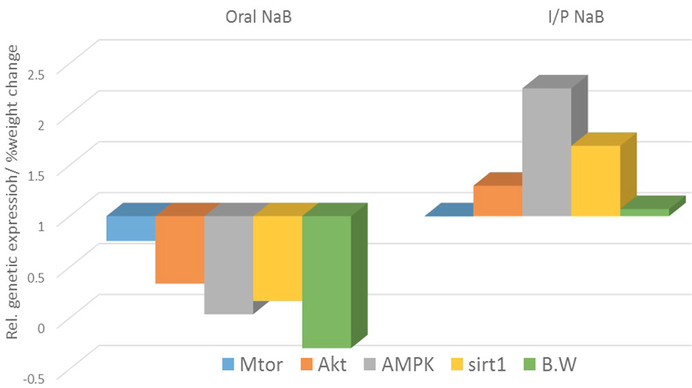
Relative genetic expression and weight change in mice treated with oral and intraperitoneal sodium buyrate.
References
- Bellaver, B., Bobermin, L.D., Souza, D.G., Rodrigues, M.D.N., de Assis, A.M., Wajner, M., Gonçalves, C.A., Souza, D.O. and Quincozes-Santos, A., 2016. Signaling mechanisms underlying the glioprotective effects of resveratrol against mitochondrial dysfunction. Biochim. biophys. Acta, 1862: 1827-1838. https://doi.org/10.1016/j.bbadis.2016.06.018[CrossRef]
- Biancotto, C., Frigè, G. and Minucci, S., 2010. Histone modification therapy of cancer. Adv. Genet., 70: 341-386. https://doi.org/10.1016/B978-0-12-380866-0.60013-7[CrossRef]
- Booth, A., Magnuson, A., Fouts, J. and Foster, M., 2015. Adipose tissue, obesity and adipokines: Role in cancer promotion. Horm. Mol. Biol. Clin. Investig., 21: 57-74. https://doi.org/10.1515/hmbci-2014-0037[CrossRef]
- Cai, H., Dong, L. Q. and Liu, F., 2016. Recent advances in adipose mTOR signaling and function: Therapeutic prospects. Trends pharmacol. Sci., 37: 303-317. https://doi.org/10.1016/j.tips.2015.11.011[CrossRef]
- Canani, R.B., Di Costanzo, M. and Leone, L., 2012. The epigenetic effects of butyrate: Potential therapeutic implications for clinical practice. Clin. Epigenet., 4: 4. https://doi.org/10.1186/1868-7083-4-4[CrossRef]
- Canani, R.B., Di Costanzo, M., Leone, L., Pedata, M., Meli, R. and Calignano, A., 2011. Potential beneficial effects of butyrate in intestinal and extraintestinal diseases. World J. Gastroenterol., 17: 1519. https://doi.org/10.3748/wjg.v17.i12.1519[CrossRef]
- Canfora, E.E., Jocken, J.W. and Blaak, E.E., 2015. Short-chain fatty acids in control of body weight and insulin sensitivity. Nat. Rev. Endocrinol., 11: 577. https://doi.org/10.1038/nrendo.2015.128[CrossRef]
- Cantó, C. and Auwerx, J., 2012. Targeting sirtuin 1 to improve metabolism: All you need is NAD+? Pharmacol. Rev., 64: 166-187. https://doi.org/10.1124/pr.110.003905[CrossRef]
- Cantó, C., Gerhart-Hines, Z., Feige, J.N., Lagouge, M., Noriega, L., Milne, J.C., Elliott, P.J., Puigserver, P. and Auwerx, J., 2009. AMPK regulates energy expenditure by modulating NAD+ metabolism and SIRT1 activity. Nature, 458: 1056-1060. https://doi.org/10.1038/nature07813[CrossRef]
- Cantoni, S., Galletti, M., Zambelli, F., Valente, S., Ponti, F., Tassinari, R., Pasquinelli, G., Galiè, N. and Ventura, C., 2013. Sodium butyrate inhibits platelet-derived growth factor-induced proliferation and migration in pulmonary artery smooth muscle cells through Akt inhibition. FEBS J., 280: 2042-2055. https://doi.org/10.1111/febs.12227[CrossRef]
- Cetrullo, S., D’Adamo, S., Tantini, B., Borzi, R.M. and Flamigni, F., 2015. mTOR, AMPK, and Sirt1: key players in metabolic stress management. Crit. Rev. Eukaryot. Gene Expr., 25: 59-75. https://doi.org/10.1615/CritRevEukaryotGeneExpr.2015012975[CrossRef]
- Chen, J., Ghazawi, F.M., Bakkar, W. and Li, Q., 2006. Valproic acid and butyrate induce apoptosis in human cancer cells through inhibition of gene expression of Akt/protein kinase B. Mol. Cancer, 5: 71. https://doi.org/10.1186/1476-4598-5-71[CrossRef]
- Crujeiras, A., Díaz-Lagares, A., Carreira, M., Amil, M. and Casanueva, F., 2013. Oxidative stress associated to dysfunctional adipose tissue: a potential link between obesity, type 2 diabetes mellitus and breast cancer. Free Radic. Res., 47: 243-256. https://doi.org/10.3109/10715762.2013.772604[CrossRef]
- El-Masry, O.S., Al-Sakkaf, K., Brown, B.L. and Dobson, P.R., 2015. Differential crosstalk between the AMPK and PI3K/Akt pathways in breast cancer cells of differing genotypes: Leptin inhibits the effectiveness of AMPK activation. Oncol. Rep., 34: 1675-1680. https://doi.org/10.3892/or.2015.4198[CrossRef]
- Gao, Z., Yin, J., Zhang, J., Ward, R.E., Martin, R.J., Lefevre, M., Cefalu, W.T. and Ye, J., 2009. Butyrate improves insulin sensitivity and increases energy expenditure in mice. Diabetes, 58: 1509-1517. https://doi.org/10.2337/db08-1637[CrossRef]
- Hardie, D.G., Schaffer, B.E. and Brunet, A., 2016. AMPK: An energy-sensing pathway with multiple inputs and outputs. Trends Cell Biol., 26: 190-201. https://doi.org/10.1016/j.tcb.2015.10.013[CrossRef]
- Kauppinen, A., Suuronen, T., Ojala, J., Kaarniranta, K. and Salminen, A., 2013. Antagonistic crosstalk between NF-κB and SIRT1 in the regulation of inflammation and metabolic disorders. Cell Signal., 25: 1939-1948. https://doi.org/10.1016/j.cellsig.2013.06.007[CrossRef]
- Khan, S. and Jena, G., 2016. Sodium butyrate reduces insulin-resistance, fat accumulation and dyslipidemia in type-2 diabetic rat: A comparative study with metformin. Chem. Biol. Interact., 254: 124-134. https://doi.org/10.1016/j.cbi.2016.06.007[CrossRef]
- Kyrylenko, S., Kyrylenko, O., Suuronen, T. and Salminen, A., 2003. Differential regulation of the Sir2 histone deacetylase gene family by inhibitors of class I and II histone deacetylases. Cell. Mol. Life Sci., 60: 1990-1997. https://doi.org/10.1007/s00018-003-3090-z[CrossRef]
- Lage, R., Diéguez, C., Vidal-Puig, A. and López, M., 2008. AMPK: A metabolic gauge regulating whole-body energy homeostasis. Trends Mol. Med., 14: 539-549. https://doi.org/10.1016/j.molmed.2008.09.007[CrossRef]
- Lee, B.C. and Lee, J., 2014. Cellular and molecular players in adipose tissue inflammation in the development of obesity-induced insulin resistance. Biochim. biophys. Acta, 1842: 446-462. https://doi.org/10.1016/j.bbadis.2013.05.017[CrossRef]
- Lee, M.J., Wu, Y. and Fried, S.K., 2013. Adipose tissue heterogeneity: implication of depot differences in adipose tissue for obesity complications. Mol. Aspects Med., 34: 1-11. https://doi.org/10.2119/molmed.2012.00308[CrossRef]
- Legrand, P. and Rioux, V., 2015. Specific roles of saturated fatty acids: Beyond epidemiological data. Eur. J. Lipid Sci. Technol., 117: 1489-1499. https://doi.org/10.1002/ejlt.201400514[CrossRef]
- Li, F., Cheng, K., Lam, K., Vanhoutte, P. and Xu, A., 2011. Cross-talk between adipose tissue and vasculature: role of adiponectin. Acta Physiol., 203: 167-180. https://doi.org/10.1111/j.1748-1716.2010.02216.x[CrossRef]
- Li, F., Yang, H., Duan, Y. and Yin, Y., 2011. Myostatin regulates preadipocyte differentiation and lipid metabolism of adipocyte via ERK1/2. Cell Biol. Int., 35: 1141-1146. https://doi.org/10.1042/CBI20110112[CrossRef]
- Li, H., Gao, Z., Zhang, J., Ye, X., Xu, A., Ye, J. and Jia, W., 2012. Sodium butyrate stimulates expression of fibroblast growth factor 21 in liver by inhibition of histone deacetylase 3. Diabetes, 61: 797-806. https://doi.org/10.2337/db11-0846[CrossRef]
- Masud, F., Shakoori, T.A., Khawaja, K.I., Ali, M. and Ameer, F., 2016. Changes in body weight and lipid profile of mice treated with sodium butyrate and metformin. Pakistan J. Zool., 48: 1343-1348.
- Mollica, M.P., Raso, G.M., Cavaliere, G., Trinchese, G., De Filippo, C., Aceto, S., Prisco, M., Pirozzi, C., Di Guida, F. and Lama, A., 2017. Butyrate regulates liver mitochondrial function, efficiency, and dynamic, in insulin resistant obese mice. Diabetes, 66: 1405-1418. https://doi.org/10.2337/db16-0924[CrossRef]
- Nieman, K.M., Romero, I.L., Van Houten, B. and Lengyel, E., 2013. Adipose tissue and adipocytes support tumorigenesis and metastasis. Biochim. biophys. Acta, 1831: 1533-1541. https://doi.org/10.1016/j.bbalip.2013.02.010[CrossRef]
- O’Neill, H.M., Holloway, G.P. and Steinberg, G.R., 2013. AMPK regulation of fatty acid metabolism and mitochondrial biogenesis: implications for obesity. Mol. cell. Endocrinol., 366: 135-151. https://doi.org/10.1016/j.mce.2012.06.019[CrossRef]
- Pant, K., Saraya, A. and Venugopal, S.K., 2017. Oxidative stress plays a key role in butyrate-mediated autophagy via Akt/mTOR pathway in hepatoma cells. Chem. Biol. Interact., 273: 99-106. https://doi.org/10.1016/j.cbi.2017.06.001[CrossRef]
- Park, J., Kim, M., Kang, S.G., Jannasch, A.H., Cooper, B., Patterson, J. and Kim, C.H., 2015. Short-chain fatty acids induce both effector and regulatory T cells by suppression of histone deacetylases and regulation of the mTOR–S6K pathway. Mucosal Immunol., 8: 80-93. https://doi.org/10.1038/mi.2014.44[CrossRef]
- Peng, L., Li, Z.R., Green, R.S., Holzman, I.R. and Lin, J., 2009. Butyrate enhances the intestinal barrier by facilitating tight junction assembly via activation of AMP-activated protein kinase in Caco-2 cell monolayers. J. Nutr., 139: 1619-1625. https://doi.org/10.3945/jn.109.104638[CrossRef]
- Price, N.L., Gomes, A.P., Ling, A.J., Duarte, F.V., Martin-Montalvo, A., North, B.J., Agarwal, B., Ye, L., Ramadori, G. and Teodoro, J.S., 2012. SIRT1 is required for AMPK activation and the beneficial effects of resveratrol on mitochondrial function. Cell Metab., 15: 675-690. https://doi.org/10.1016/j.cmet.2012.04.003[CrossRef]
- Pulliam, S.R., Pellom, S.T., Shanker, A. and Adunyah, S.E., 2016. Butyrate regulates the expression of inflammatory and chemotactic cytokines in human acute leukemic cells during apoptosis. Cytokine, 84: 74-87. https://doi.org/10.1016/j.cyto.2016.05.014[CrossRef]
- Rada-Iglesias, A., Enroth, S., Ameur, A., Koch, C.M., Clelland, G.K., Respuela-Alonso, P., Wilcox, S., Dovey, O.M., Ellis, P.D. and Langford, C.F., 2007. Butyrate mediates decrease of histone acetylation centered on transcription start sites and down-regulation of associated genes. Genome Res., 17: 708-719. https://doi.org/10.1101/gr.5540007[CrossRef]
- Ruderman, N.B., Carling, D., Prentki, M. and Cacicedo, J.M., 2013. AMPK, insulin resistance, and the metabolic syndrome. J. clin. Invest., 123: 2764-2772. https://doi.org/10.1172/JCI67227[CrossRef]
- Schmittgen, T.D. and Livak, K.J., 2008. Analyzing real-time PCR data by the comparative CT method. Nat. Protoc., 3: 1101-1108. https://doi.org/10.1038/nprot.2008.73[CrossRef]
- Sengupta, S., Peterson, T.R. and Sabatini, D.M., 2010. Regulation of the mTOR complex 1 pathway by nutrients, growth factors, and stress. Mol. Cell, 40: 310-322. https://doi.org/10.1016/j.molcel.2010.09.026[CrossRef]
- Tan, J., McKenzie, C., Potamitis, M., Thorburn, A.N., Mackay, C.R. and Macia, L., 2014. The role of short-chain fatty acids in health and disease. Adv. Immunol., 121: e119. https://doi.org/10.1016/B978-0-12-800100-4.00003-9[CrossRef]
- Terova, G., Díaz, N., Rimoldi, S., Ceccotti, C., Gliozheni, E. and Piferrer, F., 2016. Effects of sodium butyrate treatment on histone modifications and the expression of genes related to epigenetic regulatory mechanisms and immune response in European sea bass (Dicentrarchus Labrax) fed a plant-based diet. PLoS One, 11: e0160332. https://doi.org/10.1371/journal.pone.0160332[CrossRef]
- Viollet, B., Horman, S., Leclerc, J., Lantier, L., Foretz, M., Billaud, M., Giri, S. and Andreelli, F., 2010. AMPK inhibition in health and disease. Crit. Rev. Biochem. mol. Biol., 45: 276-295. https://doi.org/10.3109/10409238.2010.488215[CrossRef]
- Wu, Y., Song, P., Xu, J., Zhang, M. and Zou, M.H., 2007. Activation of protein phosphatase 2A by palmitate inhibits AMP-activated protein kinase. J. biol. Chem., 282: 9777-9788. https://doi.org/10.1074/jbc.M608310200[CrossRef]
- Xu, J., Ji, J. and Yan, X.H., 2012. Cross-talk between AMPK and mTOR in regulating energy balance. Crit. Rev. Fd. Sci. Nutr., 52: 373-381. https://doi.org/10.1080/10408398.2010.500245[CrossRef]
- Yan, H. and Ajuwon, K.M., 2015. Mechanism of butyrate stimulation of triglyceride storage and adipokine expression during adipogenic differentiation of porcine stromovascular cells. PLoS One, 10: e0145940. https://doi.org/10.1371/journal.pone.0145940[CrossRef]
- Yuan, H.X., Xiong, Y. and Guan, K.L., 2013. Nutrient sensing, metabolism, and cell growth control. Mol. Cell, 49: 379-387. https://doi.org/10.1016/j.molcel.2013.01.019[CrossRef]
- Zhang, J., Yi, M., Zha, L., Chen, S., Li, Z., Li, C., Gong, M., Deng, H., Chu, X. and Chen, J., 2016. Sodium butyrate induces endoplasmic reticulum stress and autophagy in colorectal cells: Implications for apoptosis. PLoS One, 11: e0147218. https://doi.org/10.1371/journal.pone.0147218[CrossRef]
- Zhou, J., Gao, S., Chen, J., Zhao, R. and Yang, X., 2016. Maternal sodium butyrate supplement elevates the lipolysis in adipose tissue and leads to lipid accumulation in offspring liver of weaning-age rats. Lipids Hlth. Dis., 15: 119. https://doi.org/10.1186/s12944-016-0289-1[CrossRef]
Services
Article usage statistics
Loading…