
Colon cancer often has problems of recurrence and chemotherapy resistance in the late stage. Metabolic reprogramming is one of the characteristics of colon tumors and contributes to autophagy and cell cycle progression of tumors. A ketogenic diet (KD) is a high-fat, adequate-protein, and low-carbohydrate diet with documented anti-tumor effects. However, the mechanism of KD inhibiting colon cancer remains unclear. In this study, we investigated the molecular mechanism underlying the effects of KD on cell cycle and autophagy in colon tumor-bearing mice. Our results showed that compared with the SD group, KD treatment upregulated the expressions of autophagy-related proteins LC3-II and Beclin-1 while downregulated the expression of p62 protein in CT26+ tumor-bearing mice. In parallel, the expressions of CDK4-Cyclin D1 and CDK2-Cyclin E proteins were decreased, while the expression of p21 and p16 proteins increased in KD group. The data analysis suggested that KD promoted autophagy and blocked the tumor cell cycle in the G1/S phase. In addition, the western blot showed that KD significantly downregulated the expressions of PI3K, p-Akt, p-mTOR, HDAC3, p-JAK2 and p-STAT3 proteins. In vitro results (RGFP966, an HDAC3 inhibitor, and NSC74859, a STAT3 inhibitor) also supported those of the in vivo findings. Overall, the current study demonstrated that KD induced autophagy and G1/S phase cell cycle arrest in tumors by inhibiting the activation of the PI3K/Akt/mTOR, STAT3/HDAC3 and JAK2/STAT3 signaling pathways. Together, we can conclude that KD prevents colon cancer progression by regulating autophagy and cell cycle arrest of tumor cells.
Research Article · Volume 57, Issue 1, 8 January 2025 · Open Access
Dynamic Effects of Ketogenic Diet on Autophagy and Cell Cycle in a Mouse Model of CT26+ Colon Cancer
· · · · · · · · · ·
Affiliations & Notes ⌄
- 1Institute of Biopharmaceutical Research, Liaocheng University, Liaocheng, Shandong 252000, China 2Department of Veterinary Pathology, Faculty of Veterinary and Animal Sciences PMAS-Arid Agriculture University, Rawalpindi 46000, Pakistan 3Shandong Donkey Industry, Technology Collaborative Innovation Center, Liaocheng University, Liaocheng, China 4Department of Clinical Studies, Faculty of Veterinary Sciences, Bahauddin Zakariya University, Multan 60800, Pakistan 5Department of Parasitology and Mi
Article Info ⌄
- Journal:
- Pakistan Journal of Zoology
- Published:
- 8 January 2025

From the article
- Therefore, we studied the effect of KD on autophagy and cell cycle to investigate the mechanisms underlying the KD-mediated inhibition of cell proliferation.
- Our study found that KD treatment significantly reduced the phosphorylation levels of JAK2 and STAT3, and the expression level of cell cycle-related proteins (including Cyclin D1, CDK4, Cyclin E and CDK2) and increased the expression of CDKI (including p21 and p16).
- Meanwhile, NSC74859 further confirmed that CT26+ colon cell cycle arrest was mediated by inhibiting JAK2/STAT3 signaling pathway.
- Our results demonstrated that KD-induced colon cancer cells autophagy mainly by inhibiting STAT3/HDAC3 and PI3K/Akt/mTOR signaling pathways, and KD reduced cell proliferation by inhibiting the G1/S phase transition during the cell cycle.
Selected from the article's own text.
Abstract
INTRODUCTION
Colon cancer is one of the leading causes of death among all malignant cancers . The current treatment options available for colon cancer include surgery , radiotherapy , and chemotherapy . However, these therapies have some side effects and limitations. Therefore, it is urgent to search for a novel strategy for the treatment of colon cancer. Metabolic reprogramming is a hallmark of tumor cells and critical for cell survival and proliferation . The ketogenic diet (KD) is a high-fat, low carbohydrate and appropriate protein diet. It has been reported that KD could inhibit tumor development by regulating metabolic reprogramming. Moreover, combining KD with traditional anti-cancer strategies, such as conventional chemotherapy and radiotherapy, has been considered an effective anti-tumor method therapy . KD can be used as an adjuvant treatment for gastric cancer , malignant glioma , prostate cancer and colon cancer . Although KD is increasingly used in clinical practice as an adjunctive therapy for cancer, anti-cancer mechanisms need to be further explored.
Autophagy is a lysosomal degradation pathway that widely exists in eukaryotic cells . Many factors can induce autophagy, such as hypoxia and DNA damage . Excessive autophagy can result in cell death, called autophagic cell death . A recent study has demonstrated that resveratrol inhibited colon tumor cell growth by activating autophagy via inhibiting the mammalian target of the rapamycin (mTOR) signaling pathway . Autophagy exerts different regulatory roles according to the stage of the tumor. Autophagy can inhibit tumor growth by removing damaged organelles and proteins, but once a tumor begins to develop, it can promote proliferation, invasion, and adaptability of tumor cells . Therefore, according to the characteristics of autophagy in different stages of tumor tumorigenesis, the regulation of autophagy can be used as one of the effective treatment strategies for cancer treatment.
Cell cycle is a highly regulated process critical to normal growth and division , and mainly divided into G0/G1, S and G2/M phases . Cell cycle is mainly regulated by cyclin and cyclin-dependent kinases (CDKs) in the nucleus . Cyclin interacts with the corresponding CDKs and forms Cyclin-CDK complexes, which regulate cell cycle progression . Cyclin E combines with CDK2 to promote retinoblastoma protein phosphorylation, thereby controlling the cell cycle and the normal process of DNA replication, and enabling cells to pass through G1/S phase smoothly. A previous study demonstrated that the activity of Cyclin E-CDK2 complex was increased in colon cancer . Cyclin D1 and CDK4/6 can regulate the cell cycle transition from G1 to S phase and integrate mitosis-related signals . Cyclin D1 is frequently dysregulated in many tumor types, leading to the rapid growth of tumor cells and ultimately promoting the tumor development process . Tobias Otto et al. demonstrated that overexpression of Cyclin D1 and CDK4 could promote tumor development . As an inhibitor of CDK2, p16 deletion has been shown to accelerate the cell cycle progress . Moreover, p21 specifically interferes with Cyclin D1 binding as a CDK4 inhibitor, resulting in G1 arrests and/or S phase entry . Therefore, blocking the activity of CDK2-Cyclin E and CDK4-Cyclin D1 complexes and increasing the expression of p16 and p21 proteins can effectively inhibit the cycle process of tumor cells and induce their apoptosis.
Histone deacetylases (HDACs) are involved in cell migration and invasion and are overexpressed in some cancers, such as colon cancer , breast cancer and liver cancer . HDACs inhibitors can induce tumor cell apoptosis or programmed cell death , and was used to treat neurodegenerative diseases and diabetes . HDAC3 upregulation predicted poor prognosis . RGFP966 acts as an inhibitor of HDAC3 and plays an inhibitory role in different types of cancer, such as prostate cancer and colon cancer ; . Furthermore, it has been reported that the signal transducer and activator of transcription 3 (STAT3) intersect cancer-related signal pathways and can be activated by cytokines, growth factors, and oncogene signals . It is highly expressed in tumor cells and participates in many cellular processes, such as survival, angiogenesis, cell cycle and differentiation of tumor cells . NSC74859, an inhibitor of STAT3, also known as S31-201, was reported to selectively inhibit dimerization, phosphorylation, and nuclear translocation of STAT3. It has been reported to play a tumor-suppressing role in breast, glioma and liver cancer ; ; . Targeting STAT3 has now become the main focus of drug development in cancer.
As a downstream target of phosphatidylinositol-3-kinase (PI3K) and Akt, mTOR is overexpressed in tumors. Activation of mTOR promotes tumor growth and metastasis , while mTOR inactivation inhibits tumor development in colorectal carcinoma . The PI3K/Akt/mTOR signaling pathway can regulate autophagy which is essential in tumor initiation and progression . In addition, Janus Kinases 2 (JAK2)/STAT3 signaling pathway is associated with cell cycle arrest in tumor cells .
In the present study, we explored underlying anticancer mechanisms of KD on CT26+ murine colon cancer by determining cell cycle and autophagy.
MATERIALS AND METHODS
Animals
Seven-week-old male BALB/c mice (weight approximately 20g) were purchased from Pengyue Experimental Animal Breeding (Jinan, China). All the mice were fed in a specific pathogen-free (SPF) mouse facility (temperature was maintained at 23 ± 2 ℃), 60-65% humidity, with free access to food and water, and the dark cycle of 12h/12h. The experimental protocol was reviewed and approved by the Animal Care and Use Committee of Liaocheng University (permit number 2021111030).
Tumor implantation
Approximately one million CT26+ cells were implanted subcutaneously in BALB/c mice.
Ketogenic diet treatments
Colon cancer CT26 cells were inoculated into BALB/c mice, SD and KD dietary interventions were initiated on the seventh day after tumor inoculation. The macronutrient composition of KD (% w/w) was: 1.0% carbohydrate, 69% fat, and 16.25% protein, while SD was 62.6% carbohydrate, 7% fat and 20% protein. The macronutrients of KD used in our experiment refer to our previous research .
Tissue preparation
After 15 days of tumor inoculation, mice were euthanized, and the tumor was removed. Tissue samples were partly embedded in paraffin and cut into serial transverse sections (5μm). Another part of the tumor tissue was put into liquid nitrogen immediately and stored at −80 °C .
Cell culture
The CT26+ cell line was obtained from RIKEN BioResource Center (Tsukuba, Japan). Cells were cultured in RPMI 1640 medium (Gibco, Carlsbad, CA, USA) supplemented with 10% fetal bovine serum (FBS; Gibco, Carlsbad, CA, USA) and 1% penicillin-streptomycin. All cells were cultured in a sterile incubator at 37°C and 5% CO2.
CCK8 assay
Cell viability was assessed by Cell Counting Kit-8 (CCK8) assay according to the manufacturer’s protocols . CT26 + cells were plated at a density of 5x10³ cells per well on 96-well microplates in 100 μL of culture medium. The CT26+ cells were seeded in 96-well microplates at the density of 5 × 10³ per well. RGFP966 (an HDAC3 inhibitor) and NSC74859 (a STAT3 inhibitor) were purchased from Apex Bio (Houston, USA). Cells were treated with different concentrations of RGFP966 and NSC74859, after incubation of 24h, 10 μl CCK8 reagent was added to all wells, and incubation was continued for two hours. Three replicate wells were set up for each experiment. The absorbance of each well was analyzed at 450 nm using a Microplate Reader. Wells without cells were used as blanks. Additionally, RGFP966 at 15 μM could specifically inhibit HDAC3 and has no inhibitory effect on other HDACs . NSC74859 at 100 μM could specifically inhibit STAT3 activation . Therefore, 15μM RGFP966 and 100μM NSC74859 were used as reported before.
Flow cytometry
CT26+ cells were cultured in 6-well microplates at the density of 2 × 10⁵ per well in RPMI 1640 medium for 24h. RGFP966 (15μM) and NSC74859 (100μM) were added into the cells for 24h. Cells were collected into a centrifuge tube and centrifuged at 1000 × g/min for 5 min to precipitate the cells. Centrifuged cells were suspended in PBS and adjusted the cell concentration to 1 × 10⁶/ml at 1000 × g/min for 5 min to precipitate cells. 1ml of 70 % cold ethanol was added and cells were fixed overnight. On the second day, cells were washed with the fixing solution with pre-cooled PBS before dyeing. 100 μ L of RNase A was added and the cells were placed at 37 ℃ for 30 minutes. 500 μ L propidium iodide staining solution was added to the cells and incubated in the dark room at 4℃ for 30 minutes . Red fluorescence and light were detected and analyzed using a flow cytometer set at the excitation wavelength of 488nm.
Western blotting
The total proteins from tumor cells or tissues were extracted by RIPA buffer (Beyotime, China) with 1% protease inhibitor and 1% phosphatase inhibitor. Protein extracts were separated by sodium dodecyl sulfate-polyacrylamide gel electrophoresis (SDS-PAGE) and transferred to polyvinylidene difluoride (PVDF) membranes . Then 5% nonfat milk in Tris-buffered saline solution was added to block the membranes for 2h at room temperature (RT). After that, the membranes were incubated with primary antibodies at 4℃ overnight. The membranes were rinsed three times and then incubated with anti-rabbit IgG (H+L) secondary antibody (1:1,0000, Proteintech) and anti-mouse IgG (H+L) secondary antibody (1:1,0000, Proteintech) at room temperature for 1h. After the membranes were washed three times, the proteins were visualized using a chemiluminescent detection system (Amersham Biosciences, NJ, USA). Relative expression levels of proteins in different groups were analyzed. Each experiment was repeated three times. β-actin was used as the internal control. All the antibodies used are shown in Table I.
Table I. Antibodies of Western blot experiment.
| Protein | Catalog No | Molecular weight | Manufacturers | Dilution ratio |
| PI3K | 67121-1-Ig | 110kDa | Proteintech | 1:1000 |
| Akt P-Akt | 60203-2-Ig ab38449 | 60kDa 56 kDa | Proteintech abcam | 1:5000 1:1000 |
| mTOR p- mTOR JAK2 | ab137133 ab109268 ab108596 | 289 kDa 289kDa 131kDa | abcam abcam abcam | 1:2000 1:5000 1:5000 |
| p-JAK2 | ab32101 | 120kDa | abcam | 1:5000 |
| STAT3 | 10253-2-AP | 88kDa | Proteintech | 1:2000 |
| p-STAT3 HDAC3 | ab267373 ab32369 | 88kDa 49kD | abcam abcam | 1:1000 1:5000 |
| p16 Beclin-1 p62 p21 LC3 Cyclin D1 Cyclin E CDK2 CDK4 β-actin Goat anti-rabbit Goat anti-mouse | 10883-1-AP 11306-1-AP ab109012 ab109199 14600-1-AP 60186-1-Ig 11554-1-AP ab32147 ab108357 66009-1-Ig SA00001-2 SA00001-1 | 16kDa 52-60kDa 62kDa 21kDa 14,16kDa 34kDa 47kDa 33kDa 34kDa 42kDa | Proteintech Prteintech abcam abcam Proteintech Proteintech Proteintech abcam abcam Proteintech Proteintech Proteintech | 1:4000 1:4000 1:20000 1:1000 1:2000 1:5000 1:1000 1:2000 1:2000 1:10000 1:10000 1:10000 |
Statistical analyses
All the data were statistically performed by SPSS 24.0 software. This paper used GraphPad Prism software (GraphPad Software Inc., San Diego, CA, USA) for statistical analysis and mapping. All data were expressed as mean ± standard error of the mean (SEM) in each case. Independent-samples T-test was used when two sets of data were compared. Moreover, comparisons of more than two sets of data were performed using a one-way analysis of variance (ANOVA). Significant differences between different groups were indicated as *p < 0.05, **p < 0.01, ***p < 0.001 and ns: no significance.
RESULTS
Our previous results have shown that KD inhibited the development of CT26+ colon cancer by enhancing immune response, inhibiting angiogenesis and epithelial-mesenchymal transition (EMT) . However, the effect of KD on autophagy and cell cycle is still unclear. Therefore, we studied the effect of KD on autophagy and cell cycle to investigate the mechanisms underlying the KD-mediated inhibition of cell proliferation.
KD induced autophagy in CT26+ colon cancer mouse model
Beclin-1, LC3, and p62 are regarded as autophagy-related proteins. In the early stages of cancer development, autophagy contributes to the suppression of tumor growth . However, whether KD can cause autophagy in colon cancer has not been reported. In the present study, the protein expression levels of Beclin-1, LC3 and p62 in the tumor tissues were measured by western blot analysis. Compared with the SD group, Beclin-1 and LC3 protein expression levels were significantly increased in the KD group (Fig. 1B, C, *p < 0.05 and ***p < 0.001). In contrast, the expression level of p62 was significantly decreased, as shown in Figure 1D (*p < 0.05). These results suggested that KD treatment induced the accumulation of autophagosomes in the mouse CT26+ colon cancer model.
STAT3/HDAC3 and PI3K/Akt/mTOR expressions were downregulated after KD treatment
The PI3K/Akt/mTOR pathway is closely associated with autophagy regulation . Phosphorylated STAT3 enters the nucleus and interacts with transcription factors, leading to increased transcriptional initiation. STAT3 can inhibit autophagy by recruiting HDAC3 . Thus, we detected the expression of PI3K, p-Akt, Akt, p-mTOR, mTOR, HDAC3, p-STAT3 and STAT3 proteins in CT26+ colon cancer. Our results suggested that after KD treatment, the total expression levels of Akt, mTOR and STAT3 proteins remained unchanged, while the phosphorylation levels of Akt, mTOR and STAT3 decreased significantly (Fig. 2C, D, F, *p < 0.05, **p < 0.01 and ***p < 0.001), and the expression of PI3K and HDAC3 proteins were significantly decreased (Fig. 2B, E, **p < 0.01). These results suggested that KD-regulated autophagy was related to STAT3/HDAC3 and PI3K/Akt/mTOR pathways.
STAT3/HDAC3 regulated autophagy by PI3K/Akt/mTOR pathway
To further explore the relationship between STAT3/HDAC3 and autophagy, we investigated Beclin-1, LC3, and p62 protein expression levels in CT26+ colon cancer cells with RGFP966 and NSC74859 treatments. Firstly, we examined whether RGFP966 and NSC74859 could affect the viability of CT26+ cells by a CCK8 assay. Our results revealed the dose-dependent cytotoxicity of RGFP966 and NSC74859 on CT26+ cells (Fig. 3A, B). RGFP966 at 15 μM could specifically inhibit HDAC3 and has no
inhibitory effect on other HDACs . As previously reported, low cytotoxic concentration (100 μM) NSC74859 was selected to inhibit STAT3 activation specifically for our experiments . In addition, the result of CCK8 also suggested the used doses of RGFP966 and NSC74859 are 15 and 100 μM, respectively. Thus, RGFP966 at 15μM and NSC74859 at 100 μM were used for subsequent experiments. In addition, western blot revealed that the expression of Beclin-1 and LC3 proteins increased (Fig. 3D, E, **p < 0.01 and ***p < 0.001), while the expression of p62 protein decreased (Fig. 3F, *p < 0.05 and **p < 0.01) both in RGFP966 and NSC74859 treated group. Moreover, we also found that the RGFP966 and NSC74859 treatment downregulated the expression of PI3K protein, ratio of p-Akt/Akt and ratio of p-mTOR/mTOR when compared with the control group (Fig. 3G-J, *p < 0.05, **p < 0.01, ***p < 0.001 and ns: no significance). These results suggested that the PI3K/Akt/mTOR signaling pathway was involved in autophagy regulation, inhibiting the HDAC3 and STAT3 promotes autophagy.
KD induced cell cycle arrest in G1/S phase
Some studies have indicated that the Cyclin D1-CDK 4 complex regulates cells in the G0/G1 phase, while the Cyclin E-CDK 2 complex controls cells from the G0/G1 phase to the S phase . To investigate whether KD treatment affects the cell cycle, the expressions of Cyclin E, CDK2, Cyclin D, CDK4, p16 and p21 were detected by western blot. Compared with the SD group, CDK2, CDK4, Cyclin E and Cyclin D1 expression levels were decreased (Fig. 4B-E, *p < 0.05, **p < 0.01 and ***p < 0.001), whereas p16 and p21 expressions were significantly increased in KD group (Fig. 4F, G, *p < 0.05, **p < 0.01 and ***p < 0.001). These results showed that KD treatment led to the arrest of cell cycle in G1/S phase in CT26+ colon cancer mouse model.
JAK2/STAT3 expression were downregulated after KD treatment
It has been reported that JAK2/STAT3 signaling pathway inhibition results in cell cycle arrest . Western blot results showed the total expression levels of JAK2 protein remained unchanged, while the phosphorylation levels of JAK2 significantly decreased (Fig. 5A, B, *p < 0.05) in KD treated group compared with the control group. Correspondingly, the level of phosphorylated STAT3 and cycle-associated proteins were decreased in the KD group. Thus, our results suggested that the cell cycle arrest induced by KD might be associated with the JAK2/STAT3 signaling pathway.
STAT3 induced cell cycle arrest at G1/S phase
The cell cycle was detected by western blot and flow cytometry to analyze further the effect of STAT3/HDAC3 on the cell cycle of CT26+ colon cancer. Western blot results suggested that NSC74859 treatment significantly reduced the expression of CDK4, Cyclin D1, CDK2 and Cyclin E, while increasing the expression of p21 and p16 in CT26+ colon cancer cell lines (Fig. 6B-G, *p < 0.05, **p < 0.01, ***p < 0.001 and ns: no significance). In addition, upon RGFP966 induction, the expression of p21 and p16 was increased. However, CDK4, Cyclin D1, CDK2 and Cyclin E proteins expression levels were not affected by RGFP966 treatment (Fig. 6B-G, *p < 0.05, **p < 0.01, ***p < 0.001 and ns: no significance).
In addition, flow cytometry results showed that 24h treatment with NSC74859 decreased the G0/G1 phase DNA content from 48.4% to 37% and decreased the G2/M phase DNA content from 15.6% to 4.86%, whereas the S phase DNA content increased, from 14.4% to 31.3%, when compared with the control group. Meanwhile, RGFP966 treatment decreased the G0/G1 phase DNA content from 48.4% to 23.3% and decreased G2/M phase DNA content from 15.6% to 2.16%. At the same time, the S phase DNA content decreased from 23.4% to 13.9%, compared with the control group (Fig. 6H). The results suggested that inhibition of STAT3 induced G1/S cell cycle arrest in CT26+ colon cancer cells.
DISCUSSION
We have previously shown that KD suppressed proliferation, growth, and invasion of CT26+ colon cancer via inhibiting tumor angiogenesis and altering the tumor microenvironment . Moreover, our previous research results also showed that KD inhibits the growth of CT26+ tumor volumes in vivo . However, the effects of KD on autophagy and cell cycle arrest in CT26+ colon cancer have not been reported. Our study found that (1) RGFP966 combined with NSC74859 enhances autophagy inhibition by strongly reducing HADC3 and STAT3 activation, the results showed the autophagy induced by KD in the CT26+ colon cancer mouse model was regulated by STAT3/HDAC3 and PI3K/Akt/mTOR signal pathways; (2) RGFP966 combined with NSC74859 strongly reduces HADC3 and STAT3 activation, and
we found that treatment of NSC blocked G1/S cell cycle. Therefore, the G1/S phase cell cycle arrest induced by KD was mainly regulated by JAK2/STAT3 signal pathway. As a way of programmed cell death, autophagy is a catabolic process that can degrade and recycle damaged organelles, lipids and cellular proteins , enabling some cells to survive in harsh environments . Autophagy plays different roles in different stages and types of tumor growth. At the early stage of tumor development, autophagy acts as a tumor suppressor, and in advanced stages, autophagy promotes tumor development. Some drugs can induce autophagy, leading to tumor cell death . He et al. (2020) established the neuroblastoma BALB/c-nu mouse model and concluded that KD exerts an anti-tumor effect by inducing autophagy . A previous study has shown that combining curcumin and temozolomide could induce autophagy and promote apoptosis of glioblastoma cells . PI3K/Akt/mTOR signaling pathway is a classic pathway involved in the regulation of autophagy. STAT3 also can inhibit autophagy and promote migration/invasion of primary cancers, such as liver cancer . Our research showed that KD contributed to the up-regulation of autophagy-related protein expression and down-regulation of PI3K, p-Akt, p-mTOR, p-STAT3 and HDAC3 expression, suggesting that KD could effectively reduce the growth of CT26+ tumor cells via promotion of autophagy through STAT3/HDAC3 and PI3K/AKT/mTOR signaling pathways.
The occurrence and development of tumors is a multifactor and gradual development process, which includes abnormal cell cycle regulation. Inhibiting the expression of CDK4/6 can effectively suppress the development of tumors . It has been reported that anthraquinone derivative C10 blocked the cell cycle of colon cancer cells via JAK2/STAT3 signal pathway . Bauerane induced S-phase cell cycle arrest of A549 human lung cancer cells through PI3K/Akt and STAT3 signaling pathways . Our study found that KD treatment significantly reduced the phosphorylation levels of JAK2 and STAT3, and the expression level of cell cycle-related proteins (including Cyclin D1, CDK4, Cyclin E and CDK2) and increased the expression of CDKI (including p21 and p16). Meanwhile, NSC74859 further confirmed that CT26+ colon cell cycle arrest was mediated by inhibiting JAK2/STAT3 signaling pathway. Based on the above analysis, the JAK2/STAT3 signaling pathway mainly regulated KD-induced cell cycle arrest.
Our results demonstrated that KD-induced colon cancer cells autophagy mainly by inhibiting STAT3/HDAC3 and PI3K/Akt/mTOR signaling pathways, and KD reduced cell proliferation by inhibiting the G1/S phase transition during the cell cycle. The specific mechanism of action is shown in Figure 7.
Funding
This work was supported by Natural Science Foundation of Shandong province (ZR2020QH112), Open Project of Liaocheng University Animal Husbandry Discipline (319312101-03, 319312101-25, 319312101-26 and 319462207-21), Open Project Program of State Key Laboratory of Food Science and Technology, Jiangnan University (SKLF-KF-202112), Youth Innovative Science and Technology Program of Shandong Colleges and University (2021KJ099), Shandong Rural Revitalization Science and Technology Innovation Action Plan (Key technology innovation and demonstration of Integrated development of Dong-E Black Donkey industry, 2021TZXD012) and Taishan Scholar Research Foundation (319190201). This work was also technically supported by Shandong Collaborative Innovation Center for Antibody Drugs and Engineering Research Center for Nanomedicine and Drug Delivery Systems.
IRB approval and ethical statement
All animal experiments were approved by the Research Ethics Committee of Liaocheng University (permit number 2022111030).
Data availability statement
The data used to support the findings of this study are available from the corresponding author upon request.
Statement of conflict of interest
The authors have declared no conflict of interest.
Figures
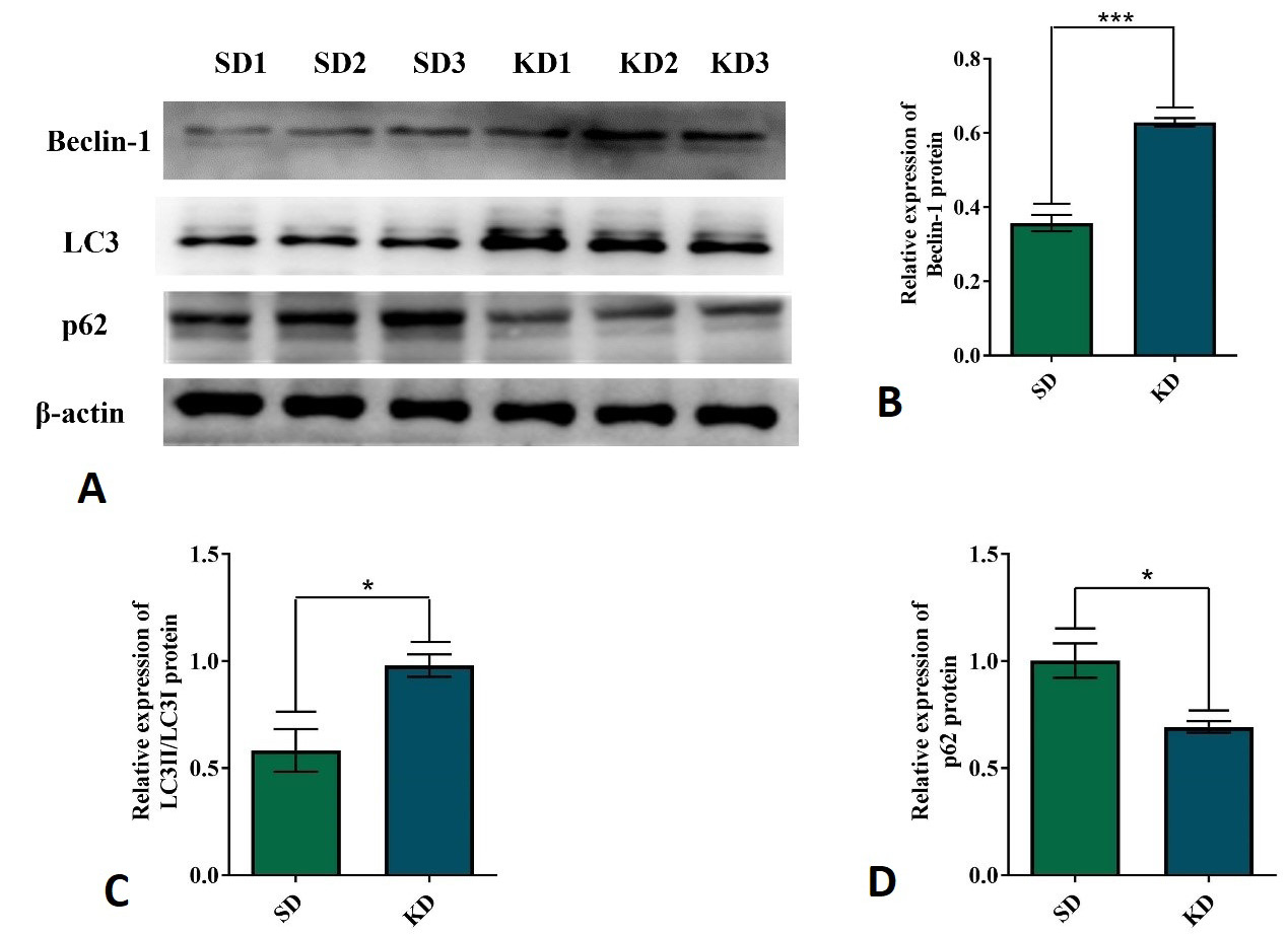
KD induced autophagy in CT26+ colon cancer mouse model. (A) The representative expression of Beclin-1, LC3, and p62 proteins in CT26+ tumor tissues and β-actin as the internal control for normalization. Quantitative analysis of Beclin-1 (B), LC3 (C), and p62 (D) proteins. Each experiment was repeated thrice, and all the data were presented as mean ± SEM in each case. Independent-sample T-test was used when different sets of data were compared. Statistically significant differences between different groups were indicated as *p < 0.05, **p < 0.01 and ***p < 0.001.
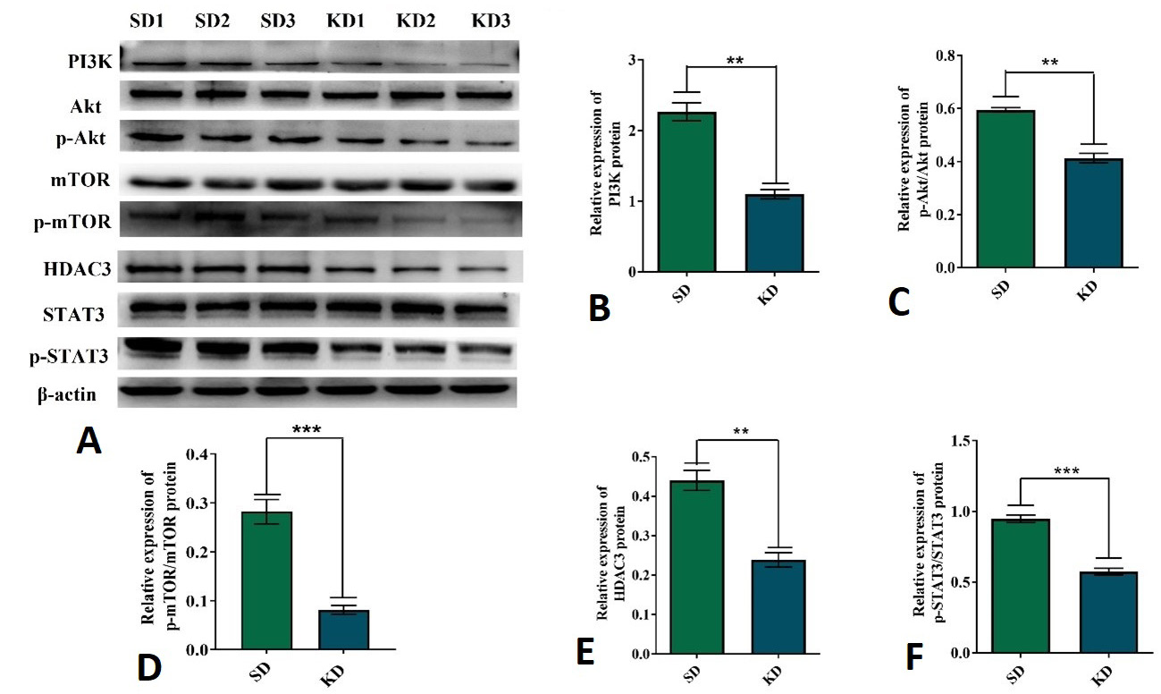
The expression of PI3K, p-Akt, Akt, p-mTOR, mTOR, p-STAT3, STAT3 and HDAC3 proteins in CT26+ colon cancer mouse model with SD and KD treatment. (A) The representative expression of PI3K, p-Akt, Akt, p-mTOR, mTOR, p-STAT3, STAT3 and HDAC3 in CT26+ tumor tissues and β-actin as the internal control for normalization. Quantitative analysis of PI3K protein (B), the ratios of p-Akt to total Akt protein (C), the ratios of p-mTOR to total mTOR protein (D), HDAC3 protein (E) and the ratios of p-STAT3 to total STAT3 protein (F). Each experiment was repeated three times, and all the data were presented as mean ± SEM in each case. Independent-sample T-test was used when different sets of data were compared. Statistically significant differences between different groups were indicated as *p < 0.05, **p < 0.01 and ***p < 0.001.
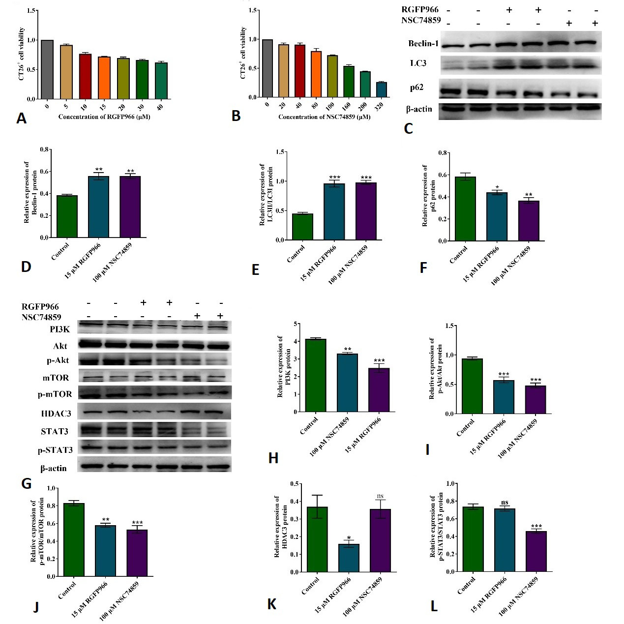
HDAC3 and STAT3 regulated the expression of PI3K, p-Akt, Akt, p-mTOR, mTOR, Beclin-1, LC3 and p62 in CT26+ colon cancer cells. The viability of CT26+ cells was detected by CCK8 with RGFP966 treatment (A) and NSC74859 treatment (B). (C) The representative expression of Beclin-1, LC3 and p62 in CT26+ cells treated with RGFP966 and NSC74859, β-actin as the internal control for normalization. Quantitative analysis of Beclin-1 (D), LC3 (E) and p62 (F) proteins in RGFP966 and NSC74859 treated groups. (G) The representative expression of PI3K, p-Akt, Akt, p-mTOR, mTOR, p-STAT3, STAT3 and HDAC3 in CT26+ colon cells treated with RGFP966 and NSC74859, β-actin as the internal control for normalization. Quantitative analysis of PI3K (H), p-Akt/Akt (I), p-mTOR/mTOR (J), HDAC3 (K) and p-STAT3/STAT3 (L) in RGFP966 and NSC74859 treated groups. Each experiment was repeated three times, and all the data were presented as mean ± SEM in each case. ANOVA was used when different sets of data were compared. Statistically significant differences between different groups were indicated as *p < 0.05, **p < 0.01 and ***p < 0.001 and ns: no significance.
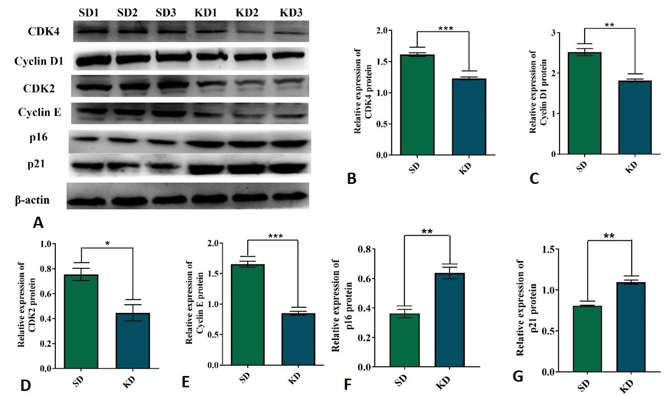
The effect of KD on the expression of Cyclins and CDKs in CT26+ colon cancer mouse model. (A) The representative expression of CDK4, Cyclin D1, CDK2, Cyclin E, p21 and p16 proteins in tumor tissues and β-actin as the internal control for normalization. Quantitative analysis of CDK4 (B) and Cyclin D1 (C), CDK2 (D) and Cyclin E (E), p16 (F) and p21 (G) proteins. Each experiment was repeated thrice, and all the data were presented as mean ± SEM in each case. Independent-sample T-test was used when different sets of data were compared. Statistically significant differences between different groups were indicated as *p < 0.05, **p < 0.01 and ***p < 0.001.
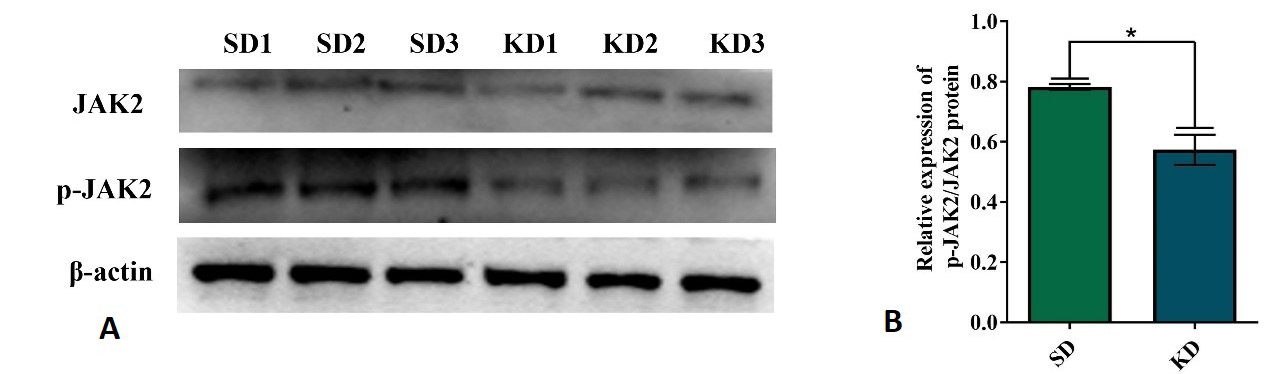
The effect of KD on the expression of JAK2 in the CT26+ colon cancer mouse model. (A) The representative expression of p-JAK2 and JAK2 protein tumor tissues and β-actin as the internal control for normalization. (B) Quantitative analysis of p-JAK2/JAK2 ratio in tumor tissues. Each experiment was repeated three times, and all the data were presented as mean ± SEM in each case. Independent-sample T-test was used when different sets of data were compared. Statistically significant differences between different groups were indicated as *p < 0.05, **p < 0.01 and ***p < 0.001.
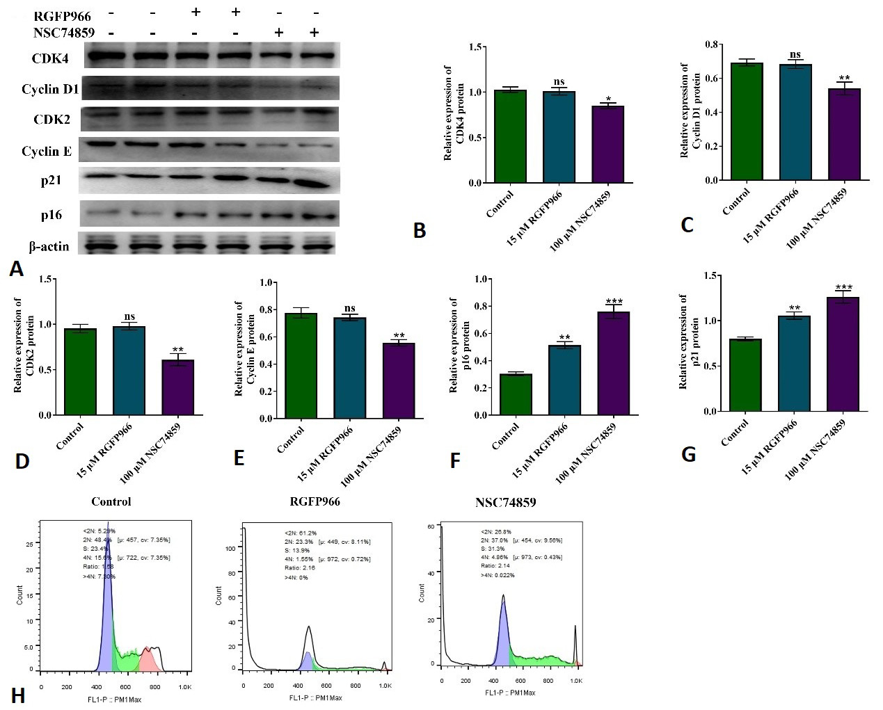
Effect of RGFP966 and NSC74859 on the cell cycle of CT26+ colon cancer cells. (A) The representative expression of CDK4, Cyclin D1, CDK2, Cyclin E, p21 and p16 in CT26+ colon cancer cells and β-actin as the internal control for normalization. Quantitative analysis of CDK4 (B), Cyclin D1 (C), CDK2 (D), Cyclin E (E), p16 (F) and p21 (G) proteins. (H) Flow cytometric analysis of CT26+ colon cancer cells treated with RGFP966 and NSC74859. Each experiment was repeated three times, and all the data were presented as mean ± SEM in each case. One-way analysis of variance (ANOVA) was used when different data sets were compared. Statistically significant differences between different groups were indicated as *p < 0.05, **p < 0.01, ***p < 0.001and ns: no significance.
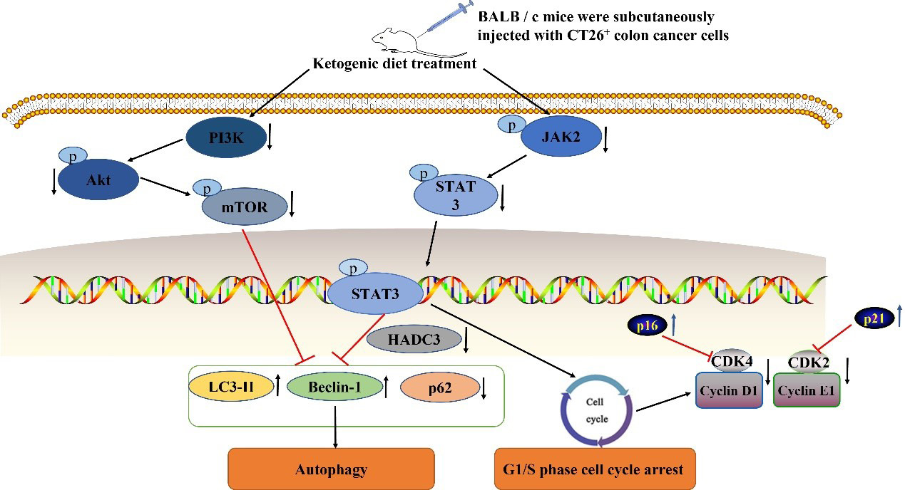
Suggested mechanism of KD on autophagy and cell cycle arrest in CT26+ tumor bearing mice.
References
- Álvarez-Fernández, M. and Malumbres, M., 2020. Mechanisms of sensitivity and resistance to cdk4/6 inhibition. Cancer Cell, 37: 514-529. https://doi.org/10.1016/j.ccell.2020.03.010[CrossRef]
- Bai, L., Zhou, H., Xu, R., Zhao, Y., Chinnaswamy, K., McEachern, D., Chen, J., Yang, C.Y., Liu, Z., Wang, M., Liu, L., Jiang, H., Wen, B., Kumar, P., Meagher, J.L., Sun, D., Stuckey, J.A. and Wang, S., 2019. A potent and selective small-molecule degrader of stat3 achieves complete tumor regression in vivo. Cancer Cell, 36: 498-511.e417. https://doi.org/10.1016/j.ccell.2019.10.002[CrossRef]
- Biondo, S., Gálvez, A., Ramírez, E., Frago, R. and Kreisler, E., 2019. Emergency surgery for obstructing and perforated colon cancer: Patterns of recurrence and prognostic factors. Tech. Coloproctol., 23: 1141-1161. https://doi.org/10.1007/s10151-019-02110-x[CrossRef]
- Chen, Q., Wang, M. and Shen, C., 2020. Bauerane induces s-phase cell cycle arrest, apoptosis, and inhibition of proliferation of a549 human lung cancer cells through the phosphoinositide 3-kinase (pi3k)/akt and signal transducer and activator of transcription 3 (stat3) signaling pathway. Med. Sci. Monit. Int. Med. J. exp. clin. Res., 26: e919558. https://doi.org/10.12659/MSM.919558[CrossRef]
- Cui, Z., Xie, M., Wu, Z. and Shi, Y., 2018. Relationship between histone deacetylase 3 (hdac3) and breast cancer. Med. Sci. Monit. Int. Med. J. exp. clin. Res., 24: 2456-2464. https://doi.org/10.12659/MSM.906576[CrossRef]
- Endo, S., Nakata, K., Ohuchida, K., Takesue, S., Nakayama, H., Abe, T., Koikawa, K., Okumura, T., Sada, M., Horioka, K., Zheng, B., Mizuuchi, Y., Iwamoto, C., Murata, M., Moriyama, T., Miyasaka, Y., Ohtsuka, T., Mizumoto, K., Oda, Y., Hashizume, M. and Nakamura, M., 2017. Autophagy is required for activation of pancreatic stellate cells, associated with pancreatic cancer progression and promotes growth of pancreatic tumors in mice. Gastroenterology, 152: 1492-1506.e1424. https://doi.org/10.1053/j.gastro.2017.01.010[CrossRef]
- Feng, X., Zhang, H., Meng, L., Song, H., Zhou, Q., Qu, C., Zhao, P., Li, Q., Zou, C., Liu, X. and Zhang, Z., 2021. Hypoxia-induced acetylation of pak1 enhances autophagy and promotes brain tumorigenesis via phosphorylating atg5. Autophagy, 17: 723-742. https://doi.org/10.1080/15548627.2020.1731266[CrossRef]
- Gartel, A.L., Serfas, M.S. and Tyner, A.L., 1996. P21 negative regulator of the cell cycle. Proceedings of the society for experimental biology and medicine. Soc. exp. Biol. Med. (New York), 213: 138-149. https://doi.org/10.3181/00379727-213-44046[CrossRef]
- Gesing, A., Jagiela, J. and Lewinski, A., 2003. Melatonin does not affect p21 expression in rat thyroid follicular cells. Neuro Endocrinol. Lett., 24: 310-313.
- Gong, C. and Xia, H., 2020. Resveratrol suppresses melanoma growth by promoting autophagy through inhibiting the pi3k/akt/mtor signaling pathway. Exp. ther. Med., 19: 1878-1886. https://doi.org/10.3892/etm.2019.8359[CrossRef]
- Gosavi, R., Chia, C., Michael, M., Heriot, A.G., Warrier, S.K. and Kong, J.C., 2021. Neoadjuvant chemotherapy in locally advanced colon cancer: A systematic review and meta-analysis. Int. J. Colorectal Dis., 36: 2063-2070. https://doi.org/10.1007/s00384-021-03945-3[CrossRef]
- Han, Y.H., Mun, J.G., Jeon, H.D., Kee, J.Y. and Hong, S.H., 2019. Betulin inhibits lung metastasis by inducing cell cycle arrest, autophagy, and apoptosis of metastatic colorectal cancer cells. Nutrients, 12: 66. https://doi.org/10.3390/nu12010066[CrossRef]
- Hanlon, M.M., Rakovich, T., Cunningham, C.C., Ansboro, S., Veale, D.J., Fearon, U. and McGarry, T., 2019. Stat3 mediates the differential effects of oncostatin m and tnfα on ra synovial fibroblast and endothelial cell function. Front. Immunol., 10: 2056. https://doi.org/10.3389/fimmu.2019.02056[CrossRef]
- He, J., Lü, L., Peng, J., Li, C., Kong, X., Zhang, J. and Peng, L., 2020. Inhibitory effect of ketogenic diet on neuroblastoma in balb/c-nu mouse models. J. Southern Med. Univ., (Nan fang yi ke da xue xue bao). 40: 1155-1164.
- He, P., Li, K., Li, S.B., Hu, T.T., Guan, M., Sun, F.Y. and Liu, W.W., 2018. Upregulation of akap12 with hdac3 depletion suppresses the progression and migration of colorectal cancer. Int. J. Oncol., 52: 1305-1316. https://doi.org/10.3892/ijo.2018.4284[CrossRef]
- Hua, H., Kong, Q., Zhang, H., Wang, J., Luo, T. and Jiang, Y., 2019. Targeting mtor for cancer therapy. J. Hematol. Oncol., 12: 71. https://doi.org/10.1186/s13045-019-0754-1[CrossRef]
- Juretschke, T. and Beli, P., 2021. Causes and consequences of DNA damage-induced autophagy. Matrix Biol. J. Int. Soc. Matrix Biol., 100-101: 39-53. https://doi.org/10.1016/j.matbio.2021.02.004[CrossRef]
- Kim, T.W., Lee, S.Y., Kim, M., Cheon, C. and Ko, S.G., 2018. Kaempferol induces autophagic cell death via ire1-jnk-chop pathway and inhibition of g9a in gastric cancer cells. Cell Death Dis., 9: 875. https://doi.org/10.1038/s41419-018-0930-1[CrossRef]
- Krishna, K., Behnisch, T. and Sajikumar, S., 2016. Inhibition of histone deacetylase 3 restores amyloid-β oligomer-induced plasticity deficit in hippocampal ca1 pyramidal neurons. J. Alzheimer’s Dis., 51: 783-791. https://doi.org/10.3233/JAD-150838[CrossRef]
- Lee, G.E., Lee, C.J., An, H.J., Kang, H.C., Lee, H.S., Lee, J.Y., Oh, S.R., Cho, S.J., Kim, D.J. and Cho, Y.Y., 2021. Fargesin inhibits egf-induced cell transformation and colon cancer cell growth by suppression of cdk2/cyclin e signaling pathway. Int. J. mol. Sci., 22: 2073. https://doi.org/10.3390/ijms22042073[CrossRef]
- Li, B., Yu, Y., Liu, K., Zhang, Y., Geng, Q., Zhang, F., Li, Y. and Qi, J., 2021. Β-hydroxybutyrate inhibits histone deacetylase 3 to promote claudin-5 generation and attenuate cardiac microvascular hyperpermeability in diabetes. Diabetologia, 64: 226-239. https://doi.org/10.1007/s00125-020-05305-2[CrossRef]
- Li, Y., Guo, F., Chen, T., Zhang, L. and Qin, Y., 2021. Anthraquinone derivative c10 inhibits proliferation and cell cycle progression in colon cancer cells via the jak2/stat3 signaling pathway. Toxicol. appl. Pharmacol., 418: 115481. https://doi.org/10.1016/j.taap.2021.115481[CrossRef]
- Lin, S.R., Fu, Y.S., Tsai, M.J., Cheng, H. and Weng, C.F., 2017. Natural compounds from herbs that can potentially execute as autophagy inducers for cancer therapy. Int. J. mol. Sci., 18: 1412. https://doi.org/10.3390/ijms18071412[CrossRef]
- Liu, B., Zhang, N., Yang, J., Sun, W., Zhang, R., Zheng, X., Wang, Z., Siebert, H.C. and Han, J., 2022. Preparation, characterization, evaluation of neuroprotective effect, and related mechanisms of phosphatidylserine emulsion in 5- and 12-week old mice. J. agric. Fd. Chem., 70: 1852-1864. https://doi.org/10.1021/acs.jafc.1c07403[CrossRef]
- Lu, X.F., Cao, X.Y., Zhu, Y.J., Wu, Z.R., Zhuang, X., Shao, M.Y., Xu, Q., Zhou Y.J., Ji, H.J., Lu, Q.R., Shi, Y.J., Zeng, Y. and Bu, H., 2018. Histone deacetylase 3 promotes liver regeneration and liver cancer cells proliferation through signal transducer and activator of transcription 3 signaling pathway. Cell Death Dis., 9: 398. https://doi.org/10.1038/s41419-018-0428-x[CrossRef]
- Luo, H., Zhai, L., Qiu, W., Liang, H., Yu, L., Li, Y., Xiong, M., Guo, J. and Tang, H., 2021. P16 loss facilitate hydroquinone-induced malignant transformation of tk6 cells through promoting cell proliferation and accelerating the cell cycle progression. Environ. Toxicol., 36: 1591-1599. https://doi.org/10.1002/tox.23155[CrossRef]
- Malvaez, M., McQuown, S.C., Rogge, G.A., Astarabadi, M., Jacques, V., Carreiro, S., Rusche, J.R. and Wood, M.A., 2013. Hdac3-selective inhibitor enhances extinction of cocaine-seeking behavior in a persistent manner. Proc. natl. Acad. Sci. U.S.A., 110: 2647-2652. https://doi.org/10.1073/pnas.1213364110[CrossRef]
- Mann, S.D., Sidhu, M.D. and Gowin, K.D., 2020. Understanding the mechanisms of diet and outcomes in colon, prostate, and breast cancer; malignant gliomas; and cancer patients on immunotherapy. Nutrients, 12: 2226. https://doi.org/10.3390/nu12082226[CrossRef]
- Mavropoulos, J.C., Isaacs, W.B., Pizzo, S.V. and Freedland, S.J., 2006. Is there a role for a low-carbohydrate ketogenic diet in the management of prostate cancer? Urology, 68: 15-18. https://doi.org/10.1016/j.urology.2006.03.073[CrossRef]
- McLeod, A.B., Stice, J.P., Wardell, S.E., Alley, H.M., Chang, C.Y. and McDonnell, D.P., 2018. Validation of histone deacetylase 3 as a therapeutic target in castration-resistant prostate cancer. Prostate, 78: 266-277. https://doi.org/10.1002/pros.23467[CrossRef]
- Otto, T. and Sicinski, P., 2017. Cell cycle proteins as promising targets in cancer therapy. Nature reviews. Cancer, 17: 93-115. https://doi.org/10.1038/nrc.2016.138[CrossRef]
- Park, K.H., Joo, S.H., Seo, J.H., Kim, J., Yoon, G., Jeon, Y.J., Lee, M.H., Chae, J.I., Kim, W.K. and Shim, J.H., 2022. Licochalcone h induces cell cycle arrest and apoptosis in human skin cancer cells by modulating jak2/stat3 signaling. Biomol. Ther., 30: 72-79. https://doi.org/10.4062/biomolther.2021.149[CrossRef]
- Plotti, F., Terranova, C., Luvero, D., Bartolone, M., Messina, G., Feole, L., Cianci, S., Scaletta, G., Marchetti, C., Di Donato, V., Fagotti, A., Scambia, G., Panici, P.B. and Angioli, R., 2020. Diet and chemotherapy: The effects of fasting and ketogenic diet on cancer treatment. Chemotherapy, 65: 77-84. https://doi.org/10.1159/000510839[CrossRef]
- Poff, A., Koutnik, A.P., Egan, K.M., Sahebjam, S., D’Agostino, D. and Kumar, N.B., 2019. Targeting the warburg effect for cancer treatment: Ketogenic diets for management of glioma. Semin. Cancer Biol., 56: 135-148. https://doi.org/10.1016/j.semcancer.2017.12.011[CrossRef]
- Poillet-Perez, L. and White, E., 2019. Role of tumor and host autophagy in cancer metabolism. Genes Dev., 33: 610-619. https://doi.org/10.1101/gad.325514.119[CrossRef]
- Qie, S. and Diehl, J.A., 2016. Cyclin d1, cancer progression, and opportunities in cancer treatment. J. mol. Med. (Berlin, Germany), 94: 1313-1326. https://doi.org/10.1007/s00109-016-1475-3[CrossRef]
- See, A.P., Han, J.E., Phallen, J., Binder, Z., Gallia, G., Pan, F., Jinasena, D., Jackson, C., Belcaid, Z., Jeong, S.J., Gottschalk, C., Zeng, J., Ruzevick, J., Nicholas, S., Kim, Y., Albesiano, E., Pardoll, D.M. and Lim, M., 2012. The role of stat3 activation in modulating the immune microenvironment of gbm. J. Neuro-oncol., 110: 359-368. https://doi.org/10.1007/s11060-012-0981-6[CrossRef]
- Shao, Y., Gao, Z., Marks, P.A. and Jiang, X., 2004. Apoptotic and autophagic cell death induced by histone deacetylase inhibitors. Proc. natl. Acad. Sci. U.S.A., 101: 18030-18035. https://doi.org/10.1073/pnas.0408345102[CrossRef]
- Siddiquee, K., Zhang, S., Guida, W.C., Blaskovich, M.A., Greedy, B., Lawrence, H.R., Yip, M.L., Jove, R., McLaughlin, M.M., Lawrence, N.J., Sebti, S.M. and Turkson, J., 2007. Selective chemical probe inhibitor of stat3, identified through structure-based virtual screening, induces antitumor activity. Proc. natl. Acad. Sci. U.S.A., 104: 7391-7396. https://doi.org/10.1073/pnas.0609757104[CrossRef]
- Singh, S.S., Vats, S., Chia, A.Y., Tan, T.Z., Deng, S., Ong, M.S., Arfuso, F., Yap, C.T., Goh, B.C., Sethi, G., Huang, R.Y., Shen, H.M., Manjithaya, R. and Kumar, A.P., 2018. Dual role of autophagy in hallmarks of cancer. Oncogene, 37: 1142-1158. https://doi.org/10.1038/s41388-017-0046-6[CrossRef]
- Sun, W., Yang, J., Liu, B., Liu, Q., Wang, T., Wang, Q., Liu, M., Li, L., Wang, Z. and Li, S., 2022. Ketogenic diet inhibits tumor growth by enhancing immune response, attenuating immunosuppression, inhibiting angiogenesis and emt in ct26 colon tumor allografts mouse model. J. Funct. Fds., 92: 105067. https://doi.org/10.1016/j.jff.2022.105067[CrossRef]
- Suski, J.M., Braun, M., Strmiska, V. and Sicinski, P., 2021. Targeting cell-cycle machinery in cancer. Cancer Cell, 39: 759-778. https://doi.org/10.1016/j.ccell.2021.03.010[CrossRef]
- Tamas, K., Walenkamp, A.M., de Vries, E.G., van Vugt, M.A., Beets-Tan, R.G., van Etten, B., de Groot, D.J. and Hospers, G.A., 2015. Rectal and colon cancer: Not just a different anatomic site. Cancer Treat. Rev., 41: 671-679. https://doi.org/10.1016/j.ctrv.2015.06.007[CrossRef]
- Tchakarska, G. and Sola, B., 2020. The double dealing of cyclin d1. Cell Cycle (Georgetown, Tex.), 19: 163-178. https://doi.org/10.1080/15384101.2019.1706903[CrossRef]
- Vaupel, P., Schmidberger, H. and Mayer, A., 2019. The warburg effect: Essential part of metabolic reprogramming and central contributor to cancer progression. Int. J. Radiat. Biol., 95: 912-919. https://doi.org/10.1080/09553002.2019.1589653[CrossRef]
- Wan, M., Zhang, L., Chen, Y., Li, Q., Fan, W., Xue, Q., Yan, F. and Song, W., 2019. Synthesis and anticancer activity evaluation of novel phenanthridine derivatives. Front. Oncol., 9: 274. https://doi.org/10.3389/fonc.2019.00274[CrossRef]
- Wang, Z., 2021. Regulation of cell cycle progression by growth factor induced cell signaling. Cells, 10: 3327. https://doi.org/10.3390/cells10123327[CrossRef]
- Weber, D.D., Aminzadeh-Gohari, S., Tulipan, J., Catalano, L., Feichtinger, R.G. and Kofler, B., 2020. Ketogenic diet in the treatment of cancer - where do we stand? Mol. Metab., 33: 102-121. https://doi.org/10.1016/j.molmet.2019.06.026[CrossRef]
- Wilson, A.J., Byun, D.S., Popova, N., Murray, L.B., L’Italien, K., Sowa, Y., Arango, D., Velcich, A., Augenlicht, L.H. and Mariadason, J.M., 2006. Histone deacetylase 3 (hdac3) and other class i hdacs regulate colon cell maturation and p21 expression and are deregulated in human colon cancer. J. biol. Chem., 281: 13548-13558. https://doi.org/10.1074/jbc.M510023200[CrossRef]
- Wu, L., Li, J., Liu, T., Li, S., Feng, J., Yu, Q., Zhang, J., Chen, J., Zhou, Y., Ji, J., Chen, K., Mao, Y., Wang, F., Dai, W., Fan, X., Wu, J. and Guo, C., 2019. Quercetin shows anti-tumor effect in hepatocellular carcinoma lm3 cells by abrogating jak2/stat3 signaling pathway. Cancer Med., 8: 4806-4820. https://doi.org/10.1002/cam4.2388[CrossRef]
- Wu, W.Y., Li, J., Wu, Z.S., Zhang, C.L. and Meng, X.L., 2011. Stat3 activation in monocytes accelerates liver cancer progression. BMC Cancer, 11: 506. https://doi.org/10.1186/1471-2407-11-506[CrossRef]
- Wullschleger, S., Loewith, R. and Hall, M.N., 2006. Tor signaling in growth and metabolism. Cell, 124: 471-484. https://doi.org/10.1016/j.cell.2006.01.016[CrossRef]
- Xu, N.W., Chen, Y., Liu, W., Chen, Y.J., Fan, Z.M., Liu, M. and Li, L.J., 2018. Inhibition of jak2/stat3 signaling pathway suppresses proliferation of burkitt’s lymphoma raji cells via cell cycle progression, apoptosis, and oxidative stress by modulating hsp70. Med. Sci. Monit. Int. Med. J. exp. clin. Res., 24: 6255-6263. https://doi.org/10.12659/MSM.910170[CrossRef]
- Xu, Z., Han, X., Ou, D., Liu, T., Li, Z., Jiang, G., Liu, J. and Zhang, J., 2020. Targeting pi3k/akt/mtor-mediated autophagy for tumor therapy. Appl. Microbiol. Biotechnol., 104: 575-587. https://doi.org/10.1007/s00253-019-10257-8[CrossRef]
- Yaghoubi, A., Khazaei, M., Avan, A., Hasanian, S.M. and Soleimanpour, S., 2020. The bacterial instrument as a promising therapy for colon cancer. Int. J. Color. Dis., 35: 595-606. https://doi.org/10.1007/s00384-020-03535-9[CrossRef]
- Yang, J., Pi, C. and Wang, G., 2018. Inhibition of pi3k/akt/mtor pathway by apigenin induces apoptosis and autophagy in hepatocellular carcinoma cells. Biomed. Pharmacother. Biomed. Pharmacother., 103: 699-707. https://doi.org/10.1016/j.biopha.2018.04.072[CrossRef]
- You, L., Wang, Z., Li, H., Shou, J., Jing, Z., Xie, J., Sui, X., Pan, H. and Han, W., 2015. The role of stat3 in autophagy. Autophagy, 11: 729-739. https://doi.org/10.1080/15548627.2015.1017192[CrossRef]
- Zanotto-Filho, A., Braganhol, E., Klafke, K., Figueiró, F., Terra, S.R., Paludo, F.J., Morrone, M., Bristot, I.J., Battastini, A.M., Forcelini, C.M., Bishop, A.J.R., Gelain, D.P. and Moreira, J.C.F., 2015. Autophagy inhibition improves the efficacy of curcumin/temozolomide combination therapy in glioblastomas. Cancer Lett., 358: 220-231. https://doi.org/10.1016/j.canlet.2014.12.044[CrossRef]
- Zhang, C., Yang, X., Zhang, Q., Guo, Q., He, J., Qin, Q., Zhu, H., Liu, J., Zhan, L., Lu, J., Liu, Z., Xu, L., Ma, J., Dai, S., Cheng, H. and Sun, X., 2014. Stat3 inhibitor nsc74859 radiosensitizes esophageal cancer via the downregulation of hif-1α. Tumour Biol. J. Int. Soc. Oncodev. Biol. Med., 35: 9793-9799. https://doi.org/10.1007/s13277-014-2207-3[CrossRef]
- Zhang, N., Liu, C., Jin, L., Zhang, R., Wang, T., Wang, Q., Chen, J., Yang, F., Siebert, H.C., and Zheng, X., 2020. Ketogenic diet elicits antitumor properties through inducing oxidative stress, inhibiting mmp-9 expression, and rebalancing m1/m2 tumor-associated macrophage phenotype in a mouse model of colon cancer. J. agric. Fd. Chem., 68: 11182-11196. https://doi.org/10.1021/acs.jafc.0c04041[CrossRef]
- Zhang, N., Liu, C., Zhang, R., Jin, L., Yin, X., Zheng, X., Siebert, H.C., Li, Y., Wang, Z., Loers, G. and Petridis, A.K., 2020. Amelioration of clinical course and demyelination in the cuprizone mouse model in relation to ketogenic diet. Fd. Funct., 11: 5647-5663. https://doi.org/10.1039/C9FO02944C[CrossRef]
- Zhao, Y., He, J., Yang, L., Luo, Q. and Liu, Z., 2018. Histone deacetylase-3 modification of microrna-31 promotes cell proliferation and aerobic glycolysis in breast cancer and is predictive of poor prognosis. J. Breast Cancer, 21: 112-123. https://doi.org/10.4048/jbc.2018.21.2.112[CrossRef]
- Zheng, K., He, Z., Kitazato, K. and Wang, Y., 2019. Selective autophagy regulates cell cycle in cancer therapy. Theranostics, 9: 104-125. https://doi.org/10.7150/thno.30308[CrossRef]
- Zhou, J., Jiang, Y.Y., Chen, H., Wu, Y.C. and Zhang, L., 2020. Tanshinone i attenuates the malignant biological properties of ovarian cancer by inducing apoptosis and autophagy via the inactivation of pi3k/akt/mtor pathway. Cell Prolif., 53: e12739. https://doi.org/10.1111/cpr.12739[CrossRef]
Services
Article usage statistics
Loading…