
HGbIV is unique truncated hemoglobin (tHb) that differs from other bacterial tHbs due to its large size, polar heme pocket residues and large cavity for exogenous ligand in the heme pocket. The crystal structures of HGbIV showed that its heme pocket can accommodate and interact with bulky PO4-3 ion. Here, we have docked different smaller molecules/ion like O2, NO, CO, CO2, NO3-1, CH3COO-1 and CN-1 in the heme pocket of HGbIV. Analysis of the structure showed that the smaller ligands docked far away from most of the heme pocket residues namely His70(B9), His71(B10), Ser97(E11) and Trp137(G8), and only PO4-3 docked at a position where it can interact with all these residues. It appears that HGbIV is not designed for transport of smaller gaseous molecule like the other heme containing proteins and it may be a phosphate sensor.
Short Communication · Volume 50, Issue 4, 16 July 2018 · Open Access
Docking of Smaller Ligands in the Heme Pocket of Hell's Gate Globin IV
· ·
Affiliations & Notes ⌄
- 1Department of Biosciences, COMSATS Institute of Information Technology (CIIT), Sahiwal, Punjab , * Corresponding author: farrukhccb@gmail.com
- 2farrukh@ciitsahiwal.edu.pk
Article Info ⌄
- Journal:
- Pakistan Journal of Zoology
- Published:
- 16 July 2018

From the article
- Superimposition of the docked PO4-3 ion in HGbIV to the real 3D structure of HGbIV showed that the ion docked at same orientation as observed in crystal structure of HGbIV.
- SO4-2 ion is a structural analogue of PO4-3 ion, it docked in the heme pocket of HGbIV relatively far (2.67 Ǻ) from heme-iron compared to the position of PO4-3 ion with binding affinity of -17.16 KJ/mol (Fig. 1B; Table I).
- Analyses of the structure showed that most of the heme pocket residues can stabilize SO4-2 ion except Ser97(E11) which is relatively far (3.34 Ǻ) away (Fig. 1B).
- Analysis of these values clearly showed that the heme pocket of HGbIV is designed for bulky ligands like PO4-3 as it is only ligand that can interact with all the active site residues namely His70(B9), His71(B10), Ser97(E11) and Trp137(G8) (Fig. 1).
Selected from the article's own text.
Abstract
Methods
The PDB files of HGbIV (4NK1) and different ligands (SO4-2, NO3-1, CH3COO-1 and CN-1, O2, NO, CO and CO2) were retrieved from RCSB Protein Data Bank (http://www.rcsb.org/pdb/home/home.do) . Chimera was used for the energy minimization of HGbIV and ligands (Petterson et al., 2004). PDBQT files were generated by using the autodock 4 and autodock vina , and Gasteiger charges were added to convert protein and all ligands into PDBQT files. By using Autodock Vina, targeted docking was performed at the heme pocket of HGbIV with the above mentioned ligands and structure was visualized by using Chimera (Petterson et al., 2004).
Results
A targeted docking strategy was used for docking a PO4-3 ion in the heme pocket of HGbIV (4NK1) where it docked with the binding affinity of -17.16 KJ/mol (Fig. 1A; Table I). Superimposition of the docked PO4-3 ion in HGbIV to the real 3D structure of HGbIV showed that the ion docked at same orientation as observed in crystal structure of HGbIV. Moreover, it shows similar interactions with heme pocket residues namely His70(B9), His71(B10), Ser97(E11) and Trp137(G8) as observed in its real 3D crystal structure of HGbIV . SO4-2 ion is a structural analogue of PO4-3 ion, it docked in the heme pocket of HGbIV relatively far (2.67 Ǻ) from heme-iron compared to the position of PO4-3 ion with binding affinity of -17.16 KJ/mol (Fig. 1B; Table I). Analyses of the structure showed that most of the heme pocket residues can stabilize SO4-2 ion except Ser97(E11) which is relatively far (3.34 Ǻ) away (Fig. 1B).
Table I.- Ligands and their binding affinity.
| S No. | Ligands | Binding affinity (kJ/mol) | Hydrogen bonds |
| 1 | PO43- | -17.16 | His70(B9), His71(B10), Ser97(E11), Trp137(G8) |
| 2 | SO42- | -17.16 | His70(B9), His71(B10), W137(G8) |
| 3 | CH3COO- | -12.13 | Ser97(E11), Trp137(G8) |
| 4 | NO3-1 | -11.30 | His70(B9) |
| 5 | CN- | -5.44 | No residue |
| 6 | CO2 | -10.04 | Ser97(E11), Trp137(G8) |
| 7 | O2 | -8.37 | Ser97(E11) |
| 8 | CO | -6.69 | Ser97(E11) |
| 9 | NO | -5.85 | Trp137(G8) |
CH3COO-1, NO3-1 and CN-1 ions docked in the heme pocket of HGbIV at a distance of 3.89, 3.11 and 3.53 Ǻ away from the heme iron with binding affinities of -12.13,-11.30 and -5.44 KJ/mol, respectively (Fig. 1C-E). Analyses of the structures showed that the docked CH3COO-1 ion is stabilized by Ser97(E11) and Trp137(G8), whereas the His70(B9) and His71(B10) are far away (> 3.2 Ǻ) from the ion. Previously, CH3COO-1 ion has been reported in a crystal structure of a tHb from Thermobifida fusca (tf-tHgb). A notable difference between the two structures is long distance of His(B10) from the docked CH3COO-1 ion in HGbIV compared to that of Tyr(B10) in tf-tHgb. On the other hand, the Trp(G8) plays a vital role in interacting and stabilizing the acetate ion in tf-tHgb as well as in HGbIV. Analysis of the docked NO3-1 ion in HGbIV showed that only His70(B9) can interact with NO3-1while the other heme pocket residues are far away (> 3.2 Ǻ) (Fig. 1D). The CN-1 ion docked very far from all the heme pocket residues so no residue can form hydrogen bonding with it (Fig. 1E).
CO2, O2, CO and NO were docked in the heme pocket of HGbIV at distance 3.87, 5.23, 4.38, 3.33, 3.51 Ǻ away from the heme iron with binding affinity -10.04, -8.37, -6.69 and -5.85 KJ/mol, respectively (Table I; Fig. 1F-I). Analyses of these structures showed that the CO2 is stabilized by interaction of Ser97(E11) and Trp137(G8), whereas CO and O2 are stabilised only by Ser97(E11). On the other hand, NO may only interact with Trp137(G8).
Discussion
This work is an extension of our previous work in which we have determined three dimensional structure of HGbIV under two different conditions . Structurally, HGbIV belongs to group II tHbs with several unique features. For instance, HGbIV is a large protein (194 amino acids) compared to other bacterial group II tHbs (120-140). Second, the heme pocket of HGbIV is more polar and host two His residues at B9 and B10 positions compared to highly conserved Phe(B9) and Tyr(B10) residues in other bacterial tHbs . Third, the crystal structure of HGbIV (4NK1) showed that the heme pocket can accommodate a bulky heme-bound phosphate ion. On the other hand, smaller ligands like CH3COO-1, NO3-1, CO2, O2, CO, NO, CN-1 and H2O have been reported in other bacterial tHbs ; ; ; ; . In this study we docked smaller ligands in the heme pocket of HGbIV.
The PO4-3, SO4-2, CH3COO-1, NO3-1, CO2, O2, CO, NO and CN-1 were docked in the heme pocket of HGbIV with the binding affinities of -17.16,- 17.16, -12.13, -11.30, -10.04, -8.37, -6.69, -5.85 and -5.44 Kj/mol, respectively (Table I). Analysis of these values clearly showed that the heme pocket of HGbIV is designed for bulky ligands like PO4-3 as it is only ligand that can interact with all the active site residues namely His70(B9), His71(B10), Ser97(E11) and Trp137(G8) (Fig. 1). On the other hand, smaller heme-iron bound molecules are not so stabilized in this pocket due to far location of the active site residues. In other group II bacterial tHbs smaller heme-bound ligands are mostly stabilized by highly conserved Tyr(B10) which is replaced by His71(B10) in HGbIV. In conclusion, it appears that heme pocket of HGbIV is specific for bulky ligands and we forsee remarkable displacement in HGbIV for smaller ligands stabilization.
Acknowledgement
We gratefully acknowledge Higher Education Commission (HEC) of Pakistan for grants to this study.
Statement of conflict of interest
Authors have declared no conflict of interest.
Figures
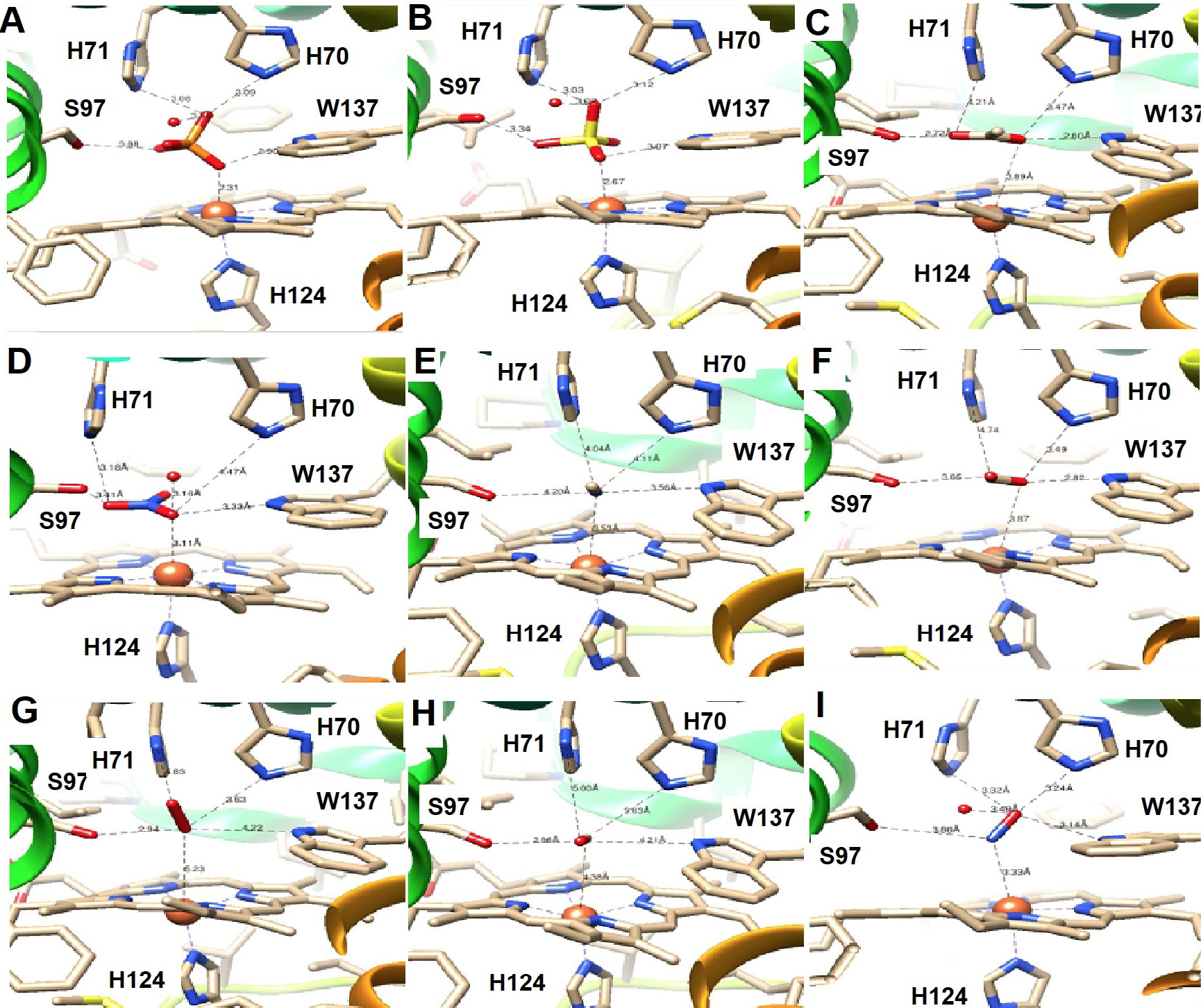
Heme pockets of HGbIV. A to I, the docked PO4-3, SO4-2, CH3COO-1, NO3-1, CN-1, CO2, O2, CO and NO in the heme pocket.
References
- Ascenzi, P. and Pesce, A., 2017. J. Biol. Inorg. Chem., 22: 1141-1150. https://doi.org/10.1007/s00775-017-1490-z[CrossRef]
- Ascenzi, P., Ciaccio, C., Gasperi, T., Pesce, A., Caporaso, L. and Coletta, M., 2017. J. Biol. Inorg. Chem., 22: 977-986. https://doi.org/10.1007/s00775-017-1476-x[CrossRef]
- Berman, H.M., Westbrook, J., Feng, Z., Gilliland, G., Bhat, T.N., Weissig, H., Shindyalov, I.N. and Bourne, P.E., 2000. Nucl. Acids Res., 28: 235-242. https://doi.org/10.1093/nar/28.1.235[CrossRef]
- Bonamore, A., Ilari, A., Giangiacomo, L., Bellelli, A., Morea, V. and Boffi, A., 2005. FEBS J., 272: 4189-4201. https://doi.org/10.1111/j.1742-4658.2005.04831.x[CrossRef]
- Bustamante, J.P., Radusky, L., Boechi, L., Estrin, D.A., Ten-Have, A. and Martí, M.A., 2016. PLoS Comput. Biol., 12: e1004701. https://doi.org/10.1371/journal.pcbi.1004701[CrossRef]
- Couture, M., Yeh, S.R., Wittenberg, B.A., Wittenberg, J.B., Ouellet, Y., Rousseau, D.L. and Guertin, M., 1999. Proc. natl. Acad. Sci. USA, 96: 11223-11228. https://doi.org/10.1073/pnas.96.20.11223[CrossRef]
- Dikshit, R.P., Dikshit, K.L., Liu, Y.X. and Webster, D.A., 1992. Arch. Biochem. Biophys., 293: 241-245. https://doi.org/10.1016/0003-9861(92)90391-9[CrossRef]
- Hou, S., Makarova, K.S., Saw, J.H., Senin, P., Ly, B.V., Zhou, Z., Ren, Y., Wang, J., Galperin, M.Y., Omelchenko, M.V., Wolf, Y.I., Yutin, N., Koonin, E.V., Stott, M.B., Mountain, B.W., Crowe, M.A., Smirnova, A.V., Dunfield, P.F., Feng, L., Wang, L. and Alam, M., 2008. Biol. Direct, 3: 26. https://doi.org/10.1186/1745-6150-3-26[CrossRef]
- Igarashi, J., Kobayashi, K. and Matsuoka, A., 2011. J. Biol. Inorg. Chem., 16: 599-609. https://doi.org/10.1007/s00775-011-0761-3[CrossRef]
- Ilari, A., Kjelgaard, P., Von-Wachenfeldt, C., Catacchio, B., Chiancone, E. and Boffi, A., 2007.Arch. Biochem. Biophys, 457: 85-94. https://doi.org/10.1016/j.abb.2006.09.033[CrossRef]
- Jamil, F., Teh, A.H., Schadich, E., Saito, J.A., Najimudin, N. and Alam, M., 2014. J. Biochem., 156: 97-106. https://doi.org/10.1093/jb/mvu023[CrossRef]
- Minaeva, E., Zalutskaya, Z., Filina, V. and Ermilova, E., 2017. PLoS One, 12: e0186851. https://doi.org/10.1371/journal.pone.0186851[CrossRef]
- Ouellet, H., Juszczak, L., Dantsker, D., Samuni, U., Ouellet, Y.H., Savard, P.Y., Wittenberg, J.B., Wittenberg, B.A., Friedman, J.M. and Guertin, M., 2003. Biochemistry, 42: 5764-5774. https://doi.org/10.1021/bi0270337[CrossRef]
- Ouellet, H., Ouellet, Y., Richard, C., Labarre, M., Wittenberg, B.A. and Guertin, M., 2002. Proc. natl. Acad. Sci. USA, 99: 5902-5907. https://doi.org/10.1073/pnas.092017799[CrossRef]
- Pesce, A., Couture, M., Dewilde, S., Guertin, M., Yamauchi, K., Ascenzi, P., Moens, L. and Bolognesi, M., 2000. EMBO J., 19: 2424-2434. https://doi.org/10.1093/emboj/19.11.2424[CrossRef]
- Pettersen, E.F., Goddard, T.D., Huang, C.C., Couch, G.S., Greenblatt, D.M., Meng, E.C. and Ferrin, T.E., 2004. J. Comput. Chem., 25: 1605-1612. https://doi.org/10.1002/jcc.20084[CrossRef]
- Rana, N., Ehsan, N., Ihsan, A. and Jamil, F., 2017. Pakistan J. Zool., 49: 1261-1265.
- Tariq, F., Khalid, Q., Sehgal, S.A., Mannan, S. and Jamil, F., 2016. Pakistan J. Zool., 48: 1805-1810.
- Trott, O. and Olson, A.J., 2010. J. Comput. Chem., 31: 455-461.
- Vinogradov, S.N., Tinajero-Trejo, M., Poole, R.K. and Hoogewijs, D., 2013. Biochim. Biophys. Acta, 1834: 1789-1800. https://doi.org/10.1016/j.bbapap.2013.03.021[CrossRef]
- Wittenberg, J.B., Bolognesi, M., Wittenberg, B.A. and Guertin, M., 2002. J. biol. Chem., 277: 871-874. https://doi.org/10.1074/jbc.R100058200[CrossRef]
Cited by 1 article
In journals published by ResearchersLinks.
Services
Article usage statistics
Loading…