
DMRT1 expression has been followed in the developing testes of three groups (Testes weight: Body weight ratios (<1%, 1%-2%, 2%-3%)) of adult mule duck using immunohistochemical staining method. The result showed that DMRT1 was mainly expressed in spermatogonia nucleus of seminiferous tubules and had no expression in Leydig cells. It was indicated that the DMRT1 expression was positively correlated with the testes development of adult mule duck.
Short Communication · Volume 54, Issue 1, 19 December 2021 · Open Access
DMRT1 Expression is Positively Correlated with the Development of Testes in Adult Mule Duck
· · · · ·
Affiliations & Notes ⌄
- 1Institute of Animal Husbandry and Veterinary Sciences, Wuhan Academy of Agricultural Sciences, Wuhan 430208, P.R. China 2Key Laboratory of Agricultural Animal Genetics, Breeding and Reproduction of Ministry of Education, Huazhong Agricultural University, Wuhan 430070, P.R. China , * Corresponding author: gongping09@foxmail.com
Article Info ⌄
- Journal:
- Pakistan Journal of Zoology
- Published:
- 19 December 2021

From the article
- The sex gland index is an indicator of sexual maturity, and the testis weight, body weight and testes index of poultry are correlated with sexual maturity and fecundity.
- Mule duck testes is abnormal and showed significant individual difference.
- The results indicated that DMRT1 expression is positively correlated with the testes weight/body weight ratios.
Selected from the article's own text.
Abstract
Material and methods
More than 50 mule ducks were raised by standard feeding process, and the feed was provided by Hua Mei Company. The mule ducks were weighed and slaughtered at the age of 300 days, and the left and right testis were taken and weighed respectively, and the growth and development of the testes were analyzed. The testes weight/body weight ratio (%) was (Gross testes weight×100) / body weight.
For histological studies, testes were placed in 4% paraformaldehyde (PFA) for more than 24 h, and 6μm thick paraffin sections were cut, and stained with hematoxylin-eosin staining (HE). The histological sections of left and right testis were mounted on poly-l-lysine-coated glass slide, dried overnight at 37°C and proceeded for Streptavidin-biotin-peroxidase complex immunohistochemical (IHC) method ; using the antibody of DMRT1 (sc 98341, Santa Cruz Biotechnology).
Results and discussion
The gross testes weight ranged from 9.4 g to 102 g, average weight of the left testis was lower than that of the right testis (Supplementary Table I). The testes weight/ body weight ratio was 1.55±0.86. The sex gland index is an indicator of sexual maturity, and the testis weight, body weight and testes index of poultry are correlated with sexual maturity and fecundity. Shen and Tong (1991) found that the testes index of Shaoxing duck was 3.16% at 16 weeks, and 3.55% at 43 weeks. This study found that the testes index was only 2.98% at 43 weeks, far lower than that of Shaoxin duck and other poultry species, and testes weight was less than that of the Khaki Campbell duck in the same period . A few individuals with high testicular index may produce sperm, which is consistent with the results of Tan et al. (1998).
In the testes weight/body weight ratio <1% sections, fewer interstitial cells between adjacent seminiferous tubule were observed, and 1-2 layer cells in typical seminiferous tubule, spermatgonia were arranged in seminiferous tubule, and no sperm cells or sperm were seen (Fig. 1). All levels of spermatogenic cells were disorganized, scattered and even filled in the entire lumen, with a large number of nuclei falling off. Part of the left testis seminiferous tubule has disappeared except spermatgonia. More than 2 layer cells in typical seminiferous tubule and a large space in the lumen were appeared in the testes weight/body weight ratio 1%-2% sections. More than 4 layer cells were observed, the permutations of spermatozoa were more regular in some testicular tissues in the testes weight/body weight ratio 2%-3% sections (Fig. 1). The occurrence of poultry sperm begins with the division of spermatogenic cells and ends with the formation of mature sperm. The structure of normal mature poultry testis has undergone significant changes during the spermatogenesis. Mature testicular seminiferous tubule has multiple layers of epithelium in different stages of spermatogenesis. The mesenchymal cells exist singly or in clusters, mainly in the larger interstitial spaces . This study found that mule duck testes is abnormal compared with general sexual maturity poultry, and showed significant individual difference. This is consistent with the testicular structure of the mule duck (Muscovy drake x Khaki Campbell) reported by Snapir et al. (1998). Li et al. (2017) screened multiple differentially expressed genes related to reproductive function in mule duck.
The IHC results showed that DMRT1 mainly expressed in seminiferous tubule, and there were significant differences in different testes weight/body weight ratio sections, with the lowest expression in the ratio <1% sections and the highest expression in ratio 2%-3% sections (Fig. 2). It has been confirmed that Dmrt1 is a necessary gene for the development of chicken embryo testis . It is speculated that Dmrt1 may play a role in the development of testicular development in mule duck. In this study, DMRT1 expression in the testis is positively correlated with the testes weight/body weight ratios, which is consistent with the results of Wang et al. (2015).
Conclusions
Mule duck testes is abnormal and showed significant individual difference. DMRT1 was mainly expressed in spermatogonia nucleus of seminiferous tubules in adult mule duck testes. The results indicated that DMRT1 expression is positively correlated with the testes weight/body weight ratios.
Acknowledgments
This research was supported the Fundamental Research Funds for the Central Universities (Grant No. 2662020DKPY004) and by National Waterfowl Industry Technology System (CARS-42-48).
Supplementary material
There is supplementary material associated with this article. Access the material online at: https://dx.doi.org/10.17582/journal.pjz/20200509030529
Statement of conflict of interest
The authors have declared no conflict of interests.
Figures
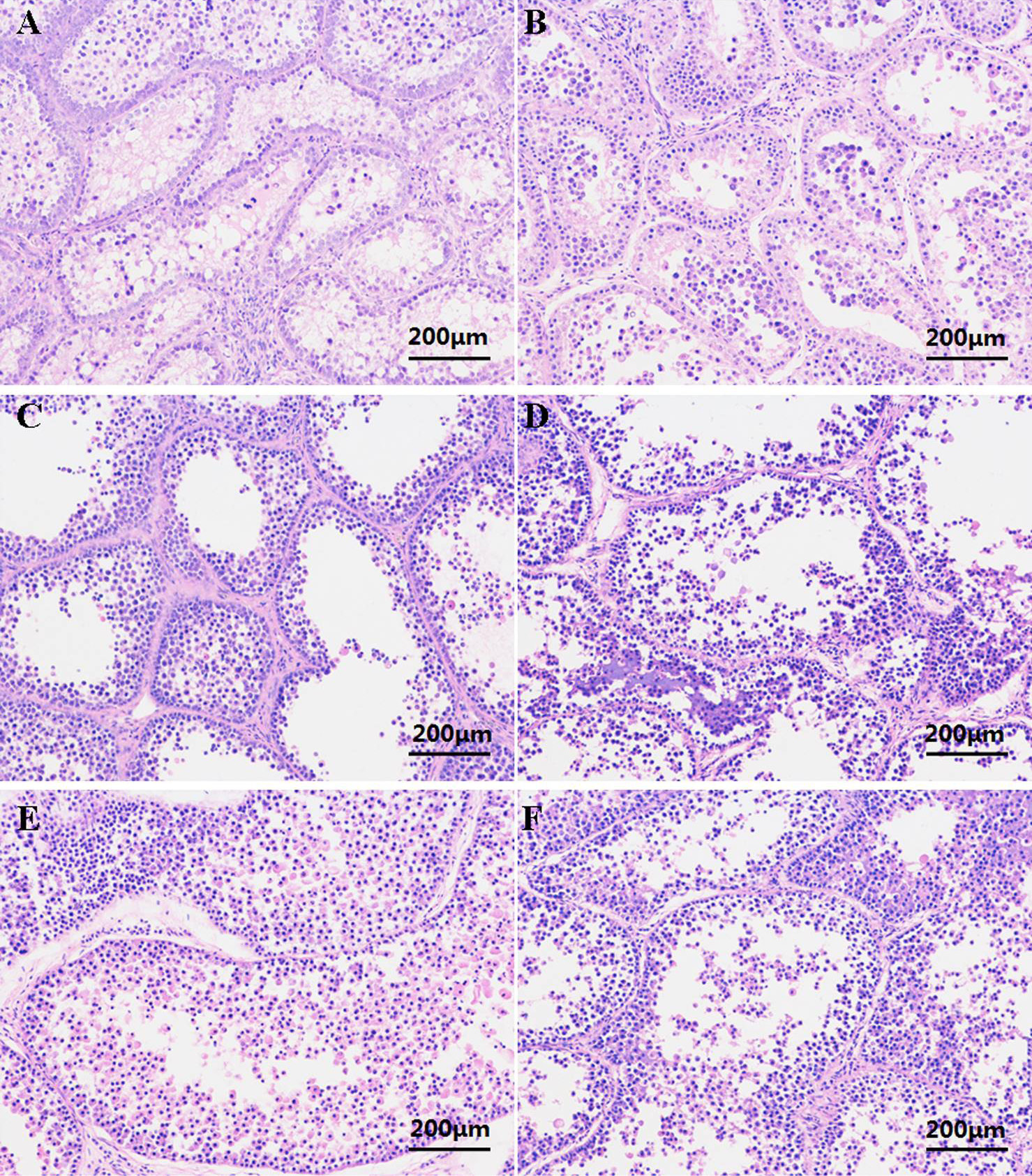
Histological structure of adult mule duck testis (200×). A and B, the testes weight/body weight ratio <1%; C and D, the testes weight/body weight ratio1-2%; E and F, the testes weight/body weight ratio 2-3%. A, C and E, the left testis; B, D and F, the right testis. Staining: H&E.
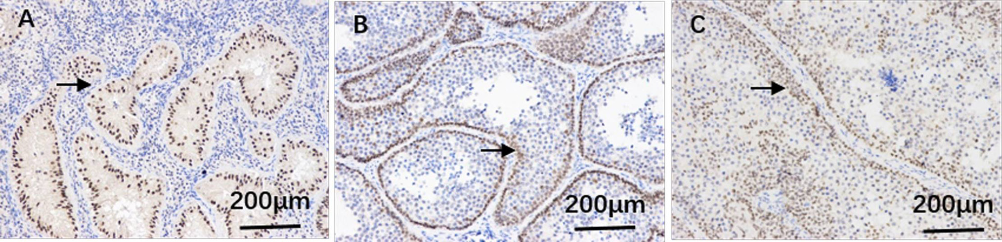
The expression of DMRT1 in adult mule duck testis (200×). A, the testes weight/body weight ratio <1%; B, the testes weight/body weight ratio 1-2%; C, the testes weight/body weight ratio 2-3%. The arrow shows the expression of DMRT1 in seminiferous tubule.
References
- Chen, S.L., Zhang, G.J., Shao, C.W., Huang, Q.F., Liu, G., Zhang, P., Song, W.T., An, N., Chalopin, D., Volff, J.N., Hong, Y., Li, Q., Sha, Z., Zhou, H., Xie, M., Yu, Q., Liu, Y., Xiang, H., Wang, N., Wu, K., Yang, C., Zhou, Q., Liao, X., Yang, L., Hu, Q., Zhang, J., Meng, L., Jin, L., Tian, Y., Lian, J., Yang, J., Miao, G., Liu, S., Liang, Z., Yan, F., Li, Y., Sun, B., Zhang, H., Zhang, J., Zhu, Y., Du, M., Zhao, Y., Schartl, M., Tang, Q. and Wang, J., 2014. Nat. Genet., 46: 253-260. https://doi.org/10.1038/ng.2890[CrossRef]
- Gong, P., Yang, Y., Lei, W.W., Feng, Y.P., Li, S.J., Peng, X.L. and Gong, Y.Z., 2012. Sex. Develop., 6: 178-187. https://doi.org/10.1159/000338471[CrossRef]
- Guan, W., Wang, Y., Hou, L., Chen, L., Li, X., Yue, W. and MA, Y., 2010. Poult. Sci., 89: 312-317. https://doi.org/10.3382/ps.2009-00413[CrossRef]
- Li, L., Miao, Z.W., Xin, Q.W., Zhu, Z.M., Zhang, L.L., Zhuang, X.D. and Zheng, N.Z., 2017. Sci. Agric. Sin., 50: 3608-3619.
- Fhamida, B.I., Satoshi, I., Yoshinobu, U., Kornsorn, S. and Yoichi, M., 2013. Jap. Poult. Sci., 50: 311-320.
- Koba, N., Ohfuji, T., Ha, Y., Mizushima, S., Tsukada, A., Saito, N. and Shimada, K., 2008. J. Poult. Sci., 45: 132-138. https://doi.org/10.2141/jpsa.45.132[CrossRef]
- Koopman, P., 2009. Trends Genet., 25: 479-481. https://doi.org/10.1016/j.tig.2009.09.009[CrossRef]
- Rigdon, R.H. and Mott, C., 1965. Pathol. Vet., 2: 553-565. https://doi.org/10.1177/030098586500200603[CrossRef]
- Shen, Y.X. and Tong, L.F., 1991. J. Zhejiang Agric. Univ., 17: 283-289.
- Smith, C.A., Roeszler, K.N., Ohnesorg, T., Cummins, D.M., Farlie, P.G., Doran, T.J. and Sinclair, A.H., 2009. Nature, 461: 267-271. https://doi.org/10.1038/nature08298[CrossRef]
- Snapir, N., Rulf, J., Meltzer, A., Gvaryahu, G., Rozenboim, I. and Robinzon, B., 1998. Br. Poult. Sci., 39: 572-574. https://doi.org/10.1080/00071669888791[CrossRef]
- Tan, J.Z., Chen, H., Song, J.J. and Liu, Y.T., 1998. Fujian J. agric. Sci., 68: 553-560. https://doi.org/10.1002/(SICI)1097-4628(19980425)68:4<553::AID-APP6>3.0.CO;2-L[CrossRef]
- Wang, Q., Weng, H., Chen, Y., Wang, C., Lian, S., Wu, X., Zhang, F. and Li, A., 2015. Br. Poult. Sci., 56: 390-397. https://doi.org/10.1080/00071668.2015.1027172[CrossRef]
- Bacha, W.J. and Bacha, L.M., 2000. Color atlas of veterinary histology, 2nd Edition. Blackwell Publishing Ltd., Oxford.
- Yang, Y., Gong, P., Feng, Y.P., Li, S.J., Peng, X.L., Ran, Z.P., Qian, Y.G. and Gong, Y.Z., 2013. Acta Biol. Hung., 64: 161-168. https://doi.org/10.1556/ABiol.64.2013.2.3[CrossRef]
- Yang, Y., Gong, P., Feng, Y.P., Yang, Y.P., Li, S.J., Peng, X.L. and Gong, Y.Z., 2012. J. Anim. Vet. Adv., 11: 561-565.
Supplementary material
Services
Article usage statistics
Loading…