
Letrozole (LET) is a triazole-containing drug that can inhibit the activity of cytochrome P450 aromatase, so as to affect the production of estrogen. In the present study, to detect the effects of letrozole on gonad differentiation, 5dph fry of carp were exposed to different concentrations of LET. At 130 dph, sex ratio was recorded. The results showed that continuous exposure to LET induced the changes of sex ratio. High concentration of LET can promote male-biased sex ratio. In order to reveal the molecular mechanism by which LET caused sex reversal, juvenile carp (120dph) were exposed to 0, 5, 25, 125, and 625μg/L LET for 15 days, and the expression profiles of seven gonad differentiation related genes (Cyp19A, Cyp19B, Arα, ERα, Dax, Foxl2, Dmrt1) and gonad histological changes were examined. The results showed that LET could inhibit oocyte growth in female and promote the development of testes in males. For the gonad differentiation related genes, the expression profiles of Cyp19A and Cyp19B show a pattern of increase in short time and low concentration, but decline in long time and high concentration. Arα has a trend of increases, ERα shows downwards trend. Dax and Foxl2 are female-biased genes, their expression profiles show similar trends to Cyp19A in ovary, but downwards trend in testis. Dmrt1 is male-biased genes, it shows a trend of increases in male and female individuals. These findings suggest that continuous exposure to LET may perturb sex differentiation in carp, the molecular mechanism not only involve inhibition of cytochrome P450 aromatase activity, but also the changes of upstream genes in sex differentiation. So, LET can be used as a reagent to control the sex differentiation of carp, but also can be used to disturb gonad differentiation, and then to reveal the molecular mechanism.
Research Article · Volume 49, Issue 6, 13 October 2017 · Open Access
Effects of Letrozole on Gonad Differentiation of Carp (Cyprinus carpio)
· · · ·
Affiliations & Notes ⌄
- 1College of Life Science, Henan Normal University, Xinxiang, Henan 453007, People’s Republic of China , * Corresponding author: 13837331530@163.com
Article Info ⌄
- Journal:
- Pakistan Journal of Zoology
- Published:
- 13 October 2017

From the article
- LET exposure also caused significant effects on the expression of other genes involved in sex differentiation.
- Continuous exposure to LET induced the changes of sex ratio in fry of carp.
- The expressions of genes related to ovarian differentiation, such as Cyp19A, Cyp19B, Dax, Foxl2 and Erα, decreased rapidly, but downstream gene such as Cyp19A, Cyp19B and Erα presented compensatory responses at short exposure time in low concentration.
- So, LET can be used as a reagent to control the sex differentiation of carp, but also can be used to disturb gonad differentiation, and then to reveal the molecular mechanism.
Selected from the article's own text.
Abstract
Introduction
Sex determination in fish is known to be significantly influenced by environmental factors, such as temperature, pH, exogenous hormones, and pesticides ; . Many of these factors are potential endocrine disruptors, which at minute levels are sufficient to cause developmental and reproductive dysfunction in numerous species ; . In recent years, numerous research efforts have been focused on man-made chemical compounds which can disrupt the endocrine system in vertebrates, including teleosts ; ; . Conazoles are a class of imidazole- or triazole-containing drugs, which have been commonly used as fungicides in agriculture and medicine . However, several azoles have adverse health effects such as carcinogenesis via CYP (e.g., CYP1A, 2B, 3A)-mediated metabolism in mammals and other non-targeted organisms, and these chemicals are also known as aromatase inhibitors (AIs) which block cytochrome P450 aromatase (P450arom) activity, resulting in a decrease in estrogen production ; ; . P450arom is the steroidogenic enzyme responsible for the conversion of androgen to estrogen. It is widely recognized that AIs play important roles in sexual differentiation and reproduction in all vertebrates studied to date ; , and the P450arom gene has been shown to be related to sex differentiation as well as gonadal development in teleost fish ; ; ; , 2015).
AIs have been widely used in studies of sex differentiation, reproduction and sex change in fish. Letrozole (LET) is a triazole-containing drug, is known to be a potent AI and acts indirectly (EPA, 2002; ; . LET can inhibit the activity of P450arom by competitively binding to the heme of the cytochrome P450 subunit, resulting in a reduction in estrogen biosynthesis in tissues . For this reason, LET has been widely used in the therapy of breast and ovarian cancer in postmenopausal women ; . It is considered a standard AI in fish ; . Some studies have shown that LET can disrupt the endocrine system of fish and cause alterations in both reproduction and development ; ; . LET was also found to inhibit oocyte growth and reduce plasma vitellogenin (VTG) levels in adult female Medaka . To date, very few studies have been carried out to determine the differences in the expression of genes related to gonadal development and gonadal sex differentiation, which could serve as biomarkers of damage in reproductive organs caused by LET. It is vital to identify the mechanism involved in the effects of LET exposure during several development stages. Genes regulating gonadal differentiation and maintenance of reproductive functions are good candidates for this purpose.
Carp is one of the most important aquaculture species in China. As females grow faster than males, the output of these fish can be increased and the economic benefit improved by controlling the sex. In practice, hormones are used to increase the number of females. In this study, fry and juvenile carp were exposed to LET at environmentally relevant concentrations (0, 5, 25, 125, 625 μg/L) for 130 or 15 days to investigate the expression of the upstream genes associated with sex development in the brain and gonads as well as their histological changes. The results of the present study will improve our understanding of the endocrine-disrupting effects of this chemical in the environment.
Materials and methods
Chemicals used
LET (purity>98%) was purchased from Beijing Dezhong-Venture Pharmaceutical Technology Development Co., Ltd. (Beijing, China). As LET has low solubility in water, LET stock solutions were prepared in acetone and diluted with acetone (Fisher Scientific, USA).
Fish and care of fish
Adult and juvenile carp Yellow River carp (Cyprinus carpio) were obtained from the aquaculture sites and maintained at the genetic laboratory (Henan normal university, Xinxiang Henan province, China) in flow-through water tanks with a constant temperature of 25 ± 1°C, three tanks per exposure concentration. Embryos were obtained by natural spawning and larvae were cultured in embryo medium following standard procedures. Fry were fed two times daily with commercial flake food.
Effect of LET on sexual differentiation of carp
On day 5 post-hatch (dph), fry were divided into several groups, held in the water containing LET at a concentration of 0, 5, 25, 125, or 625μg/L. To avoid metabolic and microbial breakdown of LET, half of the water was removed and replaced every 3 days with fresh LET-contaminated water. At 130 dph, 100 fish from each group were randomly selected for histological examination of the gonads, each concentration group was set with three replicates.
Water samples (100 mL) were randomly collected from each treatment tank every 3–5 days during the exposure period to confirm the concentration of LET. LET concentrations in the water samples were quantified by an LC system (Dionex Ultimate 3000, Thermo Scientific, San Jose, CA, USA) coupled to a triple quadruple mass spectrometer (TSQ Vantage, Thermo Scientific).
120dph juvenile carp were used to detect the expression profile of genes related to sexual differentiation. Groups of 100 fish were exposed to 0, 5, 25, 125, or 625 μg/L LET under static renewal conditions for 15 days respectively, and 50% of the test solution was renewed every three days. Two control groups were included, one is acetone and the other was kept in water without chemicals. No physical signs of negative health effects were observed, and all fish survived during the 15-day treatment period.
During the treatment period, 20 fish from each group were sacrificed at day 5, 10 and 15, respectively. Brain and gonadal tissues were dissected, the tissues were immediately stored at −80°C for gene expression analysis and stored in formaldehyde for subsequent histological observation.
Histological studies
The gonads and brains were fixed in Bouin’s solution, embedded in paraffin, and 6 µm sections were cut. The sections were then stained with hematoxylin and eosin, observed and photographs obtained under a microscope.
Quantitative real-time PCR (qPCR)
The gonads and brains of five male and female fish were homogenized and prepared at each time point. RNA isolation was performed using TRIzol reagent (Invitrogen, USA) according to the manufacturer’s protocol. Total RNA concentration was estimated by the absorbance ratio of 260/280 nm, and the quality of RNA was evaluated by 1% agarose gel electrophoresis. cDNA was obtained using a Quantitec Reverse Transcription Kit (Qiagen, Germany), according to the manufacturer’s instructions.
Real-time PCR with SYBR green detection was performed using a Light Cycle® Real-Time PCR System (Roche Applied Science), according to the operator’s guide. The transcription of functionally relevant genes (Cyp19A, Cyp19B, DMRT1, FOXL2, DAX, ERα and ARα) was analyzed. All oligonucleotide primers were designed
Table I.- Primer sequences used for gene transcription analysis.
| Gene | Primer sequence |
| Cyp19a (Cytochrome P450 aromatase 1a) | 5’-TCTCTTATGCTTCCTGTGAA-3’ 3’-ACTTACTGCGGTGTATTGT-5’ |
| Cyp19b (Cytochrome P450 aromatase 1b) | 5’-TGTCCTGAGTGGTCTGTGAACT-3’ 3’-CGGGTCTCCAGAAATCGGTAGA-5’ |
| Foxl2 (Forkhead transcriptional factor 2 Gene) | 5’-GCCTATGTCCTATACCTCCTGTC-3’ 3’-CTCATGCCGTTGTAAGTGTTCA-5’ |
| Dax (Dosage-sensitive sex reversal-adrenal hypoplasia gene on the X chromosome) | 5’-GGTGGCCGGTCCTTCTTCAAAC-3’ 3’-CCATTCACTGCAAACTGCC-5’ |
| ER-α (Estrogen receptor alpha) | 5’-TGGATAGGAGTGAAGGAGAA-3’ 3’-TGGACTGGAGCAGAATGA-5’ |
| AR-α (Androgen receptor) | 5’-CCTGCCTAATCTGCTCTG-3’ 3’-TCATCTGTCCAATCTTCCTC-5’ |
| Dmrt (Doublesex and mab-3 related transcription factor 1) | 5’-TTCGTGTCACCGCTGAAG-3’ 3’-GCTCTGAGTGATGGTAACG-5’ |
with Primer Premier 5.0. The efficiency and optimal concentration of each primer was tested using adult wild-type male and female carp cDNA (Table I). The expression profile of the target gene was normalized to that of the housekeeping gene β-actin, as the expression of β-actin had almost no influence on the effects of LET exposure according to our preliminary experiment.
Data analysis
Each sample was run in triplicate. Quantification of the expression of each gene was based on the 2−ΔΔCt method . Statistical differences between experimental samples were assessed using SPSS 13.0 software. Analysis of variance (ANOVA) followed by Dunnett’s post-hoc test were used to determine statistical differences between experimental samples. All data are expressed as means ± standard error of the mean and p<0.05 was considered statistically significant as follows: *p< 0.05, **p<0.01.
Results
Fry mortality is similar to control. No mortality was observed in juvenile carp while exposed to LET (data not shown), indicating that the concentrations of LET used in this were not acutely toxic.
Effect of LET on sex ratio
The gonads of carp were examined under a dissecting microscope to evaluate the sex at 130 dph. The results indicated that the effect of exposure to different concentration of LET on sex ratio in Yellow river carp were remarkable. The male percentage increased with increase of LET. When the concentration of LET in aquatic water was 0, 5, 25, 125, or 625 μg/L, the male percentage were 49%, 53%, 56%, 64% and 73%, respectively.
Histological changes
We chose juvenile carp for this study as their gonads had differentiated, but were not yet mature. LET was found to inhibit oocyte growth in female juvenile carp and promote the development of testes in males (Fig. 1a-d).
The results showed that primary oocytes in the ovary of the control groups were large, closely arranged, uniformly developed, with few small oocytes. However, exposure to 625 μg/L LET led to a decrease in the number of primary oocytes per unit area, the ovarian plate space was larger, the number of small early differentiated oocytes
increased, and the degree of differentiation was relatively low. In males, the control groups showed an abundance of primary sperm cells and a small number of sperm cells, however, treated males showed an abundance of sperm cells and a small number of primary sperm cells (Fig. 1d). No changes were observed in the brain (Fig. 1e-h).
Effect of LET on the expression profiles of gonad differentiation related genes
Cyp19A
Cyp19A was predominantly expressed in the ovaries, with low expression in the brain of both males and females (Fig. 2).
In the brain and testis of male carp, the expression of Cyp19A changed little at 5d, and then significantly declined in a concentration-dependent manner. In the brain and testis of female, the expression of Cyp19A slightly increased at 5d, then began to decrease at 10d and 15d. Different concentrations of LET affect the amount of decrease (Fig. 2).
Cyp19B
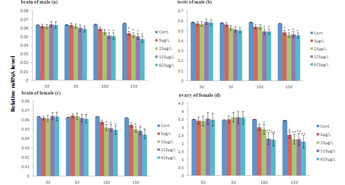
Cyp19b was predominantly expressed in the brain, with low expression in gonad (Fig. 3).
The expression of Cyp19B decreased in the brain of male carp, but increased in the brain of females at 5d, then decreased in a concentration-dependent manner. In the gonad, Cyp19B was down-regulated when exposed to LET (Fig. 3).
Foxl2
In general, treatment with LET resulted in a decrease in Foxl2 expression in the brain and gonads of both males and females (Fig. 4).
In male brains, the expression levels of Foxl2 were not significantly altered in lower concentration or at shorter time, but were significantly down-regulated in >25 μg/L groups and 5 μg/L groups at >10d. In female brains, LET exposure had no obvious effect on the expression of Foxl2 at all concentrations at 5d. Foxl2 expression was significantly down-regulated at day 10 and 15 at all concentrations. In the testes, the expression of Foxl2 was decreased with the exception of no obvious change in 5 μg/L group at 5d. In the ovary, Foxl2 was slightly up-regulated at 5d, and was significantly down-regulated at 10d and 15d (Fig. 4).
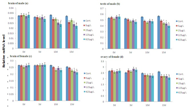
Dax
In male and female brains, exposure to LET caused a decrease to Dax expression, with the exception of less change at day 5 in the 5 μg/L and 25 μg/L treatment groups. In the testes, LET exposure down-regulated the expression of Dax in all treatment group. No effects were observed in the ovaries at 5d, however, Dax expression was inhibited by 125 μg/L and 625 μg/L group at 10d, as well as all concentration groups at 15d (Fig. 5).
Erα
In brain and gonad of female, a slightly increase in Erα expression was observed after exposure to LET for 5d, but it decreased rapidly at all groups for 10d or 15d. Acts of Erαs in male is similar to female, except that it had shown to decline at 5d. With increased treatment time, the decline trend gets slow (Fig. 6).
Arα
In males, increased levels of Arα were seen both in the brain and testes compared with the control group. In the testes of LET-exposed males, an increase in Arα mRNA was observed in the 5 μg/L and 25 μg/L groups at 10d. In the 625 μg/L group, a significant increase was observed at day 5 (Fig. 7).
The same change was observed in the brain of females. Exposure to LET caused a significant increase in Arα mRNA in female brains at all concentrations on day 5. In ovary, the expression of Arα was enhanced in a concentration-dependent manner with increased treatment time, markedly increasing to 20% (p<0.01) of the control group level at 625 μg/L (Fig. 7).
Dmrt1
No matter in the brain or the gonad, the expression of Dmrt1 was up-regulated. In male individuals, the up-regulation of Dmrt1 begins at 5d in 125 and 625 groups, at 10d and 15d in all groups. In female, the up-regulation of Dmrt1 begins at 5d, but changes are not obvious at 5d in 5 and 25 groups (Fig. 8).
Discussion
P450arom is a key enzyme which catalyzes the critical reaction that converts C19 androgens to C18 estrogens, and controls the last and rate limiting step of estrogen biosynthesis (EPA, 2002). Due to the important
role of aromatase, increasing attention has focused on the effects of AIs on normal aromatase activity and expression in fish. LET is one of the most potent of the new generation AIs, and has been widely used in medicine now . Few studies on the effects of LET on fish have been conducted. In the present study, the effects of letrozole on gonad differentiation of carp fry were detected at first. The results showed that continuous exposure to LET induced male-biased sex ratio. These results were similar to that of another AI, fadrozole, it was found to have a significant masculinization effect on many species of gonochoristic fish as well as sex-changing protogynous species ; ; ; ; ; ; ; . These two aromatase inhibitors (AIs), LET and fadrozole, were known as which block cytochrome P450 aromatase (P450arom) activity, resulting in a decrease in estrogen production. We think this is the direct cause of the male-biased sex development in carp.
In order to reveal the deep mechanism of male-biased sex development, we also analyzed the expression of genes associated with sex development in the brain and gonads and their histological changes in juvenile carp exposed to LET.
Other research has shown that fadrozole can change the expression of Cyp19 in the brain and gonads as well as the mRNA of other genes in the hypothalamic-pituitary-gonadal (HPG(L)) axis , 2008, 2009b; Zhang et al., 2008). It can also inhibit E2 biosynthesis, affect the morphology of gonads ; ; ; . LET was found to inhibit oocyte growth in adult female medaka , and these results were seen in our study. The histological results showed that LET inhibited oocyte growth in female juvenile carp and promoted the development of testes in males. The differentiation of oocytes requires a certain concentration of estrogen. Growth inhibition and the number decrease of oocyte are direct consequences of the estrogen decrease induced by LET.
LET is an aromatase inhibitors and reduces estrogen production. How does it affect sex differentiation? Many genes involve in fish gonad differentiation, and some promote ovarian development, the others is very important for testis. Our experimental results showed that in response to LET, juvenile carp showed both direct effects and compensatory adaptation. There are two P450arom isoforms in most species of teleosts, one is Cyp19A which is expressed predominantly in the ovaries, and the other form is Cyp19B which is expressed predominantly in the brain ; . Reports on the effect of xenoestrogens on the expression of Cyp19A vary with teleost species as well as developmental stage ; ; . The expression profiles of Cyp19a and Cyp19b show a pattern of stable or increase slightly in short time and low concentration, but decline in long time and high concentration. These results are basically consistent to those in other reports, which show that LET exposure in Japanese ricefish caused significant inhibition of Cyp19B expression in both the brain and ovaries of females . In our research, the inhibition of Cyp19 resulted in a reduction in the rate of endogenous estrogen biosynthesis. The expression of Cyp19 in short time were up-regulated in an attempt to maintain the normal amount of estrogen in the ovary, indicate a compensatory response to aromatase inhibition , 2007, 2009a, b; Zhang et al., 2008). In high concentration groups and long-time exposure, the involvement of the upstream regulation genes makes Cyp19 significantly down regulated. The promoter of Cyp19A contains a regulatory element called steroidogenic factor-1 (SF-1), which is considered to be regulated mainly via cyclic adenosine monophosphate-mediated signal transduction, while the Cyp19B promoter region contains estrogen response elements (EREs) and their expression is more closely tied to endogenous E2 concentration ; . Our result related to the change in expression of these two genes is in agreement with this explanation.
With regard to the hormone receptors Erα and Arα, LET showed a different mode of action. Following exposure to LET for 15 days, decrease of the Erα was seen in both the brain and gonads, the expression of Erα is closely related content of Erα. The Inhibition effects of LET on normal aromatase activity reduced estrogen and increased testosterone. LET exposure resulted in significant induction of Arα expression in the testes. The report by Sun et al. (2011) showed that Arα expression decreased at the concentration of 300 μg/L in the ovaries of female ricefish (Oryzias latipes), and did not change in the brain and testes of males . The reason for the difference between our report and others may be due to the length of exposure time, differences in LET concentrations and species of fish.
LET exposure also caused significant effects on the expression of other genes involved in sex differentiation. DMRT1 is one of the key genes in testis development, and is testis-determining gene in some fish. Wang et al. (2012) found that the expression level of Dmrt1 in Takifugu obscurus increased when exposed to LET. Our study showed that transcription of the male-predominant gene Dmrt1 was first induced in5 μg/L group at 5d, this indicated that there was a direct response at the beginning of LET exposure. Dmrt1 transcription was enhanced in the ovaries. According to our findings, exposure to LET inhibited the production of endogenous estrogen, which increased testosterone. The increase of testosterone promotes Dmrt gene expression, and the specific molecular mechanism remains to be elucidated. At present, the genome sequence of common carp has been released, preliminary analysis found that steroid receptor binding sites exist in the upstream of the Dmrt gene, which may reveal the relationship between the two genes.
Foxl2 and Dax are key genes in the development of ovary. The expression of Foxl2 was down-regulated in the brain, ovaries and testes, especially in the ovaries, which is in agreement with the findings in another report . Exposure to LET induced Dax mRNA down-regulated at >25 μg/L at 5d. The down-regulation of Foxl2 and Dax also explained the phenomenon of Oocyte growth slowing.
Conclusions
LET has direct and indirect effects on sex differentiation of carp. Continuous exposure to LET induced the changes of sex ratio in fry of carp. High concentration of LET can promote male-biased sex ratio. For juvenile carp, continuous exposure to LET could to inhibit oocyte growth in female and promote the development of testes and spermatogenesis in males. Our data show that LET exposure altered gene expression in a complex manner. The expressions of genes related to ovarian differentiation, such as Cyp19A, Cyp19B, Dax, Foxl2 and Erα, decreased rapidly, but downstream gene such as Cyp19A, Cyp19B and Erα presented compensatory responses at short exposure time in low concentration. For the genes related to testes differentiation, their expressions showed increasing trend. So, LET can be used as a reagent to control the sex differentiation of carp, but also can be used to disturb gonad differentiation, and then to reveal the molecular mechanism.
Conflict of interest statement
We declare that we have no conflict of interest.
Figures
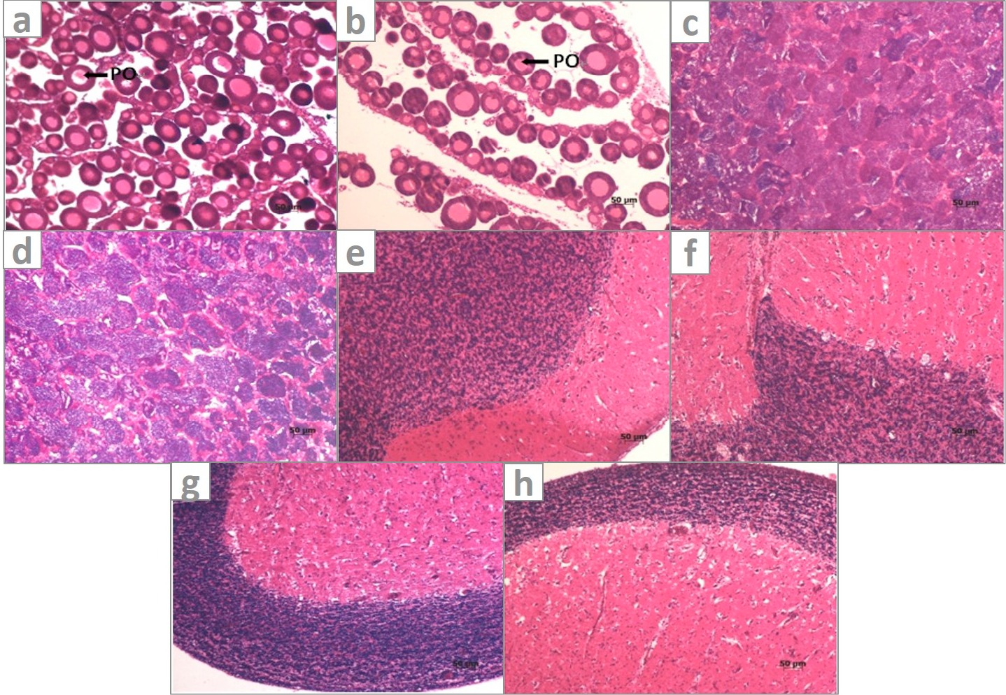
Histological structure of the gonads and brains of juvenile carp from control and the groups exposed to LET for 15d. a, Ovary from control groups showing abundance of large, closely arranged, uniformly developed primary oocytes; b, Ovary from LET(625μg/L)-treated groups showing increased prevalence of smaller early differentiated oocytes and enlarged ovarian plate space; c, Testis from control groups; d, from LET(625μg/L)-treated groups showing increased spermatogonial cells; e, brain from control male; f, brain from LET(625μg/L)-treated male; g, brain from control female; h, brain from LET(625μg/L)-treated female. PO, primary oocytes.
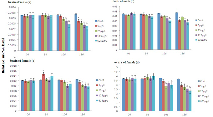
The expression of Cyp19A in the brain and gonads of juvenile carp treated with 0, 5, 25, 125, 625μg/L letrozole for 15 d. Data are presented as means ± S.E. (n=5) and normalized against the mRNA expression of β-actin. The asterisk symbols denote significant differences compared with the control group: *p< 0.05 and **p< 0.01.
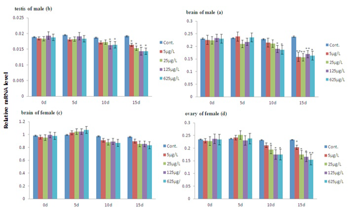
The expression of Cyp19B in the brain and gonads of juvenile carp treated with 0, 5, 25, 125, 625μg/L letrozole for 15 d. Data are presented as means ± S.E. (n=5) and normalized against the mRNA expression of β-actin. The asterisk symbols denote significant differences compared with the control group: *p< 0.05 and **p< 0.01.
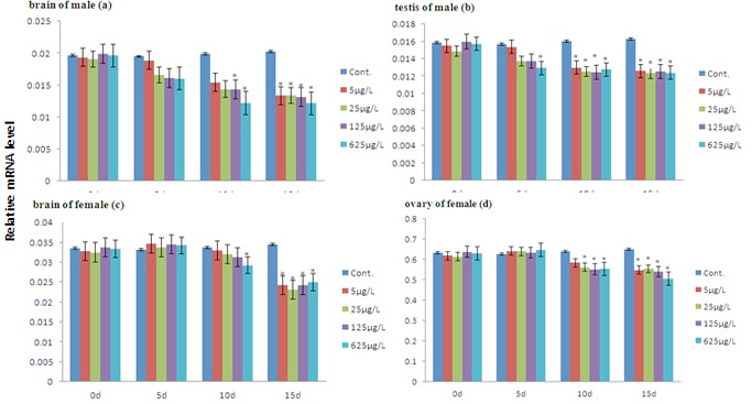
The expression of Foxl2 in the brain and gonads of juvenile carp treated with 0, 5, 25, 125, 625μg/L letrozole for 15 d. Data are presented as means ± S.E. (n=5) and normalized against the mRNA expression of β-actin. The asterisk symbols denote significant differences compared with the control group: *p< 0.05 and **p< 0.01.
The expression of Dax in the brain and gonads of juvenile carp treated with 0, 5, 25, 125, 625μg/L letrozole for 15 d. Data are presented as means ± S.E. (n=5) and normalized against the mRNA expression of β-actin. The asterisk symbols denote significant differences compared with the control group: *p< 0.05 and **p< 0.01.
The expression of Erα in the brain and gonads of juvenile carp treated with 0, 5, 25, 125, 625μg/L letrozole for 15 d. Data are presented as means ± S.E. (n=5) and normalized against the mRNA expression of β-actin. The asterisk symbols denote significant differences compared with the control group: *p< 0.05 and **p< 0.01.
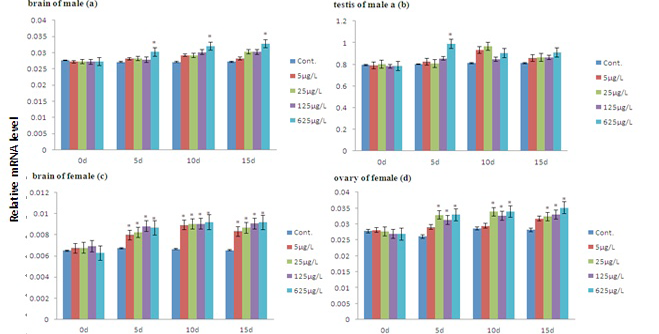
The expression of Arα in the brain and gonads of juvenile carp treated with 0, 5, 25, 125, 625μg/L letrozole for 15 d. Data are presented as means ± S.E. (n=5) and normalized against the mRNA expression of β-actin. The asterisk symbols denote significant differences compared with the control group: *p< 0.05 and **p< 0.01.
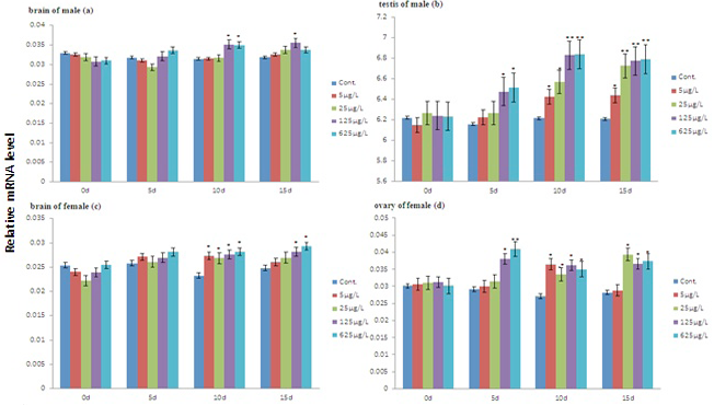
The expression of Dmrt1 in the brain and gonads of juvenile carp treated with 0, 5, 25, 125, 625 μg/L letrozole for 15 d. Data are presented as means ± S.E. (n=5) and normalized against the mRNA expression of β-actin. The asterisk symbols denote significant differences compared with the control group: *p< 0.05 and **p< 0.01.
References
- Allen, J.W., Wolf, D.C., George, M.H., Hester, S.D., Sun, G. S. Thai, F., Delker, D.A., Moore, T., Jones, C., Nelson, G., Roop, B.C., Leavitt, S., Winkfield, E., Ward, W.O. and Nesnow, S., 2006. Toxicity profiles in mice treated with hepatotumorigenic and non-hepatotumorigenic triazole conazole fungicides: propiconazole, triadimefon, and myclobutanil. Toxicol. Pathol., 34: 853-862. https://doi.org/10.1080/01926230601047816[CrossRef]
- Alam, M.A., Bhandari, R.K., Kobayashi, Y., Soyano, K. and Nakamura, M., 2006. Induction of sex change within two full moons during breeding season and spawning in grouper. Aquaculture, 255: 532-535. https://doi.org/10.1016/j.aquaculture.2006.01.008[CrossRef]
- Adkins-Regan, E., 1987. Hormones and sexual differentiation. In: Hormones and reproduction in fishes, amphibians and reptiles (eds. D.O. Norris and R.E. Jones), Plenum Press, New York, pp. 1-29. https://doi.org/10.1007/978-1-4613-1869-9_1[CrossRef]
- Ana, L.Z., Sandra, M.M., Sofia, G.S., Rocha, E., Antonio A.F.F. and Ana, M.C., 2015. Zebrafish sex differentiation and gonad development after exposure to 17α-ethinylestradiol, fadrozole and their binary mixture: A stereological study. Aquat. Toxicol., 166: 83-95. https://doi.org/10.1016/j.aquatox.2015.07.015[CrossRef]
- Ana, L.Z., Sandra, M.M., Rocha, E., Antonio A.F.F. and Ana, M.C., 2016. Development and recovery of histopathological alterations in the gonads of zebrafish (Danio rerio) after single and combined exposure to endocrine disruptors (17α-ethinylestradiol and fadrozole). Aquat. Toxicol., 175: 90-105. https://doi.org/10.1016/j.aquatox.2016.03.014[CrossRef]
- Bhandari, R.K., Higa, M., Nakamura, S. and Nakamura, M., 2004. Aromatase inhibitor induces complete sex in protogynous honeycomb grouper, Epinephelus merra. Mol. Reprod. Dev., 67: 303-307. https://doi.org/10.1002/mrd.20027[CrossRef]
- Corcoran, J., Winter, M.J. and Tyler, C.R., 2010. Pharmaceuticals in the aquatic environment: a critical review of the evidence for health effects in fish. Crit. Rev. Toxicol., 40: 287-304. https://doi.org/10.3109/10408440903373590[CrossRef]
- Colborn, T., 1993. Developmental effects of endocrine-disrupting chemicals in wildlife and humans. Environ. Hlth. Perspect., 101: 378. https://doi.org/10.1289/ehp.93101378[CrossRef]
- Dang, Y., Giesy, J.P., Wang, J.H. and Liu, C.S., 2015. Dose-dependent compensation responses of the hypothalamic-pituitary-gonadal-liver axis of zebrafish exposed to the fungicide prochloraz. Aquat. Toxicol., 160: 69-75. https://doi.org/10.1016/j.aquatox.2015.01.003[CrossRef]
- Deng, S.P., Chen, S.L., Xu, J.Y. and Liu, B.W., 2009. Molecular cloning, characterization and expression analysis of gonadal P450 aromatase in the half-smooth tongue-sole, Cynoglossus semilaevis. Aquaculture, 287: 211-218. https://doi.org/10.1016/j.aquaculture.2008.10.034[CrossRef]
- Devlin, R.H. and Nagahama, Y., 2002. Sex determination and sex differentiation in fish: an overview of genetic, physiological, and environmental influence. Aquaculture, 208: 191-364. https://doi.org/10.1016/S0044-8486(02)00057-1[CrossRef]
- Deng, S.P., Zhu, S.L., Sun, J., Wang, W.D., Wu, T.L., Chen, H.P., Shi, S.L. and Li, G.L., 2015. Foxl2 of the Hong Kong catfish (Clarias fuscus): cDNA cloning, tissue distribution and changes in gene expression towards methyltestosterone, estradiol and letrozole exposure of the fries during gonadal differentiation. Genes Genom., 37: 669-677. https://doi.org/10.1007/s13258-015-0296-z[CrossRef]
- Das, R., 2007. Effect of aromatase inhibitors on gonadal development in Oreochromis mossambicus (Peters). Dissertation, Collage of Fisheries, Mangalore, pp. 56-62.
- EPA, U.S, 2002. Draft detailed review paper on fish screening assays for endocrine disruption, Columbus, Ohio, pp. 695-705.
- Fukada, S., Tanaka, M., Matsuyama, M., Kobayashi, D. and Nagahama, Y., 1996. Isolation, characterization, and expression of cDNAs encoding the medaka (Oryzias latipes) ovarian follicle cytochrome P-450aromatase. Mol. Reprod. Dev., 45: 285-290. https://doi.org/10.1002/(SICI)1098-2795(199611)45:3<285::AID-MRD4>3.0.CO;2-O[CrossRef]
- Gen, K., Okuzama, K., Kumakura, N., Yamaguchi, S. and Kagawa, H., 2001. Correlation between messenger RNA expression of cytochrome P450 aromatase and itsenzyme activity during oocyte development in the red seabream (Pagrus major). Biol. Reprod., 65: 1186-1194. https://doi.org/10.1095/biolreprod65.4.1186[CrossRef]
- Hamilton, A. and Piccart, M., 1999. The third-generation non-steroidal aromatase inhibitors: a review of their clinical benefits in the second-line hormonal treatment of advanced breast cancer. Annls. Oncol., 10: 377-384. https://doi.org/10.1023/A:1008368300827[CrossRef]
- Haynes, B.P., Dowsett, M., Miller, W.R., Dixon, J.M. and Bhatnagar, A.S., 2003. The pharmacology of letrozole. J. Steroid Biochem., 87: 35-45. https://doi.org/10.1016/S0960-0760(03)00384-4[CrossRef]
- Howell, A., Cuzick, J., Baum, M., Buzdar, A., Dowsett, M., Forbes, J.F., Hoctin-Boes, G., Houghton, J., Locker, G.Y. and Tobias, J.S., 2005. Results of the ATAC (arimidex, tamoxifen, alone or in combination) trial after completion of 5 years’ adjuvant treatment for breast cancer. Lancet, 365: 60-62. https://doi.org/10.1016/S0140-6736(04)17666-6[CrossRef]
- Harries, J.E., Runnalls, T., Hill, E., Harris, C.A., Maddix, S., Sumpter, J.P. and Tyler, C.R., 2000. Development of a reproductive performance test for endocrine disrupting chemicals using pair-breeding fathead minnows (Pimephales promelas). Environ. Sci. Technol., 34: 3003-3011. https://doi.org/10.1021/es991292a[CrossRef]
- Higa, M., Ogasawara, K., Sakaguchi, A., Nagahama, Y. and Nakamura, M., 2003. Role of steroid hormone in sex change of Protogynous wrasse. Fish Physiol. Biochem., 28: 149-150. https://doi.org/10.1023/B:FISH.0000030505.28138.d1[CrossRef]
- Hinfray, N., Palluel, O., Turies, C., Cousin, C., Porcher, J.M. and Brion, F., 2006. Brain and gonadal aromatase as potential targets of endocrine disrupting chemicals in a model species, the zebrafish (Danio rerio). Environ. Toxicol., 21: 332-337. https://doi.org/10.1002/tox.20203[CrossRef]
- Ijiri, S., Kazeto, Y., Lokman, P.K., Adachi, S. and Yamauchi, K., 2003. Characterization of a cDNA encoding P-450 aromatase (Cyp19) from Japanese eel ovary and its expression in ovarian follicles during induced ovarian development. Gen. Comp. Endocrinol., 130: 193-203. https://doi.org/10.1016/S0016-6480(02)00589-0[CrossRef]
- Komatsu, T., Nakamura, S. and Nakamura, M., 2006. Masculinization of female golden rabbitfish Siganus guttatus using an aromatase inhibitor treatment during sex differentiation. Comp. Biochem. Physiol. Part C, 143: 402-409. https://doi.org/10.1016/j.cbpc.2006.04.015[CrossRef]
- Kroon, F.J., Munday, P.L., Westcott, D.A., Hobbs, J.P.A. and Liley, N.R., 2005. Aromatase pathway mediates sex change in each direction. Proc. R. Soc. B, 272: 1399-1405. https://doi.org/10.1098/rspb.2005.3097[CrossRef]
- Kazeto, Y., Place, A.R. and Trant, J.M., 2004. Effects of endocrine disrupting chemicals on the expression of Cyp19 genes in zebrafish (Danio rerio) juveniles. Aquat. Toxicol., 69: 25-34. https://doi.org/10.1016/j.aquatox.2004.04.008[CrossRef]
- Livak, K. and Schmittgen, T., 2001. Analysis of relative gene expression data using real-time quantitative PCR and the 2−ΔΔCt method. Methods, 25: 402-408. https://doi.org/10.1006/meth.2001.1262[CrossRef]
- Lee, Y.M., Seo, J.S., Kim, I.C., Yoon, Y.-D. and Lee, J.S., 2006. Endocrine disrupting chemicals (bisphenol A, 4-nonylphenol, 4-tert-octylphenol) modulate expression of two distinct cytochrome P450 aromatase genes differently in gender types of the hermaphroditic fish Rivulus marmoratus. Biochem. biophys. Res. Commun., 345: 894-903. https://doi.org/10.1016/j.bbrc.2006.04.137[CrossRef]
- Li, G. L., Deng, S.P., Sun, J., Wang, W.D., Shi, S.L. and Zhu, C.H., 2013. Effects of aromatase inhibitor letrozole on sex differentiation and related gene expression in Clarias fuscus. J. Fish. Sci. China, 20: 911-917. https://doi.org/10.3724/sp.j.1118.2013.00911[CrossRef]
- Lyssimachou, A., Jenssen, B.M. and Arukwe, A., 2006. Brain cytochrome P450 aromatase gene isoforms and activity levels in atlantic salmon after waterborne exposure to nominal environmental concentrations of the pharmaceutical ethynylestradiol and antifoulant tributyltin. Toxicol. Sci., 91: 82-92. https://doi.org/10.1093/toxsci/kfj136[CrossRef]
- Nakamura, M., Bhandari, R.K. and Higa, M., 2003. The role estrogens play in sex differentiation and sex changes of fish. Fish Physiol. Biochem., 28: 113-117. https://doi.org/10.1023/B:FISH.0000030495.99487.17[CrossRef]
- Periasamy, K., 2007. Effect of aromatase inhibitor on sex steroid ratio and neurogenesis of Cyprinus carpio. Dissertation, CIFE, Mumbai, pp. 132-140.
- Panter, G.H., Hutchinson, T.H., Hurd, K.S., Sherren, A., Stanley, R.D. and Tyler, C.R., 2004. Successful detection of (anti-) androgenic and aromatase inhibitors in pre-spawning adult fathead minnows (Pimephales promelas) using easily measured endpoints of sexual development. Aquat. Toxicol., 70: 11-21. https://doi.org/10.1016/j.aquatox.2004.06.007[CrossRef]
- Requena, A., Herrero, J., Landeras, J., Navarro, E., Neyro, J.L., Salvador, C., Tur, R., Callejo, J., Checa, M.A., Farre, M., Espinos, J.J., Fabregues, F., Grana-Barcia, M. and Grp, R.E.I., 2008. Use of letrozole in assisted reproduction: a systematic review and meta-analysis. Hum. Reprod. Update, 14: 571-582. https://doi.org/10.1093/humupd/dmn033[CrossRef]
- Sheehan, D.J., Hitchcock, C.A. and Sibley, C.M., 1999. Current and emerging azole antifungalagents. Clin. Microbiol. Rev., 12: 40-79.
- Smith, I.E, 1999. Aromatase inhibitors: a dose-response effect? Endocr. Relat. Cancer, 6: 245-249. https://doi.org/10.1677/erc.0.0060245[CrossRef]
- Seralini, G.E. and Moslemi, S., 2001. Aromatase inhibitors: past, present and future. Mol. Cell. Endocrinol., 178: 117-131. https://doi.org/10.1016/S0303-7207(01)00433-6[CrossRef]
- Scott, L.J. and Keam, S.J., 2006. Letrozole: in postmenopausal hormone-responsive early-stage breast cancer. Drugs, 66: 353-362. https://doi.org/10.2165/00003495-200666030-00010[CrossRef]
- Sonnenschein, C. and Soto, A.M., 1998. An updated review of environmental estrogen and androgen mimics and antagonists. J. Steroid Biochem. mol. Biol., 65: 143- 150. https://doi.org/10.1016/S0960-0760(98)00027-2[CrossRef]
- Sun, L.W., Zha, J.M., Spear, P.A. and Wang, Z.J., 2007. Toxicity of the aromatase inhibitor letrozole to Japanese medaka (Oryzias latipes) eggs, larvae and breeding adults. Comp. Biochem. Physiol. C, 145: 533-541. https://doi.org/10.1016/j.cbpc.2007.01.017[CrossRef]
- Sun, L.W., Zhao, X.L., Chi, J., Hu, X.H., Jin, Y.X. and Fu, Z.W., 2011. Transcriptional responses in the brain, liver and gonad of Japanese ricefish (Oryzias latipes) exposed to two anti-estrogens. Comp. Biochem. Physiol. C, 153: 392-401. https://doi.org/10.1016/j.cbpc.2011.01.003[CrossRef]
- Sumpter, J.P., 1998. Xenoendorine disrupters-environmental impacts. Toxicol. Lett., 102: 337-342. https://doi.org/10.1016/S0378-4274(98)00328-2[CrossRef]
- Taxvig, C., 2008. Endocrine-disrupting properties in vivo of widely used azole fungicides. Int. J. Androl., 31: 170. https://doi.org/10.1111/j.1365-2605.2007.00838.x[CrossRef]
- Tchoudakova, A., Kishida, M., Wood, E. and Callard, G.V., 2001. Promoter characteristics of two cyp19 genes differentially expressed in the brain and ovary of teleost fish. J. Steroid Biochem., 78: 427-439. https://doi.org/10.1016/S0960-0760(01)00120-0[CrossRef]
- Uchida, D., Yamashita, M., Kitano, T. and Iguchi, T., 2004. An aromatase inhibitor or high water temperature induce oocyte apoptosis and depletion of P450 aromatase activity in the gonads of genetic female zebrafish during sex-reversal. Comp. Biochem. Physiol., 137A: 11-20. https://doi.org/10.1016/S1095-6433(03)00178-8[CrossRef]
- Villeneuve, D.L., Knoebl, I., Kahl, M.D., Jensen, K.M., Hammermeister, D.E., Greene, K.J., Blake, L.S. and Ankley, G.T., 2006. Relationship between brain and ovary aromatase activity and isoform-specific aromatase mRNA expression in the fathead minnow (Pimephales promelas). Aquat. Toxicol., 76: 353-368. https://doi.org/10.1016/j.aquatox.2005.10.016[CrossRef]
- Villeneuve, D.L., Larkin, P., Knoebl, I., Miracle, A.L., Kahl, M.D., Jensen, K.M., Makynen, E.A., Durhan, E.J., Carter, B.J., Denslow, N.D. and Ankley, G.T., 2007. A graphical systems model to facilitate hypothesis-driven ecotoxicogenomics research on the teleost brain-pituitary-gonadal axis. Environ. Sci. Technol., 41: 321-330. https://doi.org/10.1021/es061739x[CrossRef]
- Villeneuve, D.L., Knoebl, I., Larkin, P., Miracle, A.L., Carter, B.J., Denslow,, N.D. and Ankley, G.T., 2008. Altered gene expression in the brain and liver of female fathead minnows Pimephales promelas Rafinesque exposed to fadrozole. J. Fish Biol., 72: 2281–2340. https://doi.org/10.1111/j.1095-8649.2008.01897.x[CrossRef]
- Villeneuve, D.L., Mueller, N.D., Martinovic, D., Makynen, E.A., Kahl, M.D., Jensen, K.M., Durhan, E.J., Cavallin, J.E., Bencic, D. and Ankley, G.T., 2009a. Direct effects, compensation, and recovery in female fathead minnows exposed to a model aromatase inhibitor. Environ. Hlth. Perspect., 117: 624-631. https://doi.org/10.1289/ehp.11891[CrossRef]
- Villeneuve, D.L., Wang, R.L., Bencic, D.C., Biales, A.D., Martinovic, D., Lazorchak, J.M., Toth, G. and Ankley, G.T., 2009b. Altered gene expression in the brain and ovaries of zebrafish (Danio rerio) exposed to the aromatase inhibitor fadrozole: microarray analysis and hypothesis generation. Environ. Toxicol. Chem., 28: 1767-1782. https://doi.org/10.1897/08-653.1[CrossRef]
- Wang, Q., Guo, Z.L., Li, Y.S., Yun, D., Huang, P. and Zhou, Z.L., 2012. Effects of letrozole on gondadal differentiation and related gene expression in gonad of Takifugu obscurus. Chinese J. Zool., 47: 16-23.
- Zarn, J.A., Bruschweiler, B.J. and Schlatter, J.R., 2003. Azole fungicides affect mammalians teroidogenesis by inhibiting sterol 14 alpha-demethylase and aromatase. Environ. Hlth. Perspect., 111: 255-261. https://doi.org/10.1289/ehp.5785[CrossRef]
- Zhang, X.W., Hecker, M., Park, J.W., ompsett, A.R., Jones, P.D., Newsted, J.L., Au, D.W., Kong, R., Wu, R.S. and Giesy, J.P., 2008a. Time-dependent transcriptional profiles of genes of the hypothalamic-pituitary-gonadal axis in medaka (Oryzias latipes) exposed to fadrozole and 17 beta-trenbolone. Environ. Toxicol. Chem., 27: 2504-2511. https://doi.org/10.1897/08-082.1[CrossRef]
Services
Article usage statistics
Loading…