
Rabies, a Lyssavirus infection of Rhabdoviridae family, is a potential neurotropic disorder affecting all mammals and humans. This infection spreads through biting of infected and/or carrier animals to healthy ones including humans. Incubation period of this infection is quite variable ranging from a few days which can last up to one year in few cases. This case report presents the diagnosis and screening of suspected rabies samples of cow and mule. Clear behavioral changes along with paralysis of tail and hind legs were noticed in the mice of both groups between 11-15 days post inoculation while all the mice were found dead between 15-18 days post inoculation in mouse inoculation test (MIT). Reverse transcriptase-polymerase chain reaction (RT-PCR) performed for both samples gave a product of 443-bp amplifying the highly conserved “N”-region gene of virus confirming the rabies infection in both cases.
Short Communication · Volume 50, Issue 6, 10 November 2018 · Open Access
Confirmation of Rabies Infection by Mouse Inoculation Test and Reverse Transcriptase-Polymerase Chain Reaction in Suspected Samples of Cow and Mule
· · · · ·
Affiliations & Notes ⌄
- 1Veterinary Research Institute, Zarrar Shaheed Road, Lahore Cantonment-13 2Department of Pathology, Faculty of Veterinary Science, University of Agriculture Faisalabad , * Corresponding author: whab2010@gmail.com
Article Info ⌄
- Journal:
- Pakistan Journal of Zoology
- Published:
- 10 November 2018

From the article
- The abnormalities in the behaviour of all the mice were observed 7 days after injection while after 10 days of injection, paralysis of the tail followed by paralysis of hind legs was observed.
- RT-PCR was conducted for the proper confirmation in which 443-bp product of highly conserved N-region gene was obtained by both samples confirming the rabies in both cow and mule samples under consideration.
- Rabies is a highly zoonotic disease which can spread by the biting of the infected animals.
- This case report uses two methods for confirmation which can prove effective in diagnosis of rabies due to less chances of getting false negative results so that early treatment may be started to get recovered from the disease.
Selected from the article's own text.
Abstract
Materials and methods
Samples of both cases were triturated separately in a tissue grinder. A 10% brain tissue suspension by weight was prepared in phosphate buffered saline (PBS) solution, for both cases separately. The triturate was then filtered using 0.45µm syringe filter in sterile conditions.
For mouse inoculation test (MIT), twenty (20) Swiss albino mice having an age of 14-21 days irrespective of their sex were selected and divided into two groups with ten mice in each group. Mouse inoculation test was performed as described by Koprowski (1996). Eight mice from group1 were injected intracerebrally, just in the middle of line between the lateral canthus of eye and ear opening, with 0.03ml of brain tissue suspension (10%) of cow. Similarly, eight mice from group 2 were injected intracerebrally with mule sample. Two mice of each group were kept as control. The guidelines of ethical use of animals were strictly followed .
All mice were given feed and water ad libitum. Mice were observed for 21 days and checked for any abnormality in fur, paralysis of legs and tail and deaths. Any mortality observed during first 24-28 h was considered as non-specific (due to any stress condition like bacterial infection etc.).
The molecular diagnosis of the samples was done by using RT-PCR. RNA isolation was performed according to the method of Tordo et al. (1996) while RT-PCR was performed according to the method of Numan et al. (2011) with slight modifications. Highly conserved ‘N’ region of the gene was amplified using the primer pair 5׳-TTTGAGACTGCTCCTTTTG-3׳ (forward) and 5׳-CCCATATAGCATCCTAC-3׳ (reverse) amplifying a product of 443-bp.
Results
Figure 1 shows mouse from the group 1 injected with cow sample at day 14 post-inoculation while Figure 2 shows mouse from group 2 injected with mule sample at day 11 post-inoculation. The abnormalities in the behaviour of all the mice were observed 7 days after injection while after 10 days of injection, paralysis of the tail followed by paralysis of hind legs was observed. All the mice were found dead between 15-18 days post injection. Nervous disorders were clearly observed in the mice of both cases.
Figure 3 shows RT-PCR results of suspected samples, taken from UV-trans illuminator with band 1 as cow sample, 2 as mule sample, 3 as positive control and 4 as negative control. Both the samples were confirmed as rabies positive because amplification of 443-bp (conserved region) of N-region gene was done and amplicons of both samples gave product of 443-bp i.e. specific for rabies against this primer set.
Discussion
Rabies, a severe viral infection, occurs as a result of biting of infected or carrier animals especially dogs and bats . As far as diagnosis of this infection at clinical level is concerned, in wild as well as domestic animals, no post-mortem lesion or clinical sign can be taken as pathognomonic regarding it due to which a laboratory diagnosis is always required to confirm the infection in a particular suspected case. Secondly, in equine species, differential diagnosis becomes difficult and different encephalitides such as equine encephalomyelitis, toxoplasmosis, leukoencephalomalacia and other diseases, cannot be completely ignored in this regard as found by Keane and Little (1987) and Peixoto et al. (2000).
Two suspected cases of rabies, one of cow and other of mule were received to rule out the cause of death. Change in the behavior and paralysis of hind legs and tail followed by death were peculiar signs of rabies in mouse inoculation test (MIT). MIT was preferred over other tests like fluorescent antibody staining technique (FAT) as it had been previously ruled out that FAT-negative samples might be MIT-positive as the strains might have the extended incubation period which could be maintained well in MIT as observed by Webster et al. (1976).
RT-PCR was conducted for the proper confirmation in which 443-bp product of highly conserved N-region gene was obtained by both samples confirming the rabies in both cow and mule samples under consideration. Numan et al. (2011) also performed RT-PCR in mule samples suspected for rabies gaining a final product of 443-bp.
Conclusion
Rabies is a highly zoonotic disease which can spread by the biting of the infected animals. Regarding its control, free range domestic animals can be protected through the adaptation of proper vaccination schedules while its control in wild animals is quite difficult. Secondly, in spite the ancient history of this infection, unavailability of proper screening facilities does not give any proper data regarding prevalence of this disease in Asia Pacific region. This case report uses two methods for confirmation which can prove effective in diagnosis of rabies due to less chances of getting false negative results so that early treatment may be started to get recovered from the disease.
Acknowledgements
We thank to the laboratory staff of Veterinary Research Institute (VRI) Lahore for their help.
Statement of conflict of interest
It is declared that there is no conflict of interests regarding the publication of this article.
Figures
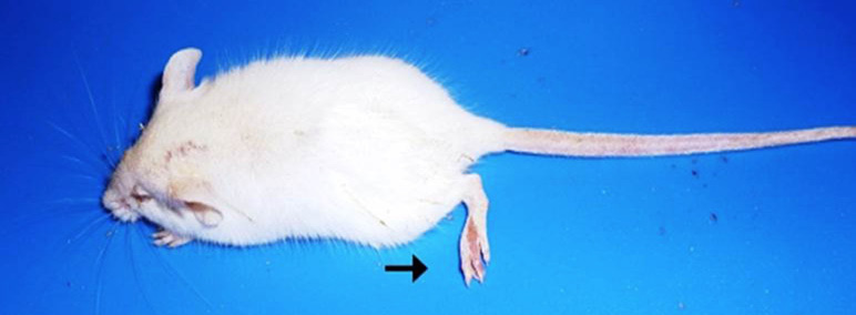
Mouse from the group 1 inoculated with suspected cow sample (Case No. 1) at day 14 post-inoculation showing paralysis of tail and hind legs along with roughness of the fur.
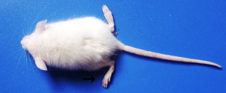
Mouse from the group 2 inoculated with suspected mule sample (Case No. 2) at day 11 post-inoculation showing more pronounced paralysis of tail and hind legs.
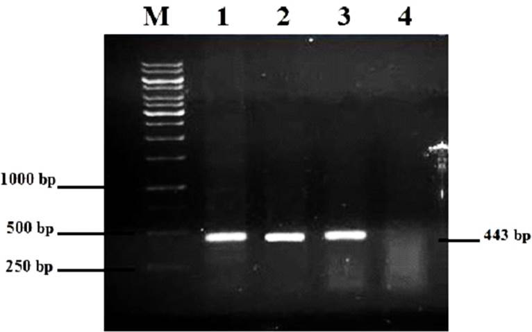
RT-PCR results of the sample. M, ladder; 1, cow sample; 2, mule sample; 3, positive control; 4, negative control.
References
- Blanton, J.D., Krebs, J.W., Hanlon, C.A. and Rupperchit, C.E., 2006. J. Am. Vet. med. Assoc., 209: 1897-1911. https://doi.org/10.2460/javma.229.12.1897[CrossRef]
- Dua, K., 2004. Veterinary ethics and jurisprudence. Kalyani Publishers, New Delhi, India.
- Durrani, U.F., Goyal, S.M., Mahmood, A.K., Hanlon, C., Moore, S., Waqas, M. and Akhtar, R., 2017. Pakistan J. Zool., 49: 1147-1149.
- Iwasaki, Y. and Tobita, M., 2002. In: Rabies (eds. A.C. Jackson and W.H. Wunner). Academic Press, San Diego, pp. 283-306.
- Jackson, A.C., 2002. In: Rabies (eds. A.C. Jackson and W.H. Wunner). Academic Press, San Diego, pp. 245-282.
- Jackson, A.C., 2003. J. Nerovirol., 9: 253-258. https://doi.org/10.1080/13550280390193975[CrossRef]
- Keane, D.P. and Little, B.L., 1987. Can. Vet. J., 28: 497-504.
- Koprowski, H., 1996. In: Laboratory techniques in rabies, 4th ed. (eds. F.X. Meslin, M.M. Kaplan and H. Koprowski). World Health Organization, Geneva, Switzerland, pp. 80-86.
- Lentz, T.L., Burrage, T.G., Smith, A.L., Crick, J. and Tignor, G.H., 1982. Science, 215: 182-184. https://doi.org/10.1126/science.7053569[CrossRef]
- Lewis, P., Fu, Y. and Lentz, T.L., 2000. Muscle Nerve, 23: 720-730. https://doi.org/10.1002/1097-4598(200010)23:10<1472::AID-MUS3>3.0.CO;2-#[CrossRef]
- Lycke, E. and Tsiang, H., 1987. J. Virol., 61: 2733-2741.
- Numan, M., Qureshi, Z.A., Shauket, M., Hashmi, H.A., Iqbal, M., Gill, Z.J., Habib, M. and Siddique, M., 2011. Res. Vet. Sci., 90: 160-162. https://doi.org/10.1016/j.rvsc.2010.04.020[CrossRef]
- Peixoto, Z.M.P., Cunha, E.M.S., Sacramento, D.R.V., Souza, M.C.A.M., da Silva, L.H.Q., Germano, P.L., Kroeff, S.S. and Kotait, I., 2000. Braz. J. Microbiol., 31: 72-75. https://doi.org/10.1590/S1517-83822000000100017[CrossRef]
- Smith, J.S., Fishbein, D.B., Rupprecht, C.E. and Clark, K., 1991. New Engl. J. Med., 324: 205-211. https://doi.org/10.1056/NEJM199101243240401[CrossRef]
- Tordo, N., Sacramento, D. and Bourhy, H., 1996. In: Laboratory techniques in rabies, 4th ed. World Health Organization, Geneva, pp. 157-174.
- Tsiang, H., Ceccaldi, P.E. and Lycke, E., 1991. J. Gen. Virol., 72: 1191-1194. https://doi.org/10.1099/0022-1317-72-5-1191[CrossRef]
- Webster, W.A., Casey, G.A. and Charlton, K.M., 1976. Can. J. Comp. Med., 40: 322-325.
Services
Article usage statistics
Loading…