
The livestock sector contributes 63.60% to agriculture and 14.97% to Pakistan’s GDP, with sheep (33.1 million) and goats (89.4 million) serving as critical sources of meat, milk, and wool, and a financial safeguards for smallholder farmers. Caseous lymphadenitis (CLA), caused by Corynebacterium pseudotuberculosis, significantly impacts productivity. The pathogen enters through skin abrasions, where phospholipase D toxin enhances vascular permeability, facilitating bacterial spread and abscess formation. Encapsulation limits antibiotic efficacy, leading to chronic infections and flock-level transmission. With no effective commercial vaccine available, this study aimed to isolate a local C. pseudotuberculosis strain from sheep and goats in the Bahawalpur district and develop an inactivated alum-adjuvanted vaccine. A total of 340 pus samples (170 sheep, 170 goats) were processed, and formalin-killed antigen preparations were administered to rabbits of age groups (0-3, 3-5, 5-7 weeks). Antibody titers increased with antigen dose, with the highest response at 1.0 ml (280 µg, 1.5×10¹⁴ CFU/ml). Rabbits aged 5–7 weeks exhibited significantly greater immune response compared to younger groups (P<0.05). Field efficacy trials in sheep and goats demonstrated maximal protection in 2–3-year-old animals (GMT=53.76), compared to <1 year (GMT=2.99) and >3 years (GMT=8.47). These findings highlight the potential of locally derived vaccines in CLA control and warrant further genomic and subunit vaccine studies.
Research Article · Volume 58, Issue 6, 25 September 2026 · Open Access
Development and Immunogenicity Evaluation of an Inactivated Corynebacterium pseudotuberculosis Vaccine in Small Ruminants
· · · · · · · ·
Affiliations & Notes ⌄
- 1Department of Microbiology, Cholistan University of Veterinary and Animal Sciences, Bahawalpur 63100, Pakistan 2Department of Medicine, Cholistan University of Veterinary and Animal Sciences, Bahawalpur 63100, Pakistan 3Department of Parasitology, Cholistan University of Veterinary and Animal Sciences, Bahawalpur 63100, Pakistan 4Veterinary Research Institute, Zarrar Shaheed Road Lahore Cantt 54810, Pakistan * Corresponding author: dr.raisahmad2068@gmail.com
Article Info ⌄
- Journal:
- Pakistan Journal of Zoology
- Published:
- 25 September 2026

From the article
- Geometric mean antibody titers observed in sheep and goat of various age groups following subcutaneous administration of 1.0 ml (280 µg biomass; >10¹⁴ CFU/ml) bacterial vaccine during the experimental trial.
- The 2–3-year age group exhibited significantly higher mean antibody titers compared with both the 3 years groups (mean difference = 44.8929 ± 1.52916, 95% CI: 41.7699–48.0159).
- The study concludes that the inactivated C. pseudotuberculosis vaccine is effective and safe against CLA in small ruminants.
- Vaccinated animals showed higher antibody titers and reduced clinical signs, indicating strong immunogenicity.
Selected from the article's own text.
Abstract
Introduction
Sheep and goats play a crucial role in Pakistan’s agricultural economy, particularly in arid and semi-arid regions such as Bahawalpur, where they provide meat, milk, wool, and financial security for smallholder farmers. Despite their importance, productivity is severely affected by CLA, a chronic disease of small ruminants caused by C. pseudotuberculosis which leads to abscess formation in lymph nodes and internal organs, resulting in weight loss, reduced milk production and wool yields, increased culling, and welfare concerns. The pathogen spreads via the skin wounds, with its virulence factor phospholipase D promoting dissemination. Encapsulation of bacteria render antibiotic treatment largely ineffective, enabling persistence and flock-level transmission .
Economically, CLA imposes major burdens worldwide. In Australia, losses are estimated at A$12–15 million annually in the sheep meat industry and an additional A$17 million in wool production, equating to roughly US$20–25 million per year. Similar productivity impact has been reported in New Zealand and Europe. Although exact estimates for Pakistan are lacking, with over 120 million small ruminants and high disease prevalence, national losses are likely in the millions of US dollars annually . These figures emphasize the urgent need for locally developed vaccines to mitigate CLA’s economic and productivity impacts.
Various vaccine approaches including attenuated strains, inactivated or toxoid bacterins, recombinant proteins, and combined formulations have been investigated with varying degrees of success. While some attenuated mutants and recombinant constructs, such as rPLD-based vaccines, have demonstrated protective responses, no formulation has achieved consistent and complete protection . A commercially available bacterin-toxoid vaccine trials in Australia reduced abscess formation but did not fully prevent infection, underscoring the limitations of existing options.
In Pakistan, the challenge is compounded by additional gaps. Commercial CLA vaccines are not readily available, and local isolates may differ antigenically from those used in reference or foreign vaccines, reducing efficacy. Moreover, immune responses to CLA vaccination vary with animal age, antigen dose, formulation, and adjuvant, yet there is little epidemiological data on age-related susceptibility and immune maturity in Pakistani sheep and goats . This knowledge gap hinders the development of targeted control strategies.
This disease poses a substantial economic burden on the livestock sector, particularly in regions such as Bahawalpur where small ruminants are central to rural livelihoods. To address this challenge, there is a pressing need for vaccines adapted to local C. pseudotuberculosis strains and production systems. Inactivated alum-adjuvanted vaccines represent a practical option, offering safety, affordability, and suitability for resource-limited settings. This study therefore isolated a local C. pseudotuberculosis strain, formulated an inactivated vaccine, and evaluated its immunogenicity and protective efficacy in sheep and goats. Complementary serological techniques, including the Indirect Haemagglutination (IHA) test, were applied to assess seroprevalence and vaccine-induced immune responses. These findings provide essential evidence to strengthen CLA control strategies and support future vaccine development in Pakistan and beyond.
Materials and Methods
Study area and sample collection
Pus samples (n=340) were aseptically collected from clinically infected sheep (n=170) and goats (n=170) in rural and Cholistan regions of Bahawalpur District, Punjab, Pakistan, including Toba Janu Wala, Kudwala Bangla, Toba Mouj Ghar, Chak 13 BC, Chak 42 DB, Chak 43 DB, and Khanqah Shareef. Standard personal protective equipment (PPE) and biosafety practices were observed during sampling. Lesion sites were disinfected with 70% alcohol prior to collection, and pus was transferred into pre-labeled sterile polythene bags as described by Khanamir et al. (2023). Samples were transported under cold chain conditions to the laboratory for isolation and identification of Corynebacterium pseudotuberculosis.
Molecular confirmation of C. pseudotuberculosis
C. pseudotuberculosis isolates were initially cultured on 5% sheep blood agar (Oxoid, UK) and were subjected to standard biochemical characterization and molecular confirmation through polymerase chain reaction (PCR) following Meng et al. (2023). Genomic DNA was extracted from 22 isolates obtained from sheep and 27 isolates from goats using the WizPrep™ gDNA Mini Kit (Cell/Tissue, Wizbiosolutions, Korea), according to the manufacturer’s protocol. Molecular confirmation was performed using modified 16S rRNA gene-specific primers: 16S-F (5′-ACCGCACTTTAGTGTGTGTG-3′) and 16S-R (5′-TCTCTACGCCGATCTTGTAT-3′). PCR amplification was conducted in a thermal cycler (C1000 Touch™, Bio-Rad, USA) under optimized conditions: An initial denaturation at 95 °C for 3 min; 40 cycles of denaturation at 95 °C for 1 min, annealing at 58 °C for 40 s, and extension at 68 °C for 90 s; followed by a final extension at 68 °C for 7 min. The amplified products were analyzed by agarose gel electrophoresis (Sub-Cell® GT System, Bio-Rad, USA) to confirm the presence of the target 16S rRNA gene fragment .
Preparation of vaccinal strain
C. pseudotuberculosis strains were cultured separately in nutrient broth (Oxoid, UK) and incubated at 37 °C for 24 h in an incubator (Isotherm® IFA-54-8, Jeio Tech, Korea). Cultures were inactivated by adding formalin to a final concentration of 0.5% (v/v) and maintained at room temperature for 24 h, as recommended in vaccine production guidelines as described by Adib et al. (2023). The inactivated bacterial suspensions were then standardized to contain 1×10⁶ CFU/mL. The suspensions were subsequently combined with a 1% alum solution (Brenntag Biosector, Denmark) as an adjuvant to enhance immunogenicity. The vaccine formulation was homogenized using a vortex mixer (Vortex-Genie® 2, Scientific Industries, USA) to ensure uniform antigen distribution. Thiomersal sodium (Sigma-Aldrich, USA) was dissolved in physiological saline and added to the formulation to achieve a final concentration of 0.01% (equivalent to 50 μg per 0.5 mL dose) as a preservative, in line with OIE recommendations for vaccine safety.
Quality control testing of the vaccine
Quality control of the vaccine was performed according to safety and sterility requirements. Stability was assessed by storing aliquots at 25 °C, 35 °C, and 45 °C for 7 days, followed by centrifugation at 400 g for 10 min in a benchtop centrifuge (Eppendorf 5702, Germany) to detect sedimentation or phase separation following the protocol of Barbosa et al. (2022). Sterility testing was performed by inoculating vaccine samples onto nutrient agar (Oxoid, UK) and MacConkey agar (Oxoid, UK) to confirm the absence of bacterial contaminants, and onto Sabouraud dextrose agar (Oxoid, UK) to exclude fungal contamination.
Potency testing and dose determination in rabbits
A total of 20 clinically healthy rabbits of uniform breed and body weight were procured and randomly allocated into four equal groups based on age: 0–3 weeks, 3–5 weeks, 5–7 weeks, and a control group (n = 5 per group). Animals were acclimatized and observed for two weeks to monitor potential morbidity or mortality following Rashed et al. (2021). Prior to vaccination, all rabbits were dewormed orally with oxfendazole syrup (Oxfenil®, Safepharm, Pakistan) at a dose of 2 mL per animal as described by Rodríguez-Durán et al. (2023). Experimental groups were immunized subcutaneously with different vaccine doses of inactivated C. pseudotuberculosis: 0.3 mL (120 μg, >10⁶ CFU/mL), 0.5 mL (220 μg, >10⁹ CFU/mL), and 1.0 mL (280 μg, >10¹⁴ CFU/mL), respectively, while the control group received sterile phosphate-buffered saline (PBS, Gibco™, USA). Vaccinations were administered using sterile disposable syringes (BD Plastipak™, USA).
Blood samples were collected aseptically from the marginal ear vein of each rabbit prior to immunization (day 0) and subsequently at days 14, 21, and 28 post-vaccination. Samples were centrifuged at 3,000 rpm for 10 min in a benchtop centrifuge (Eppendorf 5702, Germany) to obtain serum following Wu et al. (2021). Antibody responses were quantified using the indirect hemagglutination assay (IHA kit, Atlas Medical, UK), following the manufacturer’s instructions.
Efficacy testing in sheep and goats
A total of 24 sheep and 24 goats, all clinically healthy, were procured and divided into four equal groups (n = 6 per group) according to age: 1 year, 2–3 years, >3 years, and a control group. Before vaccination, fecal samples were screened for parasitic infection using standard stool examination techniques (Flotac®, Italy), and all animals were dewormed with oxfendazole (Oxfenil®, Safepharm, Pakistan) at a dose of 15 mL per animal as described by Antunes et al. (2022).
Sheep and goats in the experimental groups received the inactivated C. pseudotuberculosis vaccine subcutaneously on day 1, followed by a booster dose on day 14. Each animal was administered 1.0 mL (280 μg, >10¹⁴ CFU/mL) of the vaccine using sterile disposable syringes (BD Plastipak™, USA), while control animals received sterile phosphate-buffered saline (Gibco™, USA).
Blood samples were collected aseptically from the jugular vein before vaccination, two weeks after the primary dose, one month after the booster, and subsequently at monthly intervals up to the seventh month following Lianou et al. (2022). Serum was separated by centrifugation (Eppendorf 5702, Germany) and analyzed for antibody titers using the indirect hemagglutination assay (Atlas Medical, UK).
Well-immunized animals were later challenged with a virulent strain of C. pseudotuberculosis (>400 μg, >10¹⁸ CFU) administered orally, sprayed over superficial wounds, and distributed on grazing soil to mimic natural exposure as described by de Jesus et al. (2025). Animals were monitored daily for clinical signs of caseous lymphadenitis, and blood samples were collected on days 14, 21, and 28 post-challenge for serological evaluation.
All experimental procedures were conducted in strict accordance with the ethical guidelines for animal experimentation and were approved by the Institutional Ethical Committee of Cholistan University of Veterinary and Animal Sciences, Bahawalpur under approval # ORIC-67 dated 26-04-2021. Biosafety protocols for handling live pathogens were observed in compliance with WHO recommendations following Joseph (2021).
Assessment of antibody response to C. pseudotuberculosis in sheep and goats across field areas
Blood samples were collected from sheep and goats in the rural and Cholistani regions of District Bahawalpur, specifically from Toba Janu Wala (n= 19), Kudwala Bangla (n= 21), Toba Mouj Ghar (n= 27), Chak 13-BC (n= 21), Chak 42-DB (n= 24), Chak 43-DB (n= 27), and Khanqah Shareef (n= 31). All sampled animals were clinically healthy at the time of collection. Samples were obtained aseptically from the jugular vein using sterile disposable vacutainers (BD Vacutainer®, USA), and sera were separated by centrifugation at 3,000 rpm for 10 min (Eppendorf 5702, Germany).
To evaluate prior exposure of these populations to C. pseudotuberculosis, antibodies were detected using the IHA kit (Atlas Medical, UK). For sensitization of erythrocytes, sonicated antigen of C. pseudotuberculosis was prepared and coupled with human blood group “O” erythrocytes (supplied by the Blood Bank, BVH Bahawalpur, Pakistan) using 1% glutaraldehyde (Sigma-Aldrich, USA) as a coupling agent, following the method described by Mikailov et al. (2024) and refined according to OIE guidelines. The geometric mean antibody titers (GMT) were calculated for each group following Hussain et al. (2003).
Statistical analysis
All data were analyzed using two-way ANOVA with significance at P < 0.05. Tukey’s post hoc test identified group differences, while the Chi-square assessed seroprevalence. Antibody responses were expressed as geometric mean titers (GMTs) and compared across groups. Statistical analyses were conducted using SPSS software (version 22.0).
Results
C. pseudotuberculosis forms white, mucoid colonies on nutrient agar and exhibited β-hemolysis on sheep blood agar. Microscopic examination revealed Gram-positive rods further confirmed through biochemical assays, including a catalase test, which demonstrated bubble formation, indicative of a positive reaction characteristic of C. pseudotuberculosis (Fig. 1).
A total of 340 pus samples were collected, comprising 170 from sheep and 170 from goats, from Cholistani and rural areas of Bahawalpur, Punjab, for the detection of C. pseudotuberculosis. Significant prevalence was observed in both species, with 12.94% in sheep (χ² = 13.43, df = 6, P < 0.05) and 15.89% in goats (χ² = 12.94, df = 6, P < 0.05).
Table I shows the geometries mean antibody titer in rabbits of different age groups following such contanes vaccination with 0.3, 0.5 and 1.0 ml of bacterial vaccine. The antibody titers in rabbits increased proportionally with antigen dosage across all age groups. In the 0–3-week group, mean titers rose from 0.6960 ± 0.16 at a 0.30 mL dose
Table I. Geometric mean antibody titers in rabbits of different age groups following subcutaneous vaccination with 0.3 ml (120 µg biomass; >10⁶ CFU/ml), 0.5 ml (220 µg biomass; >109 CFU/ml) and 1.0 ml (280 µg biomass; >1014 CFU/ml) bacterial vaccine.
|
Age(weeks) |
No. of samples |
1:2 |
1:4 |
1:8 |
1:16 |
1:32 |
1:64 |
1:128 |
1:256 |
1:512 |
GMT |
|
Titer after 0.3 ml bacterial vaccine |
|||||||||||
|
0-3 |
5 |
2 |
2 |
1 |
- |
- |
- |
- |
- |
- |
3.482 |
|
3-5 |
5 |
1 |
1 |
2 |
1 |
- |
- |
- |
- |
- |
6.062 |
|
5-7 |
5 |
- |
- |
- |
2 |
3 |
- |
- |
- |
- |
24.251 |
|
Titer after 0.5 ml bacterial vaccine |
|||||||||||
|
0-3 |
5 |
1 |
1 |
1 |
2 |
- |
- |
- |
- |
- |
6.964 |
|
3-5 |
5 |
- |
- |
2 |
1 |
1 |
1 |
- |
- |
- |
18.379 |
|
5-7 |
5 |
- |
- |
- |
- |
1 |
1 |
3 |
- |
- |
84.448 |
|
Titer after 1.0 ml bacterial vaccine |
|||||||||||
|
0-3 |
5 |
- |
- |
- |
1 |
2 |
2 |
- |
- |
- |
36.758 |
|
3-5 |
5 |
- |
- |
- |
- |
1 |
1 |
1 |
2 |
- |
111.43 |
|
5-7 |
5 |
- |
- |
- |
- |
- |
- |
- |
2 |
3 |
388.02 |
Table II. Geometric mean antibody titers observed in sheep and goat of various age groups following subcutaneous administration of 1.0 ml (280 µg biomass; >10¹⁴ CFU/ml) bacterial vaccine during the experimental trial.
|
Age(year) |
No. of samples |
1:2 |
1:4 |
1:8 |
1:16 |
1:32 |
1:64 |
1:128 |
1:256 |
1:512 |
GMT |
|
Antibody titers observed in sheep |
|||||||||||
|
< 1 |
6 |
- |
- |
2 |
2 |
1 |
1 |
- |
- |
- |
17.959 |
|
2-3 |
6 |
- |
- |
- |
- |
- |
- |
1 |
2 |
3 |
322.53 |
|
> 3 |
6 |
- |
- |
- |
1 |
2 |
1 |
2 |
- |
- |
50.796 |
|
Antibody titers observed in goat |
|||||||||||
|
< 1 |
6 |
- |
- |
- |
1 |
2 |
2 |
1 |
- |
- |
45.254 |
|
2-3 |
6 |
- |
- |
- |
- |
- |
- |
- |
3 |
3 |
362.03 |
|
> 3 |
6 |
- |
- |
- |
- |
1 |
1 |
2 |
2 |
- |
114.03 |
to 7.3500 ± 0.26 at a 1.0 mL dose. A similar trend was observed in the 3–5-week group, where titers increased from 1.2320 ± 0.22 (0.30 mL dose) to 22.2860 ± 1.43 (1.0 mL dose). In the 5–7-week group, titers were markedly higher, increasing from 4.8500 ± 1.07 at 0.30 mL to 77.6040 ± 1.47 at 1.0 mL. These results demonstrate that both age and antigen dose exert strong positive effects on antibody production, with the highest titers observed in 5–7-week-old rabbits receiving the 1.0 mL dose. The humoral immune response of the prepared vaccine was evaluated by using IHA.
Efficacy trials conducted in sheep and goats demonstrated a clear age-related variation in antibody responses following vaccination. In sheep, the highest geometric mean titer (GMT) was observed in the 2–3-year age group (GMT = 322.53), followed by animals over 3 years (GMT = 50.80), while the lowest response occurred in lambs below 1 year (GMT = 17.96) (Table II). A similar trend was noted in goats, where the 2–3-year group exhibited the strongest antibody response (GMT = 362.03), compared with moderate titers in animals older than 3 years (GMT = 114.03) and the lowest in kids under 1 year of age (GMT = 45.25) (Table II). Overall, the findings indicate that young adult animals (2–3 years) mount the most robust immune response to vaccination, highlighting a significant influence of age on vaccine-induced immunogenicity.
The multiple comparison analysis of antibody titers using the Least Significant Difference (LSD) test revealed statistically significant differences (p < 0.001) among the three age groups of vaccinated sheep and goats. The 2–3-year age group exhibited significantly higher mean antibody titers compared with both the <1 year (mean difference = 51.7783 ± 1.52916, 95% CI: 48.6554–54.9013) and >3 years groups (mean difference = 44.8929 ± 1.52916, 95% CI: 41.7699–48.0159). In contrast, animals aged >3 years showed significantly higher titers than those <1 year (mean difference = 6.8854 ± 1.52916, 95% CI: 3.7624–10.0084). These results confirm that antibody responses were significantly age-dependent, with the 2–3-year group displaying the most robust immunogenicity following vaccination (Table III).
Table III. Multiple comparison of antibody titers among different age groups of vaccinated sheep and goats using the LSD test.
|
Age groups |
Mean difference |
SE |
Sig. |
95% CI |
|
|
Lower bound |
Upper bound |
||||
|
< 1 yr 2- 3 yr > 3 yr |
-51.7783± -6.8854* |
1.52916 1.52916 |
0.000 0.000 |
-54.9013 -10.0084 |
-48.6554 -3.7624 |
|
2-3 yr < 1 yr > 3 yr |
51.7783* 44.8929* |
1.52916 1.52916 |
0.000 0.000 |
48.6554 41.7699 |
54.9013 48.0159 |
|
> 3 yr < 1 yr 2- 3 yr |
-6.8854* -44.8929* |
1.52916 1.52916 |
0.000 0.000 |
3.7624 -48.0159 |
10.0084 -41.7699 |
*Indicates a statistically significant difference between the compared age groups at p < 0.05 using the LSD test.
Discussion
Caseous lymphadenitis, a chronic disease of sheep and goats, is characterized by abscess formation in lymph nodes and visceral organs . Affected animals show abscesses with whitish pus, localized alopecia, and progressive emaciation, emphasizing the need for effective control and prevention. In the present study, a significant prevalence of CLA was recorded in sheep (12.94%) and goats (15.89%) from Cholistani and rural areas of Bahawalpur, Punjab. Comparable findings have been reported globally, with varying prevalence rates across regions. In India, CLA prevalence has been documented at 4.7% in goats and 2.4% in sheep . Similarly, in Iraq, infection rates of 0.94% in sheep and 1.93% in goats have been observed . In China, a higher prevalence of 39.2% was reported in goats , whereas in Turkey, CLA prevalence was 1.1% in goats and 17% in sheep . These variations may be attributed to differences in management practices, environmental factors, diagnostic techniques, and biosecurity measures.
C. pseudotuberculosis was successfully isolated on nutrient agar and 5% sheep blood agar. On nutrient agar, colonies appeared small, white, opaque, dry, and exhibited concentric ring formation after 48 h of incubation at 37 °C, findings consistent with those reported by Syame et al. (2022). On blood agar, small to medium-sized, round, convex colonies measuring 1–3 mm in diameter were observed after 24–48 h of incubation. The colonies were cream to off-white with a smooth surface and regular margins, exhibiting weak beta-hemolysis, characterized by a narrow zone of incomplete hemolysis. Similar colonial morphology and hemolytic patterns were reported by Domínguez et al. (2021) and Torky et al. (2023). Such features are typical of C. pseudotuberculosis and help differentiate it from other coryneform bacteria. The observed variations in colony morphology across studies may be attributed to differences in media composition, incubation conditions, and strain virulence.
C. pseudotuberculosis was confirmed through biochemical tests and PCR targeting the 16S rRNA gene, which produced specific amplification bands confirming its identity (Fig. 2). These findings are consistent with those of Mohamed et al. (2025), Langova et al. (2022), and Torky et al. (2023), who also validated the 16S rRNA gene as a reliable molecular marker for accurate detection of C. pseudotuberculosis, providing greater sensitivity and specificity than conventional biochemical methods.
In the present study, formalin (0.5% at 37 °C for 24–48 h) was effectively used for the inactivation of C. pseudotuberculosis, consistent with earlier findings by Bakr et al. (2025), who reported that formalin preserves antigenic integrity necessary for eliciting host immune responses. The inactivated vaccine developed was safe and immunogenic in both sheep and goats, producing no clinical signs and inducing a strong immune response. Similar outcomes were observed by Pioquinto et al. (2023) in South Korea, where a formalin-inactivated C. pseudotuberculosis vaccine derived from local isolates conferred significant (p < 0.05) protection in Korean Native Black Goats (KNBGs), characterized by elevated CLA-specific IgG levels and absence of abscess formation. Comparable findings were also documented by Dopuđ et al. (2025) in China, where vaccinated animals exhibited improved weight gain, higher antibody titers, and reduced lesion formation post-challenge. The consistency of these results across studies indicates that formalin-inactivated vaccines maintain antigenic stability and can effectively stimulates protective immunity, representing a promising approach for CLA control in small ruminants.
The humoral immune response induced by the inactivated C. pseudotuberculosis vaccine was evaluated through an indirect hemagglutination assay (IHA) in both experimental and field trials. Vaccinated sheep and goats developed a significant antibody response, and post-challenge evaluation revealed markedly fewer or no abscesses, confirming effective protection. Similar observations were reported by Domínguez et al. (2021) and Barbosa et al. (2022), who found that inactivated vaccines produced strong humoral immunity and clinical protection against CLA. Experimental trials conducted on rabbits demonstrated a progressive increase in antibody titers across age groups 0–3, 3–5, and 5–7 weeks post-vaccination, with the highest response observed at 5–7 weeks (mean titer increase from 4.85±1.07 to 77.60±1.47). This increase reflects maturation of the immune system with age, consistent with findings by Hussain et al. (2003) and Mohamed et al. (2025), who reported age-dependent enhancement in vaccine responsiveness. None of the vaccinated animals exhibited adverse reactions, confirming the vaccine’s safety consistent with the low reactogenicity observed for formalin-inactivated formulations . Additionally, geometric mean titers varied significantly among small ruminant age groups, with animals aged 2–3 years showing higher antibody levels than those <1 year or >3 years (Mean Difference= +6.8854; 95% CI= 3.7624–10.0084). These results align with previous studies demonstrating that inactivated vaccines elicit robust humoral immunity and protection against CLA, though their efficacy may be slightly lower than live or recombinant vaccines due to limited intracellular antigen presentation . Nevertheless, their superior safety and cost-effectiveness make inactivated formulations a practical option for disease control in endemic regions.
Conclusion
The study concludes that the inactivated C. pseudotuberculosis vaccine is effective and safe against CLA in small ruminants. Isolation and identification confirmed the presence of the pathogen in Cholistani and rural areas of Bahawalpur, with spatial mapping revealing disease clustering. Vaccinated animals showed higher antibody titers and reduced clinical signs, indicating strong immunogenicity. Serological analysis highlighted widespread exposure to CLA. These findings underscore the need for routine vaccination and effective control measures to reduce the disease burden in southern Punjab.
Decelerations
Acknowledgment
The authors gratefully acknowledge the financial support provided by Higher Education Commission (HEC) of Pakistan under NRPU project (20-14949/NRPU/R and D/HEC/2021-2020) at Cholistan University of Veterinary and Animal Sciences, Bahawalpur, which made this study possible.
Funding
This work was supported by the Higher Education Commission (HEC) of Pakistan under NRPU Project/Grant No. 14949.
IRB approval
The study was reviewed and approved by the Institutional Review Board (IRB) of the respective institution. All procedures were conducted in accordance with approved ethical guidelines and regulations.
Ethical approval
The study was conducted in strict accordance with the guidelines for the care and use of laboratory animals. All procedures involving animals were reviewed and approved by the Ethical Committee of Cholistan University of Veterinary and Animal Sciences, Bahawalpur under approval # ORIC-67 dated 26-04-2021.
Generative AI and AI-assisted technology statement
The authors declare that no generative AI and AI assisted technology was used in the creation of this manuscript.
Statement of conflict of interest
The authors have declared no conflict of interests.
Figures
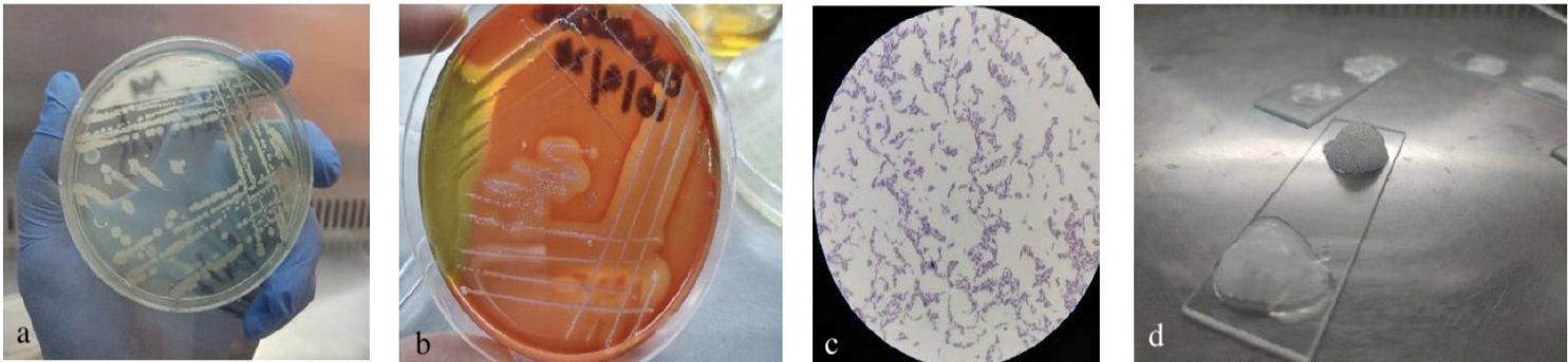
Corynebacterium pseudotuberculosis (a) displaying white, mucoid colonies on nutrient agar after 48 h of incubation at 37 °C. (b) White mucoid colonies on sheep blood agar after 48 h of incubation at 37 °C exhibiting ẞ-hemolysis, (c) Microscopic examination showing Gram-positive rods of C. pseudotuberculosis under oil immersion (100×). (d) Catalase test demonstrating bubble formation.
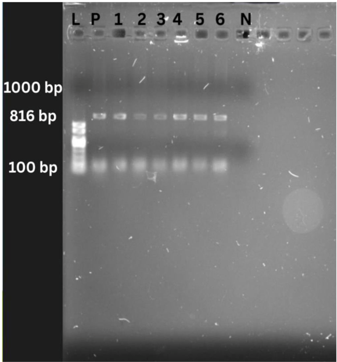
Agarose gel electrophoresis of PCR-amplified products of C. pseudotuberculosis. Lane L: 100 bp DNA ladder, Lane P: Positive control showing the expected amplification product; Lanes 1-6: Field isolates of C. pseudotuberculosis showing specific PCR bands of ~816 bp; Lane N: Negative control (no template DNA), showing no amplification. Electrophoresis was performed on a 1% agarose gel in TBE buffer at 110 V for 15 min, and DNA was visualized after ethidium bromide staining under UV illumination.
References
- Adib, N., Zahmatkesh, A., Esmaeilnejad-Ahranjani, P., Paradise, A., Khiav, L.A., Bagheri, M., Abdoli, M. and Adib, A., 2023. Effect of formalin percentage, incubation time and temperature on Clostridium chauvoei culture inactivation and immunogenicity. Anaerobe, 83: 102781. https://doi.org/10.1016/j.anaerobe.2023.102781[CrossRef]
- Antunes, M.I., Lima, M.S., Stilwell, G., Romeiras, M.I., Fragoso, L. and Madeira de Carvalho, L.M., 2022. Anthelmintic efficacy in sheep and goats under different management and deworming systems in the region of Lisbon and Tagus Valley, Portugal. Pathogen, 11: 1457. https://doi.org/10.3390/pathogens11121457[CrossRef]
- Bakr, M.B., Hashad, M.E.-S., Gomaa, I.G. and Attia, S.A., 2025. Antimicrobial susceptibility and molecular characterization of Corynebacterium pseudotuberculosis isolates recovered from caseous lymphadenitis in sheep. Egypt. J. Vet. Sci., 56: 711-723. https://doi.org/10.21608/ejvs.2025.415210.3059[CrossRef]
- Barbosa, R.D.M., Silva, A.M., Silva, C.F.D., Cardoso, J.C., Severino, P., Meirelles, L.M., Silva-Junior, A.A.D., Viseras, C., Fonseca, J. and Souto, E.B., 2022. Production technologies, regulatory parameters, and quality control of vaccine vectors for veterinary use. Technologies, 10: 109. https://doi.org/10.3390/technologies10050109[CrossRef]
- de Jesus, I.B., Droppa-Almeida, D., Ferreira, C., Nascimento, R.J.M., Portela, R.W., de Oliveira Silva, M.T., Alves, M.S.D., Dos Santos, G.S., Azevedo, V. and Padilha, F.F., 2025. Immunization of mice and goats with Corynebacterium pseudotuberculosis derived rPTS, rRBN and rCP40 recombinant proteins. World J. Microbiol. Biotechnol., 41: 1-12. https://doi.org/10.1007/s11274-025-04411-w[CrossRef]
- de Pinho, R.B., de Oliveira Silva, M.T., Bezerra, F.S.B. and Borsuk, S., 2021. Vaccines for caseous lymphadenitis: Up-to-date and forward-looking strategies. Appl. Microbiol. Biotechnol., 105: 2287-2296. https://doi.org/10.1007/s00253-021-11191-4[CrossRef]
- Domínguez, M.R., Jimenez, R. and Guerreo, J.A.V., 2021. Caseous lymphadenitis: Virulence factors, pathogenesis and vaccines. Review. Rev. Mex. Cienc. Pecu, 12: 1221-1249. https://doi.org/10.22319/rmcp.v12i4.5699[CrossRef]
- Dopuđ, M., Reil, I., Zdelar-Tuk, M., Špičić, S. and Duvnjak, S., 2025. Caseous lymphadenitis in sheep and goats–cheese glands. Vet. Stn., 56: 303-316. https://doi.org/10.46419/vs.56.3.8[CrossRef]
- El-Sebay, N.A., Abd-Elmonem, M.M., El-Sergany, E.F., Abbas, M.A., Osman, M.R. and Abd El-Moaty, D.A., 2021. Genetic characterization of Corynebacterium pseudotuberculosis isolates in Egypt. J. appl. Vet. Sci., 6: 15-21. https://doi.org/10.21608/javs.2021.154576[CrossRef]
- Gupta, S. and Pellett, S., 2023. Recent developments in vaccine design from live vaccines to recombinant toxin vaccines. Toxins, 15: 563. https://doi.org/10.3390/toxins15090563[CrossRef]
- Hussain, I., Zahoor, M., Rasool, M., Mahmood, M.S., Mansoor, M. and Riaz, M., 2003. Detection of serum antibody levels against infectious bursal disease (IBD) virus using indirect hemagglutination (IHA) test in commercial broilers. Int. J. Poult. Sci., 2; 442-445. https://doi.org/10.3923/ijps.2003.442.445[CrossRef]
- Joseph, T., 2021. Management system approach for addressing biosafety and biosecurity of emerging pathogens in a biosafety level-3 core facility. Appl. Biosaf, 26: 210-220. https://doi.org/10.1089/apb.2021.0007[CrossRef]
- Khanamir, R.A., Issa, N.A. and Abdulrahman, R.F., 2023. First study on molecular epidemiology of caseous lymphadenitis in slaughtered sheep and goats in Duhok Province, Iraq. Open Vet. J., 13: 588–598-588–598. https://doi.org/10.5455/OVJ.2023.v13.i5.11[CrossRef]
- Khattak, I., Usman, T., Swelum, A.A., Nasreen, N., Khan, A., Haider, J., Muhammad, J., Chen, C.-C. and Said, M.B., 2024. Toxoplasma gondii infection in small ruminants from Khyber Pakhtunkhwa, Pakistan: Seroprevalence, spatial distribution and associated risk factors. Vet. Parasitol. (Amst)., 47: 100979. https://doi.org/10.1016/j.vprsr.2023.100979[CrossRef]
- Langova, D., Slana, I., Okunkova, J., Moravkova, M., Florianova, M. and Markova, J., 2022. First evidence of the presence of the causative agent of caseous lymphadenitis Corynebacterium pseudotuberculosis in dairy products produced from the milk of small ruminants. Pathogens, 11: 1425. https://doi.org/10.3390/pathogens11121425[CrossRef]
- Lianou, D.T., Michael, C.K., Petinaki, E., Mavrogianni, V.S. and Fthenakis, G.C., 2022. Administration of vaccines in dairy sheep and goat farms: Patterns of vaccination, associations with health and production parameters, predictors. Vaccines, 10: 1372. https://doi.org/10.3390/vaccines10091372[CrossRef]
- Meng, W., Chen, S., Huang, L., Yang, J., Zhang, W., Zhong, Z., Zhou, Z., Liu, H., Fu, H. and He, T., 2023. Isolation, characterization, and pathogenicity assessment of Corynebacterium pseudotuberculosis biovar equi strains from alpacas (Vicugna pacos) in China. Front. Microbiol., 14: 1206187. https://doi.org/10.3389/fmicb.2023.1206187[CrossRef]
- Mikailov, M., Gunashev, S.A., Yanikova, E., Halikov, A. and Bulashev, A.K., 2024. Indirect hemagglutination assay for diagnosing brucellosis: Past, present, and future. Vet. World, 17: 811. https://doi.org/10.14202/vetworld.2024.811-819[CrossRef]
- Mohamed, M.M., El-Sergany, E.F., Osman, R.M. and El-Sebaie, N.A., 2025. Preparation of live attenuated Corynebacterium pseudotuberculosis vaccine against caseous lymphadenitis in sheep. Egypt J. Vet. Sci., pp. 1-11. https://doi.org/10.21608/ejvs.2025.382503.2832[CrossRef]
- Mustaffa, N.D.A.N., Hian, C.M., Al-Rashdan, Y., Al-Jaff, K.M., Ghazali, M.F. and Ibrahim, N.S., 2025. Comprehensive review on prevalence of caseous lymphadenitis (CLA) in dairy goats: A systematic review and meta-analysis. Vet. Integr. Sci., 23: 1-18. https://doi.org/10.12982/VIS.2025.074.[CrossRef]
- Pioquinto, J.M., Aftabuzzaman, M., Valete, E.J., Espiritu, H., Kim, S.H., Jin, S.J., Lee, G.C., Son, A.R., Jung, M. and Lee, S.S., 2023. Pilot study on risk factors associated with caseous lymphadenitis and its seasonal prevalence in the Korean native goat. Korean J. Vet. Serv., 46: 255-262. https://doi.org/10.7853/kjvs.2023.46.4.255[CrossRef]
- Rashed, W.M., Aboshanab, K.M. and Aboulwafa, M.M., 2021. Mini review on potency evaluation of rabies vaccine preparations. Arch. Pharm. Sci. Ainm. Shams Univ., 5: 265-274. https://doi.org/10.21608/aps.2021.101125.1071[CrossRef]
- Rodríguez-Durán, A., Ullah, S., Parizi, L.F., Ali, A. and da Silva Vaz Junior, I., 2023. Rabbits as animal models for anti-tick vaccine development: A global scenario. Pathogens, 12: 1117. https://doi.org/10.3390/pathogens12091117[CrossRef]
- Syame, S.M., Abuelhag, H.A., Kandil, M.M., Gaber, E. and Ebessy, E.A., 2022. Bacteriological, molecular and histopathological evaluation of four vaccines protective role against caseous lymphadenitis in sheep. Int. J. Hlth. Sci., 6: 9941-9954. https://doi.org/10.53730/ijhs.v6nS6.12561[CrossRef]
- Tarhane, S. and Büyük, F., 2024. The etiological and antimicrobial susceptibility profiles of the bacteria obtained from ovine caseous lymphadenitis cases in the Çankırı Region, Türkiye. Life, 14: 1078. https://doi.org/10.3390/life14091078[CrossRef]
- Torky, H.A., Saad, H.M., Khaliel, S.A., Kassih, A.T., Sabatier, J.-M., Batiha, G.E.-S., Hetta, H.F., Elghazaly, E.M. and De Waard, M., 2023. Isolation and molecular characterization of Corynebacterium pseudotuberculosis: Association with proinflammatory cytokines in caseous lymphadenitis pyogranulomas. Animals, 13: 296. https://doi.org/10.3390/ani13020296[CrossRef]
- Wu, P.X., Cui, X.J., Cao, M.X., Lv, L.H., Dong, H.M., Xiao, S.W., Liu, J.Z. and Hu, Y.H., 2021. Evaluation on two types of paramyosin vaccines for the control of Haemaphysalis longicornis infestations in rabbits. Parasit. Vectors, 14: 309. https://doi.org/10.1186/s13071-021-04812-4[CrossRef]
Services
Article usage statistics
Loading…