
Cypermethrin (CYP), a class II pyrethroid insecticide employed in domestic and agricultural pest management, was investigated for its toxicopathological effects in albino rats. This study further evaluated the individual and combined ameliorative potential of ascorbic acid (AA) and selenium (Se). Comparative biometric analysis revealed no statistically significant impact of treatments on final body weight and relative organ weights, although a trend towards reduced weight gain across treatment groups was observed. Biochemical analysis demonstrated elevated levels of hepatic aspartate aminotransferase (AST), alanine aminotransferase (ALT), and alkaline phosphatase (ALP) and lipid (Cholesterol) parameters, alongside altered antioxidant enzyme activities catalase (CAT), superoxide dismutase (SOD) and reduced glutathione (GSH) levels in the CYP-exposed group. Conversely, post-treatment with the Se+AA mixture resulted in increased GSH levels, while individual and combined antioxidant administration exhibited protective effects on AST, ALT, ALP, Cholesterol, CAT, and SOD activities. Histopathological examination of pulmonary and cerebral tissues from CYP-treated animals corroborated these biochemical alterations, revealing tissue damage including congested blood capillaries, alveolar collapse, neuronal vacuolation, necrosis, and hemorrhages, indicative of CYP-induced toxicity. In conclusion, the indiscriminate application of pesticides such as CYP presents significant health risks, and dietary supplementation with antioxidants may offer a strategy to mitigate associated toxicological burdens.
Research Article · Volume 58, Issue 6, 25 September 2026 · Open Access
Ascorbic Acid and Selenium Co-administration Confer Synergistic Amelioration of Cypermethrin Induced Toxicopathology in Albino Rats
· · ·
Affiliations & Notes ⌄
- 1Department of Zoology, Wildlife and Fisheries, Faculty of Sciences, University of Agriculture, Faisalabad 38040, Punjab, Pakistan 2Integrated Pest Management Laboratory, Department of Entomology, University of Agriculture, Faisalabad 38040, Punjab, Pakistan 3Department of Plant Sciences, College of Agricultural and Marine Sciences, Sultan Qaboos University, Al-Khoud 123, Muscat, Oman * Corresponding author: nawazrajpoot65@gmail.com, a.nawaz1@squ.edu.om, kaynaatakbar1999@gmail.com
Article Info ⌄
- Journal:
- Pakistan Journal of Zoology
- Published:
- 25 September 2026

From the article
- CYP exposure induced significant oxidative stress and histopathological damage in the lungs and brain tissues of adult albino rats.
- The toxicity was evidenced by increased lipid peroxidation, marked inhibition of antioxidant enzymes (SOD, CAT, POD, and GSH), and significant alterations in biochemical markers including ALP, AST, ALT, urea, uric acid, creatinine, and cholesterol.
- Notably, treatment with ascorbic acid and selenium either individually or in combination exerted a protective effect by mitigating oxidative stress and supporting the recovery of normal histological and biochemical profiles.
- These preliminary results support the therapeutic potential of ascorbic acid and selenium in counteracting CYP-induced toxicity and underscore the need for further studies to explore their application in clinical or environmental health contexts.
Selected from the article's own text.
Abstract
Introduction
Pesticides are widely used to control organisms that pose threats to crops, human health, and animals . However, their extensive application has led to a significant increase in pesticide-related toxicity. Globally, over three million cases of pesticide poisoning are reported annually, many of which result in fatal outcomes . Among these, synthetic pyrethroids have emerged as a major class due to their high stability and effectiveness against agricultural pests. These compounds are synthetic analogs of naturally occurring pyrethrins found in chrysanthemum and daisy plants (Modupe et al., 2023). Cypermethrin (CYP), a type II synthetic pyrethroid, is frequently used in agriculture for the control of pests affecting cereals, vegetables, and fruit crops. Its mechanism involves the disruption of neuronal function by delaying the closure of voltage-gated sodium channels, thereby prolonging sodium ion influx and neuronal depolarization . At elevated exposures, this action leads to neurotoxicity, with symptoms ranging from mild sensory disturbances to severe neurological impairments. The widespread and prolonged use of CYP increases the risk of human exposure through occupational routes (e.g., manufacturing, handling, or spraying) as well as through dietary intake from contaminated food and water sources (Abd El-Hameed and Mahmoud, 2020; . Studies have reported alarmingly high levels of CYP residues in vegetables (75.8%) and fruits (95.3%), posing a serious threat of bioaccumulation and entry into the human food chain . Additionally, CYP exposure has been linked to adverse gastrointestinal effects, dermal irritation, and the inhibition of butyryl cholinesterase (BChE) an enzyme crucial for normal nervous system function and a common target for insecticidal toxicity ; Kasuba et al., 2022).
Cypermethrin (CYP) readily permeates the blood-brain barrier, inducing oxidative stress and dopaminergic neurotoxicity. Its lipophilic nature contributes to a spectrum of adverse effects, including cardiotoxicity, reproductive toxicity, nephrotoxicity, hepatotoxicity, and immunotoxicity (Yuksel et al., 2023). A primary mechanism of neurotoxicants, including CYP, involves the disruption of antioxidant defense systems and the generation of excessive reactive oxygen species (ROS) within the brain, culminating in oxidative stress . Oral exposure to CYP has been associated with severe cellular agglutination, hyperplasia, and pulmonary necrosis. Furthermore, CYP exposure can lead to respiratory pathologies and pulmonary edema via proteinaceous exudation, neurological damage characterized by glial cell proliferation in the brain, as well as hepatic tissue disorganization, including sinusoidal dilation and hepatocellular necrosis ; .
Vitamins enhance enzymatic activities, gene expression and neurological functions . Therefore, they may be able to restore implications caused by xenobiotics . Vitamin C is well well-known powerful antioxidant that can protect the body against damage caused by free radicals during normal metabolism and exposure to toxins and carcinogens . According to Erkan et al. (2021) Vitamin C (ascorbic acid) is a hydrophilic antioxidant that can reduce oxidative stress and environmental toxins. By protecting cells from free radicals, vitamin C can be used as an antioxidant to reduce oxidative stress in cells. Additionally, it prevents per-oxidative damage to the bio membrane and free radical scavengers from quenching the aqueous phase of free radicals in extracellular fluids .
Selenium (Se), a potent antioxidant, is an essential micronutrient integral to the function of glutathione peroxidase (GSH-Px) and seleno-protein P. These enzymes play a crucial role in scavenging reactive oxygen species (ROS), thereby protecting cellular membrane lipids and macromolecules from oxidative damage . Beyond its direct antioxidant properties, Se has been shown to modulate neurotransmitter systems by increasing gamma-aminobutyric acid (GABA) and glutathione levels, and to exert anti-inflammatory effects by significantly reducing the levels of malondialdehyde (MDA), tumor necrosis factor-alpha (TNF-α), and interleukin-1 beta (IL-1β) . As a catalytic component, Se forms the active site of approximately 20 eukaryotic selenoproteins, many of which possess redox regulating, primarily antioxidant, activities. Notably, Se, through its incorporation into these selenoproteins, may also function as a chemoprotective agent and participate in metabolic processes critical for maintaining the body’s defense mechanisms and overall integrity .
The present study revealed the CYP-induced histopathological and biochemical alterations in the brain and lungs of adult albino rats and synergistic ameliorative potential of ascorbic acid and selenium against CYP -mediated toxicity.
Materials and Methods
This research was executed in adherence to the ethical guidelines established by the institutional bioethics committee (IBC) of the University of Agriculture, Faisalabad, Pakistan (Ethical approval code: D.No.3741/ORIC UAF, dated December 12, 2023). The experimental procedures were carried out at the animal housing facility within the Department of Clinical Medicine and Surgery, Faculty of Veterinary Sciences, University of Agriculture, Faisalabad, Pakistan. Forty adult male and female albino rats, aged 90 ± 10 days and weighing 110-145g, were utilized in this study. The assessment of cypermethrin toxicity in these animals was conducted under controlled laboratory conditions, maintaining a 12:12 h light-dark cycle at a constant temperature of 25°C.
Chemicals
The study utilized cypermethrin (100 g/L EC, manufacturer unspecified), ascorbic acid (Sigma-Aldrich), and selenium (Sigma-Aldrich) as active compounds. The dose of cypermethrin was selected based on previously published studies with slight modifications ; ; . Cypermethrin was administered at a dosage of 30 mg/kg body weight (equivalent to 1/8th of the reported LD50) and ascorbic acid at 20 mg/kg body weight, both formulated in corn oil. Selenium was dissolved in water and administered at a concentration of 0.2 mg/kg body weight.
Experimental design
Forty albino rats were stratified by body weight and randomly assigned to five experimental groups (n = 8 per group, comprising 4 males and 4 females each). Initial body weights were recorded for all subjects. Group I (negative control) received only corn oil. Group II (positive control) was administered CYP at a dosage of 30 mg/kg body weight daily for five consecutive days. Groups III, IV, and V were initially treated with CYP (30 mg/kg body weight daily for five days) followed by a three-day post-treatment with ascorbic acid (20 mg/kg body weight), selenium (0.2 mg/kg body weight), and a combination of ascorbic acid and selenium (20 mg/kg + 0.2 mg/kg body weight), respectively. No mortality was observed across any experimental group. After the treatment periods (day 5 for Groups I and II; day 8 for Groups III, IV, and V), final body weights were recorded after 8 days for each group. Subsequently, the animals were anesthetized via chloroform inhalation and subjected to necropsy. Cardiac puncture was performed to collect 3 ml of blood into gel separator tubes, which were then maintained at 25°C for 1 h to facilitate serum separation. Serum samples were carefully aliquoted into labeled Eppendorf tubes and stored at -20°C pending biochemical analysis .
Histopathological studies
Following necropsy, lung and brain tissues were excised from the albino rat specimens and immediately fixed in 10% neutral buffered formalin. These tissues were subsequently processed for histopathological analysis according to the methodology outlined by Bancroft and Gamble (2008). Morphological alterations within the tissue architecture were then assessed via light microscopy (HD 1500 T, Meiji Techno, Japan) .
Serum analysis
Serum levels of alanine transaminase (ALT), aspartate transaminase (AST), alkaline phosphatase (ALP), cholesterol, creatinine and uric acid were determined using commercially available assay kits (alanine assay kit catalogue MAKOO1, AST assay activity kit catalogue MAKO55, ALP activity kit AP0100, creatinine assay kit (Gunduz et al., 2021).
Oxidative stress
Assessment of oxidative stress markers was performed using commercially available kits from Sigma-Aldrich to quantify the concentrations of superoxide dismutase (SOD) (CS0009), peroxidase (POD) (MAK092), catalase (CAT) (CAT100), and reduced glutathione (GSH) (MAK437), following the manufacturer’s protocols.
Statistical analysis
The collected data was subjected to statistical analysis employing a one-way analysis of variance (ANOVA) based on a completely randomized design. Significant differences between the control and treatment groups were determined at a probability level of p < 0.05 using the Minitab statistical software package.
Results
Body and organ weight
Comparative biometric analysis in female albino rats revealed a statistically non-significant effect of the applied treatments on final body weight and relative body weight, although a trend towards reduced weight was observed across all treatment groups compared to the control. A significant reduction in absolute brain weight was evident in female rats treated with CYP+AA (1.25 ± 0.026 g), CYP+Se (0.89 ± 0.137 g), and CYP+AA+Se (0.75 ± 0.14 g) compared to the control group (1.56 ± 0.098 g). Conversely, no significant difference in absolute brain weight was observed between the CYP-treated female rats and the control group. Analysis of relative brain weight indicated significantly higher values in the CYP (0.95 ± 0.02) and CYP+AA (0.83 ± 0.03) treated female rats, while CYP+Se (0.78 ± 0.07) and CYP+AA+Se (0.92 ± 0.19) treated females showed no significant variation compared to the control group (0.90 ± 0.04). Absolute lung weight remained statistically unaffected by all treatments, except for the CYP+AA+Se combination (0.68 ± 0.089 g), which exhibited a significant decrease. Mean comparison of relative lung weight demonstrated no significant impact of individual CYP (0.80 ± 0.071) and CYP+AA (0.83 ± 0.109) treatments compared to the control (0.83 ± 0.019). However, post-treatment with Se (0.78 ± 0.06) and AA+Se (0.77 ± 0.17) following CYP exposure resulted in a significant reduction in relative lung weight compared to the control group (Table I).
Comparative biometric analysis in male albino rats also demonstrated a statistically non-significant effect of the applied treatments on final body weight and relative body weight, although a trend towards reduced weight was observed across all treatment groups compared to the control. A significant increase in absolute brain weight was evident in male rats treated with CYP+AA+Se (1.69 ± 0.034 g), while a significant reduction was observed in the CYP+Se treated group (1.49 ± 0.078 g) compared to the control group (1.57 ± 0.02 g). Conversely, no significant alteration in absolute brain weight was observed between the CYP-treated male rats and the control group. Analysis of relative brain weight indicated a significant reduction in the CYP+Se (0.88 ± 0.09) and CYP+AA+Se (0.94 ± 0.02) treated male rats, while CYP (1.13 ± 0.26) and CYP+AA (1.08 ± 0.18) treated males showed no significant variation compared to the control group (1.01 ± 0.02). A significant increase in absolute lung weight was observed in the CYP+AA (1.79 ± 0.09 g) and CYP+AA+Se (1.50 ± 0.10 g) treated male rats, while CYP (1.41 ± 0.11 g) and CYP+AA (1.07 ± 0.04 g) treatments showed no significant difference compared to the control (1.11 ± 0.15 g). Relative lung weight remained statistically unaffected by all treatments in male rats (Table I).
Biochemical alterations
Biochemical analysis in female albino rats revealed a significant elevation in serum AST, ALP, ALT, and cholesterol levels in the CYP-treated group when compared to the control group. Post-treatment with CYP+AA, CYP+Se, and CYP+AA+Se resulted in a significant reduction in AST (85.90 U/L, 85.90 U/L, 78.25 U/L), ALP (57.22 U/L, 59.51 U/L, 52.64 U/L), ALT (55.30 U/L, 56.48 U/L, 47.66 U/L), and cholesterol (82.58 mg/dL, 83.33 mg/dL, 74.62 mg/dL) levels compared to the CYP-treated group (AST: 104.14 U/L; ALP: 73.24 U/L; ALT: 77.07 U/L; Cholesterol: 92.93 mg/dL) (Table II). The CYP-treated group also exhibited significantly elevated levels of creatinine (0.86 mg/dL), and uric acid (20.86 mg/dL) compared to the control group (creatinine: 0.49 mg/dL; uric acid: 5.39 mg/dL). Post-treatment with CYP+AA, CYP+Se, and CYP+AA+Se led to a reduction in creatinine levels to 0.74 mg/dL, 0.73 mg/dL, and 0.64 mg/dL, respectively, when compared to the CYP-treated group (Table II).
Biochemical analysis in male albino rats also revealed a significant elevation in serum AST, ALP, ALT, and cholesterol levels in the CYP-treated group when compared to the control group. Post-treatment with CYP+AA, CYP+Se, and CYP+AA+Se resulted in a significant reduction in AST (87.07 U/L, 85.90 U/L, 72.37 U/L), ALP (53.78 U/L, 64.08 U/L, 45.77 U/L), ALT (54.72 U/L, 54.13 U/L, 41.77 U/L), and cholesterol (82.70 mg/dL, 81.82 mg/dL, 73.11 mg/dL) levels compared to
Table I. Effect of cypermethrin (CYP), ascorbic acid (AA), and selenium (Se), administered alone or in different combinations on absolute and relative body, brain, and lung weights in adult albino rats. Significant reductions in brain weight were observed in CYP-treated females, CYP+Se, and CYP+AA+Se groups compared to controls. The female relative brain weights were significantly lower in CYP+Se than in CYP, while relative brain weights of males were significantly reduced in CYP+Se and CYP+AA+Se compared to CYP alone. Female lung weights were significantly reduced in CYP+AA+Se than in the control.
|
Parameters |
Control |
CYP |
CYP+AA |
CYP+Se |
CYP+AA+ Se |
|
Female rats |
|||||
|
Initial weight (mg/kg) |
126.29±0.87a |
134.17±3.01a |
126.57±21.29a |
151.98±4.09a |
133.04±0.79a |
|
Final weight (mg/kg) |
151.64±11.02a |
145.26±0.63a |
142.16±17.69a |
167.55±0.89a |
148.97±3.25a |
|
Weight gain (g) |
25.36±10.95a |
11.09±2.47a |
13.59±5.07a |
15.56±3.70a |
15.93±2.55a |
|
Brain Wt.(g) |
1.38±0.04ab |
||||
|
Lungs Wt.(g) |
1.18± 0.14a |
1.175±0.09a |
1.26± 0.16a |
0.77±0.11ab |
|
|
Brain relative Wt. (%) |
|||||
|
Lungs relative Wt. (%) |
|||||
|
Male rats |
|||||
|
Initial weight (mg/kg) |
112.80±5.53a |
138.76± 2.08a |
128.52± 1.73a |
151.25± 21.37a |
194.67± 0.97a |
|
Final weight (mg/kg) |
147.96±3.27a |
159.74± 6.48a |
155.47±5.86a |
179.31±32.63a |
206.41± 2.02a |
|
Weight gain (g) |
35.15±7.97a |
20.98± 4.85a |
26.95± 5.11a |
28.06±11.26a |
11.74± 1.24a |
|
Brain (g) |
1.57± 0.02bc |
1.64±0.02ab |
1.52±0.01b |
1.49±0.078c |
1.69± 0.034a |
|
Lung’s weight (g) |
1.11± 0.15c |
1.41±0.11bc |
1.07±0.04c |
1.79±0.09a |
1.50± 0.10ab |
|
Brain relative Wt. (%) |
1.01± 0.02a |
1.13± 0.26a |
1.08± 0.18a |
0.88± 0.09b |
0.94±0.02b |
|
Lungs relative Wt. (%) |
0.74± 0.05a |
0.97± 0.24a |
0.79± 0.11a |
1.07± 0.12a |
1.01± 0.04a |
*Means that do not share a letter are significantly different.
Table II. Effect of cypermethrin (CYP), ascorbic acid (AA), and selenium (Se), administered alone or in different combinations on oxidative stress parameters in albino rats. CYP significantly elevated all parameters compared to controls in both sexes. Co-treatment with AA or Se significantly reduced enzyme and cholesterol levels, though values often remained elevated relative to controls. Combined AA and Se co-treatment led to further reductions, with AST, ALT, cholesterol and creatinine in males and uric acid in females approaching or reaching non-significant differences from control levels.
|
Parameter |
Control |
CYP |
CYP+AA |
CYP+Se |
CYP+AA+ Se |
|
|
Male |
AST (U/l) |
63.54±1.02 d |
108.25±0.59 a |
87.07±0.59 b |
85.90±1.18 b |
72.37±1.02 c |
|
ALT (U/l) |
32.95±0.59 d |
67.66±0.59 a |
54.72±1.02 b |
54.13±0.59 b |
41.77±0.59 c |
|
|
ALP (U/l) |
32.04±1.14 e |
77.81±1.14 a |
53.78±1.14 c |
64.08±1.14 b |
45.77±1.14 d |
|
|
Cholesterol (mg/dl) |
59.60±0.67 d |
92.17±1.10 a |
82.70±0.55 b |
81.82±1.33 b |
73.11±1.00 c |
|
|
Uric acid (mg/dl) |
3.46±0.76d |
19.64±0.23a |
12.57±0.26b |
11.58±0.69b |
8.34±0.20c |
|
|
Creatinine (mg/dl) |
0.52±0.01c |
0.86±0.02a |
0.71±0.01b |
0.74±0.01b |
0.60±0.01c |
|
|
Female |
AST (U/l) |
61.19±0.59 d |
104.14±1.02 a |
85.90±0.59 b |
85.90±1.18 b |
78.25±0.59 c |
|
ALT (U/l) |
35.89±0.59 d |
77.07±1.18 a |
55.30±1.18 b |
56.48±1.02 b |
47.66±1.02 c |
|
|
ALP (U/l) |
35.47±1.14 d |
73.24±1.14 a |
57.22±1.14 bc |
59.51±1.14 b |
52.64±1.14 c |
|
|
Cholesterol (mg/dl) |
67.42±0.58 d |
92.93±1.10 a |
82.58±0.79 b |
83.33±0.22 b |
74.62±0.44 c |
|
|
Uric acid (mg/dl) |
5.39±0.69a |
20.86±0.63a |
11.69±1.08b |
11.18±0.59c |
7.90±0.35d |
|
|
Creatinine (mg/dl) |
0.49±0.66d |
0.86±0.02a |
0.74±0.00b |
0.73±0.01bc |
0.64±0.01c |
*Means that do not share a letter are significantly different.
Table III. Effect of cypermethrin (CYP), ascorbic acid (AA), and selenium (Se), administered alone or in combination on the oxidative stress parameters in albino rats. CYP significantly decreased all oxidative stress markers compared to controls. Co-treatment with Vit C or Se partially restored enzyme activities and GSH levels, while combined Vit C+Se administration led to near-complete normalization, with most parameters showing non-significant differences from controls.
|
Treatment |
Control |
CYP |
CYP+Vit C |
CYP+ Se |
CYP+Vit C+Se |
|
|
Male |
CAT (U/min) |
2.72±0.02a |
1.83±0.09c |
2.10±0.03b |
2.33±0.03b |
2.64±0.01a |
|
SOD (U/min) |
6.91±0.09a |
3.75±0.06c |
4.17±0.02b |
4.38±0.07b |
6.87±0.07a |
|
|
POD (U/min) |
10.8±0.16a |
7.40±0.26d |
8.18±0.02c |
8.43±0.03c |
9.43±0.05b |
|
|
GSH (mg/dl) |
6.89±0.13a |
3.08±0.03c |
4.17±0.01b |
4.51±0.02b |
6.71±0.17a |
|
|
Female |
CAT (U/min) |
2.75±0.03a |
1.83±0.04c |
2.13±0.02b |
2.22±0.05b |
2.68±0.02a |
|
SOD (U/min) |
6.86±0.11a |
3.70±0.05d |
4.16±0.02c |
4.41±0.04c |
6.36±0.05b |
|
|
POD (U/min) |
10.86±0.16a |
8.84±0.05d |
8.15±0.02c |
8.41±0.04c |
9.31±0.11b |
|
|
GSH (mg/dl) |
6.88±0.13a |
3.06±0.03c |
4.19±0.01b |
4.51±0.02b |
6.71±0.17a |
*Means that do not share a letter are significantly different.
the CYP-treated group (AST: 108.25 U/L; ALP: 77.81 U/L; ALT: 67.66 U/L; Cholesterol: 92.17 mg/dL) (Table II). The CYP-treated group also exhibited significantly elevated levels of creatinine (0.86 mg/dL) and uric acid (19.64 mg/dL) compared to the control group (creatinine: 0.52 mg/dL; uric acid: 3.46 mg/dL). Post-treatment with CYP+AA, CYP+Se, and CYP+AA+Se led to a reduction in creatinine levels to 0.71 mg/dL, 0.74 mg/dL, and 0.60 mg/dL, respectively, when compared to the CYP-treated group (Table II).
Oxidative stress impacts
Analysis of variance in female albino rats revealed a significant reduction in the levels of CAT (1.83 U/min), SOD (3.70 U/min), POD (8.84 U/min), and GSH (3.06 mg/dL) in the CYP-treated group compared to the control group (CAT: 2.75 U/min; SOD: 6.86 U/min; POD: 10.86 U/min; GSH: 6.88 mg/dL). The group treated with CYP+AA+Se exhibited a significant difference in these antioxidant parameters compared to the control group. Furthermore, within the treatment groups, rats administered CYP+AA and CYP+Se showed a significant difference in these parameters when compared to the cypermethrin-treated group (Table III).
Analysis of variance in male albino rats also revealed a significant reduction in the levels of CAT (1.83 U/min), SOD (3.70 U/min), POD (7.40 U/min), and GSH (3.08 mg/dL) in the CYP-treated group compared to the control group (CAT: 2.72 U/min; SOD: 6.91 U/min; POD: 10.8 U/min; GSH: 6.89 mg/dL). The group treated with CYP+AA+Se exhibited a significant difference in these antioxidant parameters compared to the control group. Furthermore, within the treatment groups, rats administered CYP+AA and CYP+Se showed a significant difference in these parameters when compared to the cypermethrin-treated group (Table III).
Histological alterations
Brain
Histopathological examination of the cerebral cortex in the control group revealed normal cytoarchitecture with well-defined, rounded neuronal cells (n) (Fig. 1A). In contrast, the male CYP-treated group exhibited neuronal cell necrosis (nc) and focal gliosis (g) (Fig. 1B). The female CYP-treated group displayed focal demyelination (d)of nerve fibers, hemorrhages (hm), and focal gliosis (g) (Fig. 1B). Male rats post-treated with CYP+AA showed neuronal cell necrosis (nc), while the CYP+Se group presented with neuronal cell degeneration (nd) (Fig. 1C, D). In female rats, post-treatment with CYP+AA and CYP+Se resulted in hemorrhage (hm) in the neocortex, broadened gaps (b) around veins, and focal hyperplasia (hp) of cerebral gliocytes (Fig. 2C, D). Notably, the female CYP+AA+Se treated group showed only slight focal hyperplasia (hp), indicating a potential protective effect conferred by the combined antioxidant administration (Fig. 2E).
Lungs
Histological examination of pulmonary tissue from control rats revealed normal lung architecture, characterized by intact bronchioles, alveolar sacs, alveolar ducts, alveoli, and pneumocytes. In contrast, male rats exposed to CYP exhibited focal inflammatory cell infiltration (i) and non-caseating granulomas containing multinucleated giant cells (gt) (Fig. 3B). Female rats treated with CYP showed hemorrhages (hm) and focal to diffuse thickening of the alveolar epithelial walls (t) (Fig. 4B). The group post-treated with the combination of ascorbic acid and selenium (CYP+AA+Se) displayed only slight diffuse thickening of the alveolar epithelial walls (t), suggesting a protective effect against CYP-induced alterations (Fig. 3E). However, female rats in the CYP+AA+Se group exhibited acidophilic infiltration (ai) of granulocytes surrounding arterioles (Fig. 4E).
Discussion
Pyrethroid accumulation in tissues has been associated with the generation of ROS, leading to oxidative stress and contributing to the enhanced toxicity of these compounds by impairing tissue function . In the present study, administration of CYP at a dose of 30 mg/kg body weight resulted in a significant reduction (P < 0.05) in body weight gain in both male and female rats compared to the control group (Table I). These findings are consistent with previous studies reporting significantly reduced body weight gain in CYP-treated rats after 2 and 4 weeks of exposure ; . The decline in body weight may be attributed to CYP’s effecton the gastrointestinal tract, leading to reduced appetite and nutrient absorption ; , or may result from its direct systemic toxicity. Mahat et al. (2020) also suggested that the loss of lipids, proteins, and other essential macromolecules could contribute to the observed body mass reduction, indicating the severity of cypermethrin-induced toxicity . Similarly, Desai et al. (2015) proposed that weight loss could stem from either the direct cytotoxic effects of pesticides on somatic cells or their indirect interference with central nervous system functions that regulate appetite, fluid balance, and endocrine activity . Interestingly, no significant changes were observed in the absolute or relative weights of the brain and lungs in CYP-treated rats of either sex compared to controls. This observation aligns with findings by Sangha et al. (2011), who reported non-significant alterations in the weights of vital organs, including the heart and lungs, following 2 and 4 weeks of CYP exposure.
The biochemical analysis revealed a significant increase in serum levels of AST, ALT, ALP, cholesterol, creatinine, and uric acid in cypermethrin (CYP)-treated rats compared to the control group (Table II), consistent with previous studies ; ; . Co-administration of selenium and ascorbic acid alongside CYP exhibited a protective effect, significantly improving the tested biochemical parameters relative to the CYP-only group ; . The elevation in serum AST, ALT, and ALP levels may reflect hepatic dysfunction, potentially resulting from hepatocellular damage or altered membrane permeability that disrupts enzyme regulation ; . These enzymatic alterations, as well as other changes in serum constituents observed in CYP-intoxicated rats, are indicative of systemic toxicity . The increase in serum cholesterol levels could be attributed to cypermethrin-induced hepatic dysfunction, as supported by the concurrent rise in AST and ALP, possibly due to increased membrane permeability or impaired lipid metabolism . Additionally, CYP is known to affect neurotransmitter levels such as GABA and dopamine, which have been associated with elevated AST levels ; . Hepatic necrosis or acute liver injury may further explain the marked increase in AST, particularly under severe toxic conditions . ALT, a more liver-specific enzyme, is often elevated due to hepatocellular damage caused by toxicants, drugs, or viral infections, with degenerative liver changes contributing to elevated serum ALT and AST in CYP-exposed rats ; . In the present study, levels of antioxidant markers including GSH, CAT, SOD, and POD were significantly reduced in CYP-treated rats compared to controls, while co-treatment with ascorbic acid and selenium mitigated these reductions in both sexes (Table III), highlighting their protective role. These findings are consistent with reports showing that CYP significantly (P < 0.05) increases TBARS activity while reducing levels of GSH, and the activities of GST, GPx, CAT, and SOD, particularly in brain tissues following 30 days of exposure ; ; ; ; ; . The oxidative stress induced by CYP may lead to lipid peroxidation, damage to cellular structures, apoptosis, reduced total GSH levels, and DNA fragmentation , 2021). Similarly, Afolabi et al. (2019) reported elevated oxidative stress markers and altered GSH activity in rats intoxicated with pyrethroids.
According to previous findings, cypermethrin-induced oxidative stress results in an increased level of GST in plasma, while its activity is reduced in tissue organs, suggesting a compartment-specific response to oxidative damage . CAT activity was also diminished following CYP exposure, likely due to the impaired generation of superoxide anions and ROS . Elevated ROS production is a key contributor to oxidative stress and appears to drive the changes observed in SOD and CAT activities in red blood cells (RBCs) after pesticide exposure. The antioxidant defense mechanisms including the dismutation of superoxide (O₂⁻) by SOD and the subsequent breakdown of hydrogen peroxide (H₂O₂) by CAT help mitigate oxidative damage .
A decrease in SOD activity may reflect its excessive utilization in response to high levels of superoxide radicals generated under oxidative stress conditions. Similarly, increased levels of hydrogen peroxide in tissues may be linked to a reduction in GSH activity, as GSH plays a central role in peroxide detoxification . In repeated short-term cypermethrin toxicity trials, serum levels of ALP, ALT, AST, lactate dehydrogenase (LDH), and glucose were elevated, whereas CAT and SOD activities, cytochrome P450 content, and glycogen stores were significantly reduced, indicating systemic toxicity and disrupted metabolic balance . The present findings demonstrate that CYP exposure induces significant histopathological alterations in brain architecture. Histological analysis of brain tissues from control animals revealed a normal organization of neurocyte nuclei, as well as intact structures in the hypothalamus, hippocampal layers, and cerebral cortex in both male and female albino rats. In contrast, CYP-treated rats exhibited marked neurotoxic and genotoxic effects, including vascular congestion and degenerative changes in neural tissue, indicating substantial brain damage ; ; . However, co-administration of selenium and ascorbic acid alongside CYP resulted in notable histological improvement. Specifically, brain sections from this group showed recovery of neurocyte nuclei and restoration of cerebral cortex integrity (Fig. 1E), highlighting the protective and ameliorative effects of these antioxidants against CYP-induced neurotoxicity.
Histological examination of lung tissues from control rats showed normal architecture with no observable alterations in the bronchioles, alveolar sacs, alveolar ducts, alveoli, or pneumocytes, consistent with previous reports . The alveolar cells in the control group appeared thin, squamous, and flat, indicative of healthy pulmonary structure. In contrast, CYP-treated rats exhibited histopathological changes characterized by the presence of macrophages within the alveolar walls (Fig. 3B), with some migrating into the lumen. Additionally, clusters of cuboidal secretory cells were observed near the septal junctions, reflecting tissue response to toxic insult . Histological observations in rats post-treated with CYP + AA and CYP + selenium (Se) showed varying degrees of damage, including focal gliosis (g), neuronal cell necrosis (nc), hemorrhage (h), focal hyperplasia (Hp), and neuronal degeneration (nd) in brain tissue. Lung tissues also exhibited hemorrhages, along with focal or diffuse thickening of the alveolar epithelial walls (t). Notably, rats treated with a combination of CYP + AA + Se showed only mild histopathological alterations in both sexes, indicating a potential synergistic protective effect. These findings are consistent with earlier studies reporting the ameliorative role of antioxidants against pesticide-induced tissue damage ; .
Conclusion
CYP exposure induced significant oxidative stress and histopathological damage in the lungs and brain tissues of adult albino rats. The toxicity was evidenced by increased lipid peroxidation, marked inhibition of antioxidant enzymes (SOD, CAT, POD, and GSH), and significant alterations in biochemical markers including ALP, AST, ALT, urea, uric acid, creatinine, and cholesterol. These findings highlight the potential health risks of CYP exposure, with implications for both animal and human health. Notably, treatment with ascorbic acid and selenium either individually or in combination exerted a protective effect by mitigating oxidative stress and supporting the recovery of normal histological and biochemical profiles. The combination therapy demonstrated the most pronounced ameliorative impact, suggesting a synergistic antioxidant role. These preliminary results support the therapeutic potential of ascorbic acid and selenium in counteracting CYP-induced toxicity and underscore the need for further studies to explore their application in clinical or environmental health contexts.
Declarations
Acknowledgement
We sincerely appreciate the constructive insights and continuous support of our colleagues throughout the completion of this study.
Funding
The study received no external funding.
IRB approval and ethical statement
The study was conducted in compliance with the ethical standards of Institutional bioethics committee (IBC), University of Agriculture, Faisalabad, Pakistan (D. No. 3742/ ORIC UAF, dated 10 December 2023). Thestudy was compiled with all the relevant legislations and experimental research was approved by the IBC on 10 December 2023.
Availability of data and material
The data used and analyzed during this project are available from the corresponding authors on reasonable request.
Generative AI and AI-assisted technology statement
The authors declare that no generative AI and AI assisted technology was used in the creation of this manuscript.
Statement of conflict of interest
The authors have declared no conflict of interest.
Figures
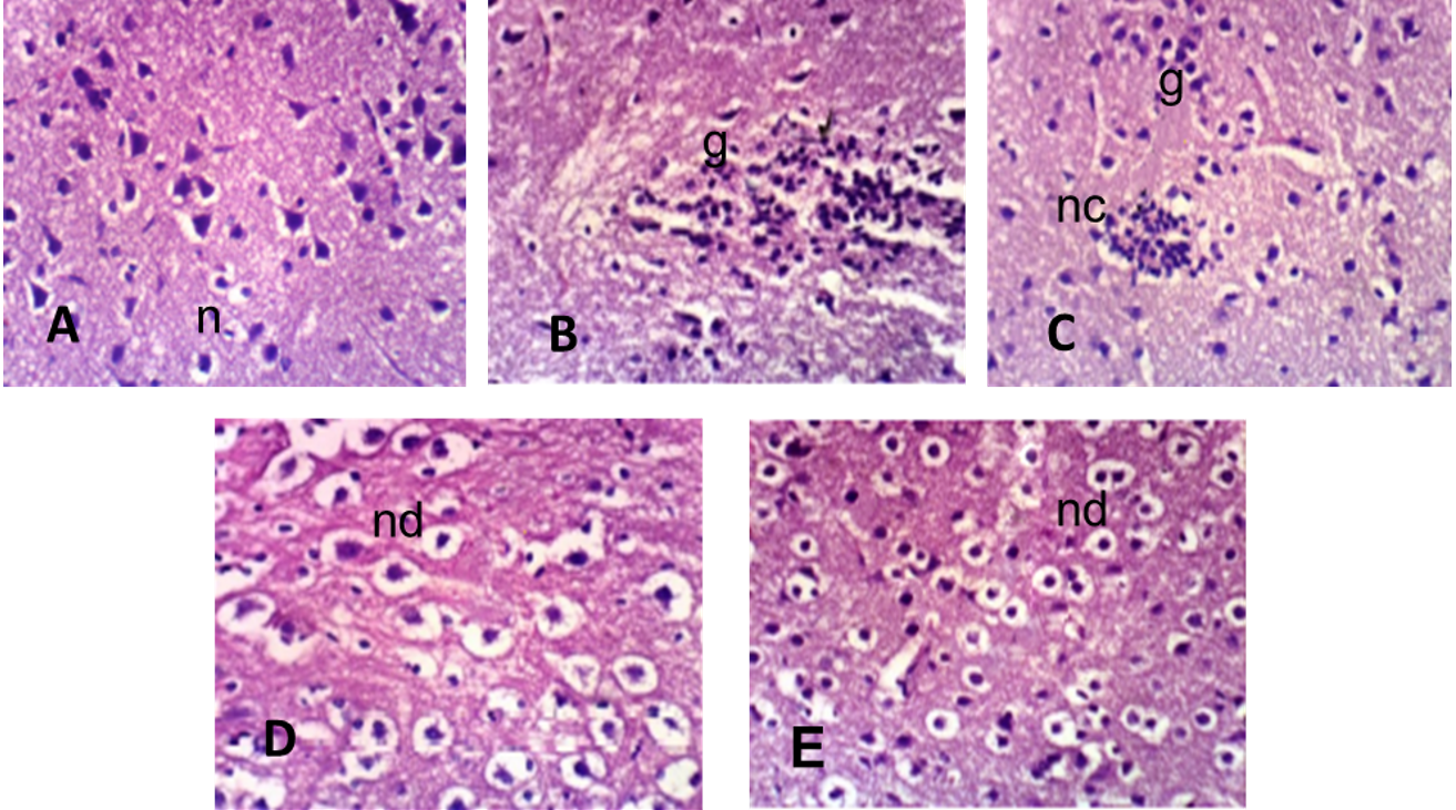
Brain section of male albino rats (A) control group shows normal morphology of cerebral cortex with normal rounded neuronal cells (n) (B) group treated with cypermethrin showed neuronal cell necrosis (nc), focal gliosis (g) (C) group treated with CYP+AA showed neuronal cell necrosis (nc) (D) group treated with CYP+Se showed neuronal cell degeneration (nd) were observed (E) the group with protective effect of ascorbic acid with selenium against cypermethrin shows almost no histopathological alterations.
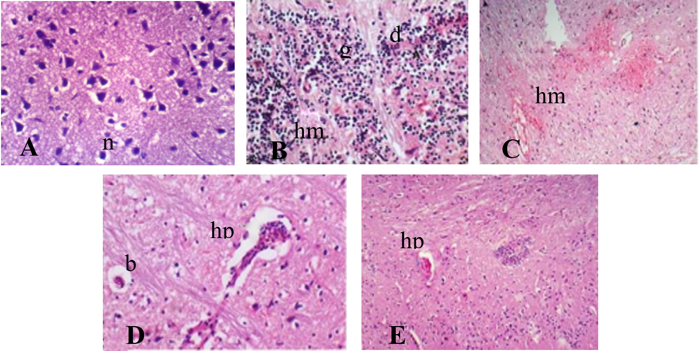
Brain section of female albino rats (A) the control group shows normal morphology of cerebral cortex with normal rounded neuronal cells (n) (B) the group treated with cypermethrin shows focal demyelination (d) of nerve fibers, hemorrhages (hm), focal gliosis (g) (C) the group treated with CYP+ AA shows hemorrhage (hm) in neocortex (D) group treated with CYP+Se shows broadened gap (b) around veins, focal hyperplasia (hp) of cerebral gliocytes (E) the group shows slight focal hyperplasia (hp) with protective effect of ascorbic acid + selenium.
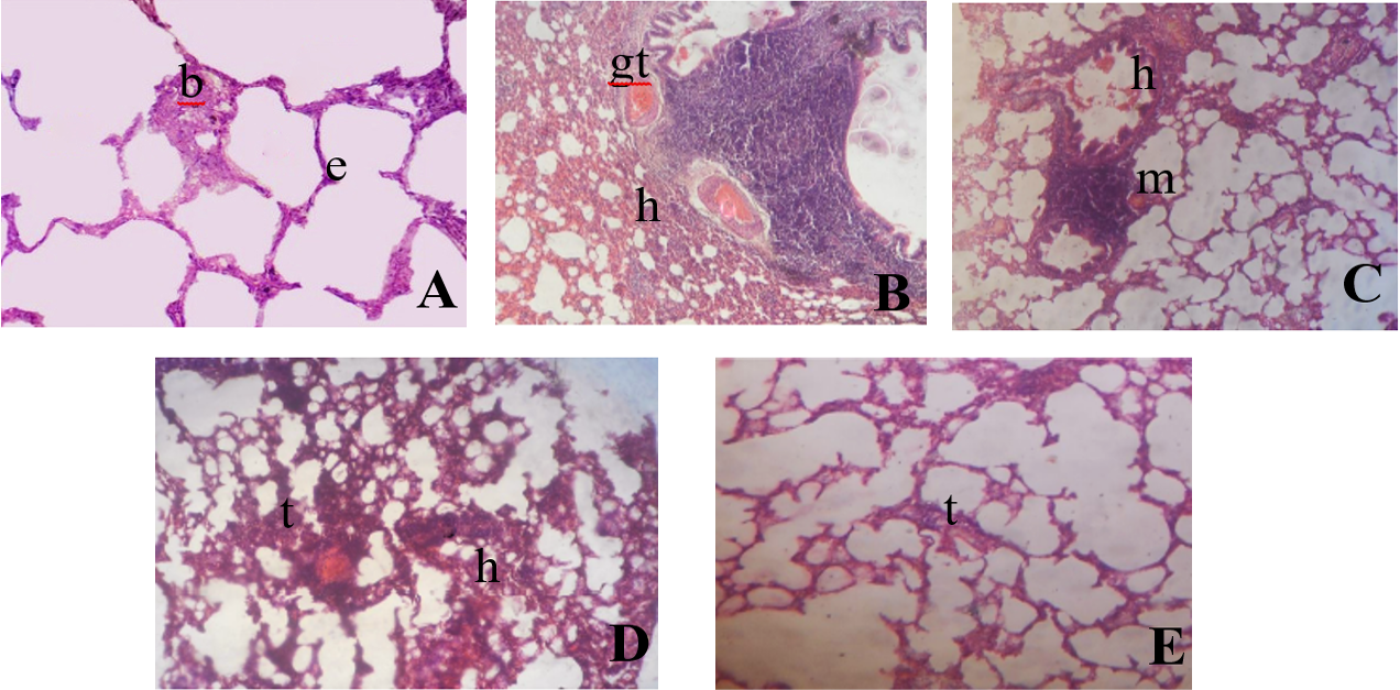
Lung section of male albino rat (A) the control group shows pulmonary parenchyma with normal bronchiolar epithelium (br) and normal epithelial wall (e) (B) group treated with cypermethrin shows haemorrhages (h), focal infiltration (i), non-caseating granuloma with presence of giant cells (gt) (C) the group treated with CYP+AA shows haemorrhages (h), intraluminal aggregation of mucous materials (m) mixed with desquamated epithelium (D) the group treated with CYP+ Se shows haemorrhages (h), focal or diffuse thickened epithelial walls (t) of pulmonary alveolus (E) group with protective effect of ascorbic acid with selenium against cypermethrin shows slightly diffuse thickened epithelial walls (t) of pulmonary alveolus.
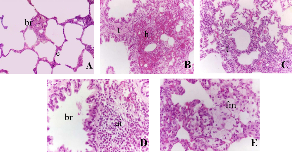
Lung section of female albino rat (A) control group shows pulmonary parenchyma with normal bronchiolar epithelium (br) and normal epithelial wall (e) (B) group treated with cypermethrin shows haemorrhages (h), focal or diffuse thickened epithelial walls (t) of pulmonary alveolus (C) the group treated with CYP+AA shows abnormal distribution of cellular elements, thickened epithelial walls (t) of pulmonary alveolus (D) the group treated with CYP+ Se shows marked dilatation in bronchioles, slightly damaged the parenchyma of lungs (E) group with protective effect of ascorbic acid with selenium against cypermethrin shows acidophilia infiltration (ai) of granulocytes around arterioles.
References
- Abd-El-Hameed, A.M. and Mahmoud, H.S., 2020. Cypermethrin induced apoptosis and testicular toxicity by upregulation of p53 in the brain and testis of male rats is alleviated by Sesame oil. J. Taibah Univ. Sci., 14: 1342–1349. https://doi.org/10.1080/16583655.2020.1822057[CrossRef]
- Abdou, H.M., Hussien, H.M. and Yousef, M.I., 2012. Deleterious effects of cypermethrin on rat liver and kidney: Protective role of sesame oil. J. environ. Sci. Hlth. B, 47: 306–314. https://doi.org/10.1080/03601234.2012.640913[CrossRef]
- Afifi, O.K. and Embaby, A.S., 2016. Histological study on the protective role of ascorbic acid on cadmium induced cerebral cortical neurotoxicity in adult male albino rats. J. Microsc. Ultrastruct, 4: 36–45. https://doi.org/10.1016/j.jmau.2015.10.001[CrossRef]
- Afolabi, O.K., Aderibigbe, F.A., Folarin, D.T., Arinola, A. and Wusu, A.D., 2019. Oxidative stress and inflammation following sub-lethal oral exposure of cypermethrin in rats: Mitigating potential of epicatechin. Heliyon, 5. https://doi.org/10.1016/j.heliyon.2019.e02274[CrossRef]
- Ali, H.F.H., El-Sayed, N.M., Ahmed, A.A., Hanna, P.A. and Moustafa, Y.M., 2020. Nano selenium ameliorates oxidative stress and inflammatory response associated with cypermethrin-induced neurotoxicity in rats. Ecotoxicol. environ. Saf., 195: 110479. https://doi.org/10.1016/j.ecoenv.2020.110479[CrossRef]
- Al-Omar, M.S., Naz, M., Mohammed, S.A., Mansha, M., Ansari, M.N., Rehman, N.U. and Khan, R.A., 2020. Pyrethroid-induced organ toxicity and anti-oxidant-supplemented amelioration of toxicity and organ damage: The protective roles of ascorbic acid and α-tocopherol. Int. J. environ. Res. Publ. Hlth., 17: 6177. https://doi.org/10.3390/ijerph17176177[CrossRef]
- Amir, N. and Amir, N., 2015. The effect of cypermethrin on Jambal Roti to AST and ALT levels the Wistar Rat (Rattus norvegicus). Int. J. PharmTech Res., 8: 235-240.
- Arafa, M.H., Mohamed, D.A. and Atteia, H.H., 2015. Ameliorative effect of N-acetyl cysteine on alpha-cypermethrin-induced pulmonary toxicity in male rats. Environ. Toxicol., 30: 26–43. https://doi.org/10.1002/tox.21891[CrossRef]
- Baghcheghi, Y., Razazpour, F., Seyedi, F., Arefinia, N. and Hedayati-Moghadam, M., 2024. Exploring the molecular mechanisms of PPARγ agonists in modulating memory impairment in neurodegenerative disorders. Mol. Biol. Rep., 51: 945. https://doi.org/10.1007/s11033-024-09850-6[CrossRef]
- Bancroft, J.D. and Gamble, M., 2008. Theory and practice of histological techniques. Elsevier Health Sciences.
- Desai, K.R., Moid, N., Patel, P.B. and Highland, H.N., 2015. Protective efficacy of Allium sativum on deltamethrin induced toxicity in reproductive tissues of male mice. Int. J. Pharm. Sci. Res., 6: 1711.
- Doseděl, M., Jirkovský, E., Macáková, K., Krčmová, L.K., Javorská, L., Pourová, J. and Oemonom, 2021. Vitamin C sources, physiological role, kinetics, deficiency, use, toxicity, and determination. Nutrients, 13: 615. https://doi.org/10.3390/nu13020615[CrossRef]
- Elblehi, S.S., Oda, S.S., Tohamy, H.G. and Elmanakhly, E.S.M., 2015. Protective effect of vitamin E and selenium combination on cypermethrin-induced toxicity in male rats. Alex. J. Vet. Sci., 47. https://doi.org/10.5455/ajvs.203149[CrossRef]
- Eraslan, G., Kanbur, M., Silici, S., Altinordulu, S. and Karabacak, M., 2008. Effecs of cypermethrin on some biochemical changes in rats: The protective role of propolis. Exp. Anim., 57: 453–460. https://doi.org/10.1538/expanim.57.453[CrossRef]
- Erkan, M., Aydin, Y., Yilmaz, B.O. and Yildizbayrak, N., 2021. Protective effects of vitamin C against fluoride toxicity. Toxicology, pp. 435–445. https://doi.org/10.1016/B978-0-12-819092-0.00043-1[CrossRef]
- Evans, M.D. and Cooke, M.S., 2009. Oxidative damage to nucleic acids. Springer Science and Business Media.
- Giray, B., Gürbay, A. and Hincal, F., 2001. Cypermethrin-induced oxidative stress in rat brain and liver is prevented by Vitamin E or allopurinol. Toxicol. Lett., 118: 139–146. https://doi.org/10.1016/S0378-4274(00)00277-0[CrossRef]
- Gündüz, A., Yalçın, E. and Çavuşoğlu, K., 2021. Combined toxic effects of aflatoxin B2 and the protective role of resveratrol in Swiss albino mice. Sci. Rep., 11: 1–14. https://doi.org/10.1038/s41598-021-95879-7[CrossRef]
- Hao, F., Bu, Y., Huang, S., Li, W., Feng, H. and Wang, Y., 2023. Effects of pyrethroids on the cerebellum and related mechanisms: A narrative review. Crit. Rev. Toxicol., 53: 229–243. https://doi.org/10.1080/10408444.2023.2229384[CrossRef]
- Hussien, H.M., Abdou, H.M. and Yousef, M.I., 2013. Cypermethrin induced damage in genomic DNA and histopathological changes in brain and haematotoxicity in rats: the protective effect of sesame oil. Brain Res. Bull., 92: 76–83. https://doi.org/10.1016/j.brainresbull.2011.10.020[CrossRef]
- Joshi, S.C., Bansal, B. and Jasuja, N.D., 2011. Evaluation of reproductive and developmental toxicity of cypermethrin in male albino rats. Toxicol. Environ. Chem., 93: 593–602. https://doi.org/10.1080/02772248.2010.537441[CrossRef]
- Kašuba, V., Tariba, L.B., Lucić, V.A., Katić, A., Kopjar, N., Micek, V. and Žunec, S., 2022. Evaluation of toxic effects induced by sub-acute exposure to low doses of α-cypermethrin in adult male rats. Toxics, 10: 717. https://doi.org/10.3390/toxics10120717[CrossRef]
- Khan, A.M., Raina, R., Dubey, N. and Verma, P.K., 2018. Effect of deltamethrin and fluoride co-exposure on the brain antioxidant status and cholinesterase activity in Wistar rats. Drug Chem. Toxicol., 41: 123–127. https://doi.org/10.1080/01480545.2017.1321009[CrossRef]
- Khan, B.A., Nadeem, M.A., Nawaz, H., Amin, M.M., Abbasi, G.H., Nadeem, M. and Ayub, M.A., 2023. Pesticides: Impacts on agriculture productivity, environment, and management strategies. In: Emerging contaminants and plants: Interactions, adaptations and remediation technologies. Springer, pp. 109–134. https://doi.org/10.1007/978-3-031-22269-6_5[CrossRef]
- Khan, S.M., Sobti, R.C. and Kataria, L., 2005. Pesticide-induced alteration in mice hepato-oxidative status and protective effects of black tea extract. Clin. Chim. Acta, 358: 131–138. https://doi.org/10.1016/j.cccn.2005.02.015[CrossRef]
- Kuban, W. and Daniel, W.A., 2021. Cytochrome P450 expression and regulation in the brain. Drug Metab. Rev., 53: 1–29. https://doi.org/10.1080/03602532.2020.1858856[CrossRef]
- Mahat, S., Jha, C.B., Shrestha, S. and Koirala, S., 2020. Effect of vitamin E on cypermethrin induced toxicity in cerebrum of wistar albino rats: A histological study. J. Karnali Acad. Hlth. Sci., 3: 1–12. https://doi.org/10.3126/jkahs.v3i1.28457[CrossRef]
- Manna, S., Bhattacharyya, D., Basak, D.K. and Mandal, T.K., 2004. Single oral dose toxicity study of a-cypermethrin in rats. Indian J. Pharmacol., 36: 25.
- Matić, M.M., Paunović, M.G., Milošević, M.D., Ognjanović, B.I. and Saičić, Z.S., 2021. Hematoprotective effects and antioxidant properties of β-glucan and vitamin C against acetaminophen-induced toxicity: An experimental study in rats. Drug Chem. Toxicol., 44: 302–309. https://doi.org/10.1080/01480545.2019.1587451[CrossRef]
- Mohafrash, S.M.M., Hassan, E.E., El-Shaer, N.H. and Mossa, A.T.H., 2021. Detoxification gene expression, genotoxicity, and hepatorenal damage induced by subacute exposure to the new pyrethroid, imiprothrin, in rats. Environ. Sci. Pollut. Res., 28: 33505–33521. https://doi.org/10.1007/s11356-021-13044-z[CrossRef]
- Mossa, A.T.H., Heikal, T.M., Belaiba, M., Raoelison, E.G., Ferhout, H. and Bouajila, J., 2015. Antioxidant activity and hepatoprotective potential of Cedrelopsis grevei on cypermethrin induced oxidative stress and liver damage in male mice. BMC Complement. Altern. Med., 15: 1–10. https://doi.org/10.1186/s12906-015-0740-2[CrossRef]
- Muthuviveganandavel, V., Muthuraman, P., Muthu, S. and Srikumar, K. 2008. A study on low dose cypermethrin induced histopathology, lipid peroxidation and marker enzyme changes in male rat. Pestic Biochem. Physiol., 91: 12–16. https://doi.org/10.1016/j.pestbp.2007.11.010[CrossRef]
- Nair, R.R., Abraham, M.J., Lalithakunjamma, C.R., Nair, N.D. and Aravindakshan, C.M., 2011. A pathomorphological study of the sublethal toxicity of cypermethrin in Sprague Dawley rats. Int. J. Nutr. Pharmacol. Neurol. Dis., 1: 179. https://doi.org/10.4103/2231-0738.84211[CrossRef]
- Namdev, N., Rai, G. and Mahobiya, P., 2023. Protective role of ashwagandha and quercetin over cypermethrin induced hypothyroidism: A hematological study in mice. Adv. Zool. Bot., 11: 103–111. https://doi.org/10.13189/azb.2023.110202[CrossRef]
- Oda, S.S. and El-Maddawy, Z.K., 2012. Protective effect of vitamin E and selenium combination on deltamethrin-induced reproductive toxicity in male rats. Exp. Toxicol. Pathol., 64: 813–819. https://doi.org/10.1016/j.etp.2011.03.001[CrossRef]
- Oladele, J., Adewale, O., Oyewole, O., Gbolagbade, A. and Oyeleke, M., 2020. Assessment of the protective effects of vitamin C and E on cypermethrin-induced nephrotoxicity and electrolyte imbalance in wistar rats. J. Basic appl. Res. Biomed., 6: 1–6. https://doi.org/10.51152/jbarbiomed.v6i1.1[CrossRef]
- Oshatunberu, M.A., Oladimeji, A., Sawyerr, O.H. and Raimi, M.O., 2023. Search what you can’t see-evaluation pestic residues grain sold sel mark. S. Niger. Curr. Res. Publ. Hlth., 3: 10–36. https://doi.org/10.31586/crph.2023.566[CrossRef]
- Raina, R., Verma, P.K., Pankaj, N.K., Kant, V. and Prawez, S., 2010. Protective role of L-ascorbic acid against cypermethrin-induced oxidative stress and lipid peroxidation in Wistar rats. Toxicol. environ. Chem., 92: 947–953. https://doi.org/10.1080/02772240903174781[CrossRef]
- Rashid, U., Qureshi, I.Z., Jan, S., Khalid, T. and Khan, D.A., 2023. Protective effects of selenium against cypermethrin induced hepatorenal damage in Sprague Dawley rats. World J. Biol. Pharm. Hlth. Sci., 22: 284–298. https://doi.org/10.30574/wjbphs.2023.13.1.0293[CrossRef]
- Sangha, G.K., Kaur, K. and Khera, K.S., 2013. Cypermethrin induced pathological and biochemical changes in reproductive organs of female rats. J. environ. Biol., 34: 99.
- Sangha, G.K., Kaur, K., Khera, K.S. and Singh, B., 2011. Toxicological effects of cypermethrin on female albino rats. Toxicol. Int., 18: 5. https://doi.org/10.4103/0971-6580.75844[CrossRef]
- Sankar, P., Telang, A.G. and Manimaran, A., 2012. Protective effect of curcumin on cypermethrin-induced oxidative stress in Wistar rats. Exp. Toxicol. Pathol., 64: 487–493. https://doi.org/10.1016/j.etp.2010.11.003[CrossRef]
- Sayim, F., Yavasoglu, N.Ü.K., Uyanikgil, Y., Aktug, H., Yavasoglu, A. and Turgut, M., 2005. Neurotoxic effects of cypermethrin in Wistar rats: A haematological, biochemical and histopathological study. J. Hlth. Sci., 51: 300–307. https://doi.org/10.1248/jhs.51.300[CrossRef]
- Shaikh, N.I. and Sethi, R.S., 2020. Exposure to chlorpyrifos and cypermethrin alone or in combination induces developmental abnormalities and lung damage in animal models: A review. J. Ent. Zool. Stud., 8: 1923–1928. https://doi.org/10.22271/j.ento.2020.v8.i5aa.7769[CrossRef]
- Shaikh, N.I. and Sethi, R.S., 2021. Differential gene expression profile of mice lung following chronic dietary exposure to chlorpyrifos and/or cypermethrin. J. Hum. Reprod. Sci., 7: 99. https://doi.org/10.22271/j.ento.2021.v9.i1b.8139[CrossRef]
- Sharma, P., Huq, A.U. and Singh, R., 2014. Cypermethrin-induced reproductive toxicity in the rat is prevented by resveratrol. J. Hum. Reprod. Sci., 7: 99. https://doi.org/10.4103/0974-1208.138867[CrossRef]
- Sheikh, N., Javed, S., Asmatullah, A., Ahmad, K.R., Abbas, T. and Iqbal, J., 2014. Histological changes in the lung and liver tissues in mice exposed to pyrethroid inhalation. Walailak J. Sci. Technol., 11: 843–849.
- Shilpakar, O. and Karki, B., 2021. Cypermethrin poisoning manifesting with prolonged bradycardia: A case report. Toxicol. Rep., 8: 10–12. https://doi.org/10.1016/j.toxrep.2020.12.005[CrossRef]
- Shuklan, P., Raj, A., Chauhan, K., Madan, P. and Rani, S., 2023. Systematic toxicity of cypermethrin and alterations in behavior of albino rats. ACS Omega, 8: 14766–14773. https://doi.org/10.1021/acsomega.3c00817[CrossRef]
- Singh, A., Mudawal, A., Shukla, R.K., Yadav, S., Khanna, V.K., Sethumadhavan, R. and Parmar, D., 2015. Effect of gestational exposure of cypermethrin on postnatal development of brain cytochrome P450 2D1 and 3A1 and neurotransmitter receptors. Mol. Neurobiol., 52: 741–756. https://doi.org/10.1007/s12035-014-8903-6[CrossRef]
- Tongjai, P., Hongsibsong, S. and Sapbamrer, R., 2021. Open access research article.
- Yavasoglu, A., Sayim, F., Uyanikgil, Y., Turgut, M. and Karabay-Yavasoglu, N.Ü., 2006. The pyrethroid cypermethrin-induced biochemical and histological alterations in rat liver. J. Hlth. Sci., 52: 774–780. https://doi.org/10.1248/jhs.52.774[CrossRef]
- Yousef, M.I., Awad, T.I. and Mohamed, E.H., 2006. Deltamethrin-induced oxidative damage and biochemical alterations in rat and its attenuation by Vitamin E. Toxicology, 227: 240–247. https://doi.org/10.1016/j.tox.2006.08.008[CrossRef]
- Yüksel, Y., Aslan, E., Tosun, M., Altunbaş, K., Akkaya, Ö.Ö., Demirel, H.H. and Pektaş, M.B., 2023. Protective effects of resveratrol on permethrin-induced fetotoxicity in rats. Anatol. J. Bot., 7: 21–28. https://doi.org/10.30616/ajb.1241886[CrossRef]
- Zalata, A., Atwa, A., Badawy, A.E-N., Aziz, A., El- Baz, R., Elhanbly, S. and Mostafa, T., 2013. Tumor necrosis factor-α gene polymorphism relationship to seminal variables in infertile men. Urology, 81: 962–966. https://doi.org/10.1016/j.urology.2013.01.026[CrossRef]
- Ziada, R.M., Nahas, A.A., Farag, A.and Kotb, G.A., 2020. Protective efficacy of combined administration of vitamins c and curcumin on cypermethrin-induced oxidative stress in male albino rats. Egypt. Acad. J. biol. Sci. F Toxicol. Pest Contr., 12: 109–118. https://doi.org/10.21608/eajbsf.2020.113976[CrossRef]
- Zuo, M., Ye, M., Lin, H., Liao, S., Xing, X., Liu, J. and Ren, X., 2024. Mitochondrial dysfunction in environmental toxicology: Mechanisms, impacts, and health implications. Chem. Res. Toxicol., 37: 1794–1806. https://doi.org/10.1021/acs.chemrestox.4c00328[CrossRef]
Services
Article usage statistics
Loading…