
Agriculture is the mainstay of Pakistan’s economy, and livestock accounts for around 14.63% of the national GDP. Of all major diseases of livestock, caseous lymphadenitis (CLA), which is an infection caused by Corynebacterium pseudotuberculosis, causes serious economic losses and threats to sheep and goats’ health and economic values. The present study explored the prevalence, genetic characterization, and phylogenetic analysis of C. pseudotuberculosis in small ruminants of Bahawalpur district. The pus samples (n=90) from infected animals were cultured on 5% sheep blood agar and identified biochemically. DNA was isolated, and the pld gene was amplified by PCR and sequenced (GenBank accession numbers PX069784 and PX069785). BLAST sequence analysis showed 99% similarity with Norwegian, Portuguese, Chinese, and Egyptian isolates. Phylogenetic analysis was performed by the neighbor-joining method in MEGA (v.11.0). Comparative modeling of proteins and epitope mapping of local isolates (CUVAS3 and CUVAS4) with the reference strain (MIC6-CP019769) showed minimal structural and antigenic differences. Statistical comparison by using SPSS (version 22.0) revealed significantly higher infection rate in sheep (8.89%) compared to goat (6.67%) (χ² = 5.874, df = 1, p = 0.0154). These results show significant genetic associations between local and global C. pseudotuberculosis strains, also emphasize to develop better diagnostic and prevention methods to counter CLA in high-risk areas.
Research Article · Volume 58, Issue 6, 28 September 2026 · Open Access
Molecular Characterization and Phylogenetic Profiling of Corynebacterium pseudotuberculosis Isolates from Small Ruminants in Bahawalpur, Pakistan
· · · · · · · · · ·
Affiliations & Notes ⌄
- 1Department of Microbiology, Cholistan University of Veterinary and Animal Sciences, Bahawalpur 63100, Pakistan 2Department of Medicine, Cholistan University of Veterinary and Animal Sciences, Bahawalpur 63100, Pakistan 3Department of Parasitology, Cholistan University of Veterinary and Animal Sciences, Bahawalpur 63100, Pakistan 4Veterinary Research Institute, Zarrar Shaheed Road, Lahore Cantt 54810, Pakistan * Corresponding author: dr.raisahmad2068@gmail.com
Article Info ⌄
- Journal:
- Pakistan Journal of Zoology
- Published:
- 28 September 2026

From the article
- BLAST analysis showed 98-100% sequence identity with global C. pseudotuberculosis isolates from Norway, Portugal, China, and Egypt, reflecting high genetic conservation across different geographic regions.
- Alignment with the reference strain MIC6-CP019769 (Table II) revealed 95% identity and 100% similarity with minor gaps, suggesting moderate conservation and slight variations between local isolates and the reference strain.
- Comparative sequence analysis of local isolates CUVAS3 and CUVAS4 with the reference strain MIC6 identified multiple amino acid substitutions, including a substitution at Q37 unique to CUVAS4, suggesting potential antigenic variation (Table III).
- This study detected the prevalence of C. pseudotuberculosis at 33% in small ruminants of Bahawalpur; pld gene sequencing, strain comparison, gene mapping, and epitope analysis confirmed its very close similarity with global strains.
Selected from the article's own text.
Abstract
Introduction
Sheep and goats are affected by a contagious chronic disease called caseous lymphadenitis (CLA), and its sporadic cases have also recorded in other species including cattle, buffaloes, and camels . The disease presents a threat to economic loss in livestock-based livelihoods and zoonotic health risk to humans. CLA decreases productivity, arrests growth, induces chronic disorders, and escalates mortality in infected herds, ultimately resulting in heavy economic losses globally. In developing nations such as Pakistan, wherein livestock is a pillar of rural economies, the effect of CLA is most harmful. The illness not only lowers animal productivity but also worsens poverty and food inadequacy in susceptible populations. Outbreaks have been reported to have an increased prevalence of CLA in the developing world, thus deserving region-specific control measures as informed by targeted epidemiological and molecular studies .
Causative agent, Corynebacterium pseudotuberc-ulosis biovar ovis, is a Gram-positive facultative intracellular bacterium. Clinically, CLA presents with abscessation of superficial lymph nodes, subcutaneous tissues, and internal organs . These abscesses often become chronic lesions that lower growth rates and reproductive efficiency, causing substantial losses in flock health and market value. The bacterium’s virulence determinant, phospholipase D (PLD), increases vascular permeability, promotes tissue invasion, and plays a role in disease progression. Molecular determinants of pathogenesis are still need to look after, even after the identification of virulence mechanism .
Traditional diagnostic methods, like phenotypic and biochemical tests, are usually not sufficiently sensitive and specific to detect the organism accurately, as Corynebacterium species share both genetic and phenotypic homology. Molecular diagnostic reagents, especially PCR assays for the pld gene of 203 bp, have proven more sensitive and reproducible for C. pseudotuberculosis detection . Moreover, rpoB gene sequencing, encoding for the β-subunit of RNA polymerase, has successfully differentiate C. pseudotuberculosis from closely related species .
The current study aims at conducting molecular characterization and phylogenetic study of C. pseudotuberculosis isolates collected from infected sheep and goats in Bahawalpur. The outcomes will show the genetic diversity, virulence, and evolutionary pathways of indigenous strains towards better diagnostic, preventive, and control strategies for the CLA in Pakistan and elsewhere.
Materials and Methods
Study area and sample collection
A total of ninety pus samples were aseptically collected from superficial abscesses of sheep (n= 45) and goats (n= 45) clinically suspected of CLA from the Bahawalpur district and adjoining Cholistani regions. The sampling sites included 42/DB, Alif 13/BC, Pakkabarah, Qainchi Mor, Toba Janu Wala, and Toba Kakrala areas characterized by semi-arid conditions and high livestock density. Prior to sampling, the abscess surface was disinfected with 70% ethanol to prevent contamination. Approximately 1 mL of purulent exudate was aspirated using a sterile 5 mL disposable syringe (Becton Dickinson, USA) or collected with sterile cotton swabs (HiMedia Laboratories, India). Each sample was transferred into sterile transport tubes and immediately stored at 4°C for further processing.
Isolation and Identification of the organism
The samples were initially cultured in nutrient broth, followed by streaking on nutrient agar, and final growth was observed on 5% sheep blood agar (Oxoid™, UK) and incubated at 37°C for 24-48 h under microaerophilic conditions. Microscopic examination following Gram staining (HiMedia™, India) and biochemical identification was conducted using the standard microbiological procedures following the protocols of Bergey (1994). The modified CAMP test, performed with Staphylococcus aureus ATCC 25923 (Thermo Fisher Scientific™, USA.
Molecular identification of C. pseudotuberculosis
Genomic DNA was extracted from 18 confirmed isolates using the WizPrep™ gDNA Mini Kit (Cell/Tissue; Wizbiosolutions, Seongnam, South Korea) following the manufacturer’s protocol. Briefly, bacterial pellets were obtained by centrifugation at 7,500 rpm for 10 min (Eppendorf 5810R, Germany), resuspended in GT1 buffer, and subjected to lysis with Proteinase K and GT2 buffer at 56°C for 10 min. DNA was purified through silica-membrane spin columns and eluted in 100 µL of elution buffer. The DNA quality and concentration were evaluated using a NanoDrop™ 2000 spectrophotometer (Thermo Fisher Scientific™, USA). Samples showing a 260/280 absorbance ratio between 1.8 and 2.0 were considered high quality and stored at −20°C until further use.
Molecular confirmation of the C. pseudotuberculosis was achieved by amplifying the phospholipase D (pld) gene (203bp) using conventional PCR. Each 25 µL reaction mixture contained 13 µL of 2× WizPure™ PCR Master Mix (Wizbiosolutions, South Korea), 1 µL of each primer (Macrogen Inc., Seoul, South Korea) at a working concentration of 10 pmol/µL, 4 µL of template DNA (20 ng/µL), and 6 µL of nuclease-free water. The primer sequences used were:
PLD-F: 5′-ATAAGCGTAAGCAGGGAGCA-3′
PLD-R1: 5′-ATCAGCGGTGATTGTCTTCC-3′
PCR amplification was performed in a Bio-Rad C1000 Thermal Cycler (Bio-Rad Laboratories, USA) under the following conditions: Initial denaturation at 95 °C for 3 min; 40 cycles of denaturation at 95 °C for 1 minute, annealing at 58°C for 40 sec, and extension at 68 °C for 90 sec; followed by a final extension at 68 °C for 7 min following the protocol of El-Sebay et al. (2021). The amplified products were electrophoresed on 1.5% agarose gel (Sigma-Aldrich™, USA) stained with ethidium bromide and visualized under UV light using a Syngene G:BOX Gel Documentation System (Syngene, Cambridge, UK).
Molecular characterization and evolutionary analysis of target gene
Representative PCR products were purified and sequenced using the Sanger method at GeneTech Pvt. Ltd. (Karachi, Pakistan) following Ali et al. (2024). The obtained nucleotide sequences were aligned using BioEdit (v.7.0.5.3.) and compared to global reference sequences via the NCBI BLASTn tool as described by Javanbakht and Hajiyan (2024). The validated pld gene consensus sequences were submitted to the NCBI GenBank database. Phylogenetic tree was constructed using the Neighbor-Joining method based on the maximum composite likelihood model in MEGA (version 11.0) following Liu et al. (2023). This analysis revealed the genetic diversity, evolutionary relationships, and regional clustering of Pakistani isolates in comparison with international strains.
Strain comparison, epitope and gene mapping
In order to quantify genetic similarity and conservation, pairwise comparisons between local isolates (CUVAS3 and CUVAS4) and the reference strain MIC6-CP019769 have been conducted using NCBI BLASTn as described by Al-Ethafa et al. (2025). Epitope mapping for identification of antigenic regions within the protein sequences was done using IEDB software following Shi et al. (2023). Mapping of the gene of isolates has been carried out to visualize sequence organization and confirm the accuracy of alignment against the reference strain.
Statistical analysis
The differences in groups were assessed using the Chi-square (χ²) test, and degree of freedom was determined with p < 0.05. This statistical analysis was done by SPSS (v.26.0) software.
Results
Ninety pus samples were collected from clinically suspected CLA cases of sheep (n = 45) and goats (n = 45) from different rural and Cholistani areas of Bahawalpur. samples (n=17) were identified as C. pseudotuberculosis with an overall prevalence of 18.9%. Species-wise distribution showed that goats had a significantly greater infection rate (13/45; 28.9%) as compared to sheep (4/45; 8.9%) (χ²= 5.874, df= 1, p= 0.0154). This difference suggests potential host-specific vulnerability and higher risk of exposure in goats under existing management and environmental conditions.
Initial culturing of the pus samples in the nutrient broth showed mixed bacterial growth. subculturing on nutrient agar produced uniform, white, mucoid, smooth and convex colonies with a dry surface typical of C. pseudotuberculosis (Fig. 1A, B). Final streaking on 5% sheep blood agar showed small (approximately 2 mm in diameter), dry, grayish-white, and non-motile, surrounded by a narrow zone of complete β-hemolysis (Fig. 1C). However, Gram staining showed Gram-positive, short and curved bacilli arranged in a V-shaped configurations (Fig. 1D, E).
C. pseudotuberculosis isolates were confirmed by Biochemical testing. All (n=17) isolates tested positive for catalase by producing bubbles (Fig. 1D, E). Collectively, the biochemical profiles were consistent with previously reported characteristics of C. pseudotuberculosis. Figure 2 shows the higher prevalence in goats than in sheep. These findings suggest that there may be some species-specific differences in susceptibility to C. pseudotuberculosis.
DNA was successfully isolated from all isolates employing the WizPrep™ g DNA Mini Kit according to the manufacturer’s protocol. DNA concentration and purity were determined on a NanoDrop™ spectrophotometer (Thermo Fisher Scientific™, USA). A260/A280 ratios between 1.8 and 2.0 considered optimal for subsequent molecular applications. DNA concentrations were heterogeneous ranging from 3.8 ng/µL to 444.5 ng/µL. Sample 12 revealed the greatest concentration of DNA (444.5 ng/µL), while the lowest (3.8 ng/µL) was recorded in Sample 6 (Supplementary Table I).
Amplification of the pld gene with regular PCR yielded clear amplicons of the right size (203 bp) in all 17 isolates (Fig. 3A). The clear, sharp bands verified molecular identity of C. pseudotuberculosis. There was no amplification in negative controls to ensure specificity and reliability of the assay.
Sanger sequencing of the pld gene produced high-quality chromatograms with well-defined peaks, indicating precise nucleotide resolution. The sequences were deposited in the NCBI GenBank database with accession numbers PX069784 and PX069785 following Fukuda et al. (2021). BLAST analysis showed 98-100% sequence identity with global C. pseudotuberculosis isolates from Norway, Portugal, China, and Egypt, reflecting high genetic conservation across different geographic regions.
Phylogenetic tree constructed with Neighbor-Joining procedure in MEGA (v.11.0) grouped Pakistani isolates (PX069784 and PX069785) together with the reference strain MIC6 (GenBank accession: CP019769.1:24150-24262), with a high bootstrap value of 99% (Fig. 3B, C).
Table I. Pairwise amino acid alignment of the CUVAS3 with CUVAS4, showing 97% identity and similarity showing no gaps, indicating high sequence conservation.
|
Strain/ID |
Start position |
Sequence alignment |
End position |
|
CUVAS3 |
1 |
MAIMLPVGNAAAAPVVHNPASTANRPVYAIAHRVLTTH |
38 |
|
CUVAS4 |
13 |
.....................................Q |
50 |
Table II. Pairwise amino acid alignment of the CUVAS3 and CUVAS4 with MIC6_CP019769 (Reference strain), showing 95% identity and 100% similarity showing less gaps, indicating moderate sequence conservation.
|
Strain/ID |
Start position |
Sequence alignment |
End position |
|
MIC6_CP019769 |
1 |
MAIMLPVGNAAAAPVVHNPASTANRPVYAIAHRVLTTH |
37 |
|
CUVAS3 |
1 |
MAIMLPVGNAAAAPVVHNPASTANRPVYAIAHRVLTTH |
38 |
|
CUVAS4 |
13 |
.....................................Q |
50 |
Table III. Amino acid variations in predicted pld epitopes (E1: pink, E2: green, E3: yellow) comparing MIC6-CP-19769 with Corynebacterium pseudotuberculosis CUVAS3 and CUVAS4. Dots indicate identity; substitution (Q37) suggest antigenic drift. Epitope regions were predicted using IEDB BepiPred, and strain differences identified by Clustal Omega alignment.
|
Epitope region Position |
C1E |
C2E |
C3E |
||||||||||||
|
2 |
4 |
6 |
8 |
10 |
12 |
14 |
16 |
18 |
20 |
25 |
27 |
30 |
34 |
37 |
|
|
MIC6-CP-19769 |
A |
M |
P |
G |
A |
A |
P |
V |
N |
A |
R |
V |
I |
V |
H |
|
CUVAS3 |
. |
. |
. |
. |
. |
. |
. |
. |
. |
. |
. |
. |
. |
. |
. |
|
CUVAS4 |
. |
. |
. |
. |
. |
. |
. |
. |
. |
. |
. |
. |
. |
. |
Q |
This genetic association corroborates the evolutionary relationship between local and global strains. Minor nucleotide differences found in the Pakistani isolates can imply regional genetic adaptation.
Pairwise amino acid alignment between CUVAS3 and CUVAS4 (Table I) showed 97% identity and similarity with no gaps, indicating strong sequence conservation. Alignment with the reference strain MIC6-CP019769 (Table II) revealed 95% identity and 100% similarity with minor gaps, suggesting moderate conservation and slight variations between local isolates and the reference strain.
Structural modeling of the pld protein using the SWISS-MODEL and visualization in UCSF Chimera (v.1.19) revealed a well-folded tertiary structure consistent with known pld protein conformations. A point mutation was identified at residues ASP121A OD2 and PRO18A CG (highlighted in Fig. 4) as described by Rodrigues et al. (2021) which could potentially influence protein stability or immune recognition. Epitope mapping via IEDB BepiPred (v.1.0) predicted three major linear B-cell epitopes (C1E, C2E, and C3E) located between residues 2–37. Comparative sequence analysis of local isolates CUVAS3 and CUVAS4 with the reference strain MIC6 identified multiple amino acid substitutions, including a substitution at Q37 unique to CUVAS4, suggesting potential antigenic variation (Table III).
Gene mapping of C. pseudotuberculosis MIC6 by Proksee (v.1.3) and Artemis (v.0.4.1) software generated circular and linear genomic maps (Fig. 5). These maps described coding sequences (CDS), tRNAs, rRNAs, GC content, and GC skew and gave a visual impression of genome organization following the protocol of Galià-Camps et al. (2024). Combination of molecular, phylogenetic, and structural information validated identification of C. pseudotuberculosis in the Bahawalpur area and demonstrated its intimate genetic association with global strains, highlighting a call for a better disease surveillance system and molecular tracking to become aware of regional strain evolution and epidemiology in small ruminants.
Discussion
Current study verified C. pseudotuberculosis as the causative agent of CLA in small ruminants in the Bahawalpur district of Pakistan, reaffirming the continued relevance of this pathogen to the livestock economy of the region. The reported prevalence rate of 33% (18 positive
isolates in 90 samples) indicates the endemic status of CLA in southern Punjab. This prevalence is significantly higher than the 12.6% described in Russia but similar to Brazil (30%) and the United States (42.2%) . It is also higher than that from South Africa (25%) , India (16.5%) , Turkey (25%) , and New Zealand (28%) . These regional variations might be due to differences in diagnostic strategies, husbandry, density of animals, weather conditions, and biosecurity protocols, which all affect disease transmission patterns.
Phylogenetic reconstruction of the pld gene validated that all the isolates belonged to the C. pseudotuberculosis biovar ovis clade, as previously established by Hiller et al. (2024), who described the same pattern of clustering based on pld gene sequences in sheep and goats. The high bootstrap supports (90–100%) in the present study validate the strength of these genetic associations. Interestingly, the Bahawalpur isolates showed small mutations indicative of local genetic diversity, potentially due to environmental selection pressures or host-specific variation. Their clustering with Brazilian and South Korean strains is indicative of possible global routes of dissemination or genetic exchange through livestock trade, as suggested by de Pinho et al. (2021).
Epitope mapping of the pld protein identified three predominant antigenic clusters (C1E, C2E, and C3E), with minor amino acid variations, specifically at Q37 in isolate CUVAS4. These changes might not necessarily impact protein function but may have an impact on antigenic variability and host immune recognition. Similar results were reported by Zhou et al. (2021), who identified comparable mutations in C. pseudotuberculosis isolates from China and correlated them with regional adaptation rather than enhanced virulence.
The use of pld gene sequencing gave satisfactory species-level identification (98–100% similarity with reference strains deposited in GenBank), in accordance with those of Terab et al. (2021). Nevertheless, as pointed out by Carella et al. (2025), this one-gene strategy restricts the ability to differentiate between closely related species, e.g., Corynebacterium ulcerans, thus limiting the ability for subspecies-level differentiation. Future studies should thus include multilocus sequence typing (MLST) and whole-genome sequencing (WGS) to attain higher discriminatory power and improved insight into the genomic epidemiology of C. pseudotuberculosis.
The gene mapping carried out in this investigation, with emphasis on coding sequences, tRNAs, and GC content, is useful to understand local strain evolution following Zhang et al. (2024). Combination of genomic and phylogenetic information with epidemiological monitoring would greatly strengthen control measures specific to CLA-endemic areas such as Bahawalpur. These observations thus validate the high prevalence and genetic conservation of C. pseudotuberculosis in Pakistan but pointing out the necessity of the development of novel molecular tools and better farm management to help combat the impact of CLA on small ruminants.
Conclusion
This study detected the prevalence of C. pseudotuberculosis at 33% in small ruminants of Bahawalpur; pld gene sequencing, strain comparison, gene mapping, and epitope analysis confirmed its very close similarity with global strains. A novel point mutation reflected regional adaptation, while the phylogenetic analysis showed high genetic conservation. These findings indicate the global transmission potential of the pathogen and call for improved genomic approaches, such as MLST and WGS, for enhanced surveillance and control of CLA.
Declarations
Acknowledgment
The authors gratefully acknowledge the financial support provided by Higher Education Commission (HEC) of Pakistan under NRPU project (20-14949/NRPU/R&D/HEC/2021-2020) at Cholistan University of Veterinary and Animal Sciences, Bahawalpur, which made this study possible.
Funding
This work was supported by the Higher Education Commission (HEC) of Pakistan under NRPU Project/Grant No.14949.
IRB approval
The study was reviewed and approved by the Institutional Review Board (IRB) of the respective institution with approved ethical guidelines and regulations.
Ethical approval
The study was conducted in strict accordance with the guidelines for the care and use of laboratory animals. All procedures involving animals were reviewed and approved by the Ethical Committee of Cholistan University of Veterinary and Animal Sciences, Bahawalpur under approval # ORIC-67 dated 26-04-2021.
Supplementary material
There is supplementary material associated with this article. Access the material online at: https://dx.doi.org/10.17582/journal.pjz/20251124144613
Generative AI and AI-assisted technology statement
The authors declare that no generative AI and AI assisted technology was used in the creation of this manuscript.
Statement of conflict of interest
The authors have declared no conflict interests.
Figures
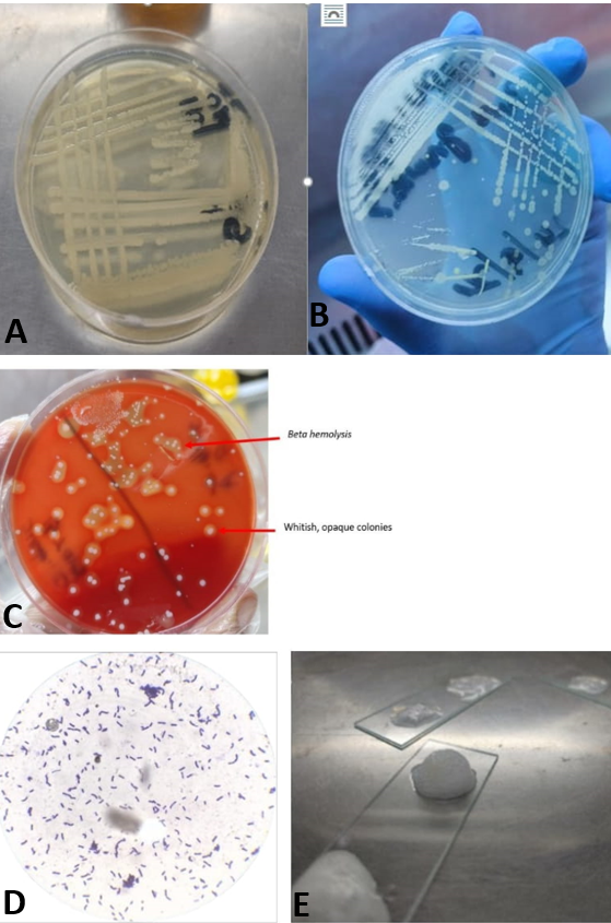
Corynebacterium pseudotuberculosis: A, B, white mucoid characteristics colonies observed on nutrient agar plates after in cubation at 37 oC for 48 h, C, purifying colonies on hemolysis; D, microscopic examination; E, bubble fromations.
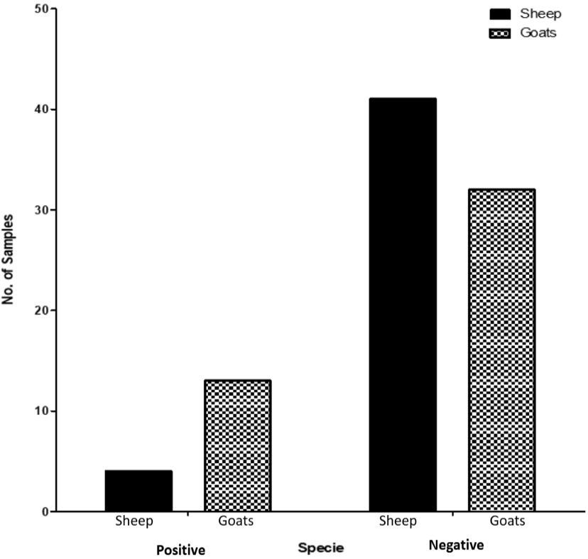
Comparative analysis of C. pseudotuberculosis positive and negative samples of sheep and goats.
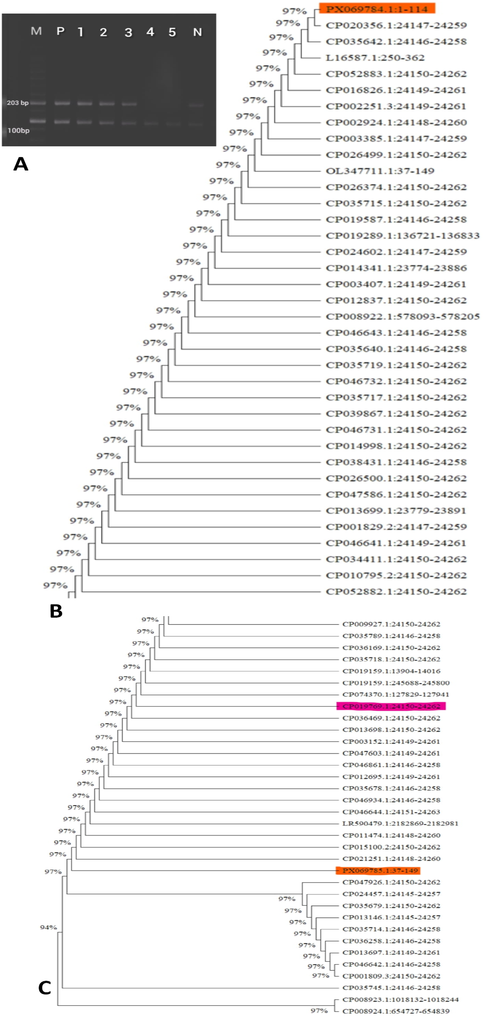
A, Agarose gel electrophoresis showing PCR amplification 1.5% Agarose Gel of the pld gene (203 bp) in Corynebacterium pseudotuberculosis isolates. M(Ladder): 100 bp DNA ladder, M: 1-3 and 6: positive samples showing expected 203 bp band, M:4 and 5 showing Negative samples. P, Positive Control; N, Negative Control. B, C, Phylogenetic tree of reference strain of C. pseudotuberculosis CP019769.1:24150-24262 (in pink) isolates constructed by the neighbor-joining method showing 97% similarity with the PX069784.1:1-114 and PX069785.1:37-149 (in red).
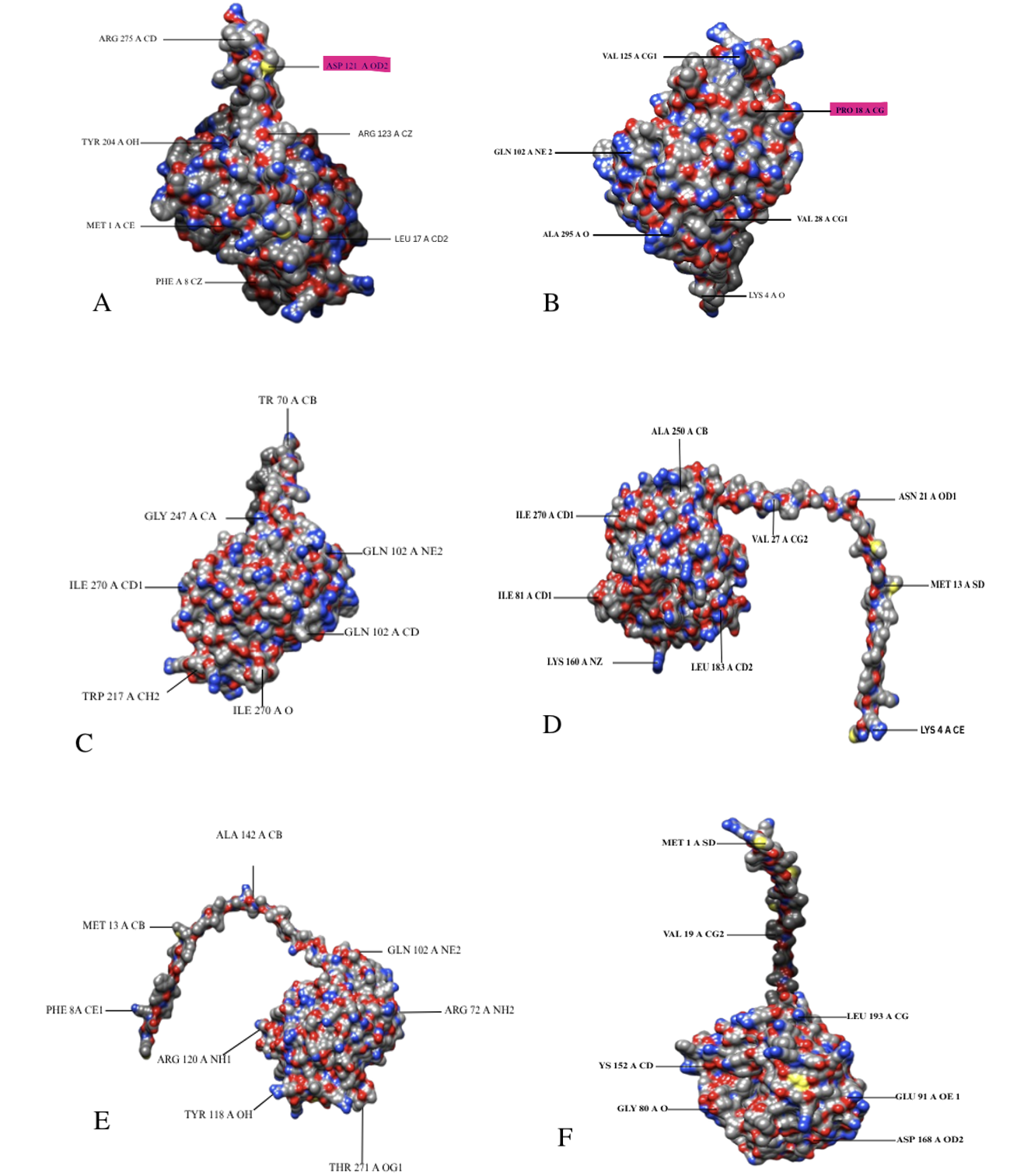
3D structural model of the PLD protein (A-F) from Corynebacterium pseudotuberculosis, highlighting residues of interest. Point mutations involving ASP 121 A OD2 and PRO 18 A CG residues are indicated, suggesting potential sites of structural and antigenic variation among strains.
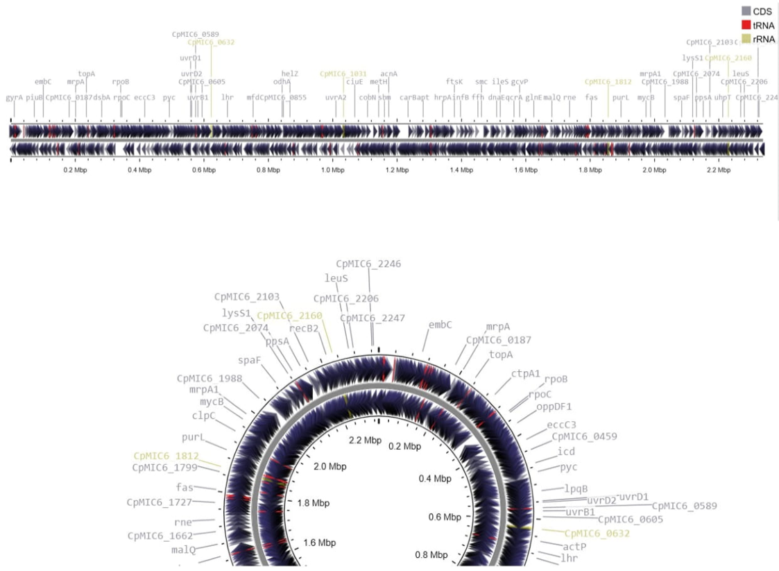
Gene map of the reference strain C. pseudotuberculosis MIC6 (CpMIC6). The circular representation highlights coding sequences (CDSs), tRNAs, and rRNAs distributed across the genome, with notable genes and loci annotated. The inner track indicates GC content and GC skew, providing insights into genomic organization and structural features.
References
- Al-Ethafa, L.F., Almialy, A.J., Gharban, H.A., Essa, I.a.M. and Al-Eqabi, S.R., 2025. First molecular phylogenetic and serological insights into Listeria monocytogenes infection in aborted ewes in Iraq: A cross-border comparative analysis. Vet. World, 18: 1899. https://doi.org/10.14202/vetworld.2025.1899-1910[CrossRef]
- Ali, M.M., Gul, M., Imran, M., Ijaz, M., Azeem, S., Ullah, A. and Yaqub, H.M.F., 2024. Molecular identification and genotyping of hepatitis E virus from Southern Punjab, Pakistan. Sci. Rep., 14: 223. https://doi.org/10.1038/s41598-023-50514-5.[CrossRef]
- Bakr, M.B., Hashad, M.E.-S., Gomaa, I.G. and Attia, S.A., 2025. Antimicrobial susceptibility and molecular characterization of Corynebacterium pseudotuberculosis isolates recovered from caseous lymphadenitis in sheep. Egypt J. Vet. Sci., 56: 711-723. https://doi.org/10.21608/ejvs.2025.415210.3059[CrossRef]
- Bergey, D.H., 1994. Bergey’s manual of determinative bacteriology, Lippincott Williams and Wilkins.
- Carella, A., Carroll, K.C. and Munson, E., 2025. An update on novel taxa and revised taxonomic status of bacteria isolated from human clinical specimens described in 2024. J. clin. Microbiol., e01068-01025. https://doi.org/10.1128/jcm.01068-25[CrossRef]
- de Pinho, R.B., de Oliveira Silva, M.T., Bezerra, F.S.B. and Borsuk, S., 2021. Vaccines for caseous lymphadenitis: Up-to-date and forward-looking strategies. Appl. Microbiol. Biotechnol., 105: 2287-2296. https://doi.org/10.1007/s00253-021-11191-4[CrossRef]
- Dopuđ, M., Reil, I., Zdelar-Tuk, M., Špičić, S. and Duvnjak, S., 2025. Caseous lymphadenitis in sheep and goats, cheese glands. Vet. Stn., 56: 303-316. https://doi.org/10.46419/vs.56.3.8[CrossRef]
- El-Khalfaoui, N., El-Amiri, B., Cabaraux, J.F., Chentouf, M., Raes, M., Marcotty, T. and Kirschvink, N., 2024. Rearing management and its impact on Caseous lymphadenitis in sheep. Animals, 14: 1504. https://doi.org/10.3390/ani14101504[CrossRef]
- El-Sebay, N.A., Abd Elmonem, M.M., El-Sergany, E.F., Abbas, M.A., Osman, M.R. and Abd El-Moaty, D.A., 2021. Genetic characterization of Corynebacterium pseudotuberculosis isolates in Egypt. J. appl. Vet. Sci., 6: 15-21. https://doi.org/10.21608/javs.2021.154576[CrossRef]
- Farhan, H.E., Yousseff, F.M. and Helal, M.E., 2023. Bacteriological and clinicopathological studies of Corynebacterium pseudotuberculosis isolated from caseous lymphadenitis in sheep. J. Adv. Vet. Res., 13: 584-593.
- Fernandes, M.d.C.R., Neto, A.A.M., Lemos, E.I.B., Silva, J.I.A., da Costa Soares, M.E., Silva, D.F., da Silveira Pereira, J.C., Feij, F.M.C., Bezerra, A.C.D.S. and Borsuk, S., 2023. Occurrence of caseous lymphadenitis in goats from a slaughterhouse in the Brazilian Northeast. Acta Vet. Bras., 17. https://doi.org/10.21708/avb.2023.17.4.11899[CrossRef]
- Fukuda, A., Kodama, Y., Mashima, J., Fujisawa, T. and Ogasawara, O., 2021. DDBJ update: Streamlining submission and access of human data. Nucleic Acids Res., 49: D71-D75. https://doi.org/10.1093/nar/gkaa982[CrossRef]
- Galià-Camps, C., Pegueroles, C., Turon, X., Carreras, C. and Pascual, M., 2024. Genome composition and GC content influence loci distribution in reduced representation genomic studies. BMC Genom., 25: 410. https://doi.org/10.1186/s12864-024-10312-3[CrossRef]
- Hiller, E., Hörz, V. and Sting, R., 2024. Corynebacterium pseudotuberculosis: Whole genome sequencing reveals unforeseen and relevant genetic diversity in this pathogen. PLoS One, 19: e0309282. https://doi.org/10.1371/journal.pone.0309282[CrossRef]
- Javanbakht, H. and Hajiyan, R., 2024. Molecular characterization of haemoparasites genus Lankestrella (Apicomplexa: Coccidia) in two species of lizards from Iran. J. Wildl. Biodiv., 8: 192-202.
- Khanamir, R.A., Issa, N.A. and Abdulrahman, R.F., 2023. First study on molecular epidemiology of caseous lymphadenitis in slaughtered sheep and goats in Duhok Province, Iraq. Open Vet. J., 13: 588–598. https://doi.org/10.5455/OVJ.2023.v13.i5.11[CrossRef]
- Li, S.J. and Wu, Q., 2022. Endobronchial ultrasound-guided transbronchial needle aspiration in intrathoracic lymphadenopathy with extrathoracic malignancy. World J. clin. Cases, 10: 13227. https://doi.org/10.12998/wjcc.v10.i36.13227[CrossRef]
- Liu, Y., Wang, Y., Wang, Y., Mai, H., Chen, Y., Zhang, Y., Ji, Y., Cong, X. and Gao, Y., 2023. Phylogenetic analysis of HA and NA genes of influenza A viruses in immunosuppressed inpatients in Beijing during the 2018–2020 influenza seasons. Virol. J., 20: 101. https://doi.org/10.1186/s12985-023-02067-2.[CrossRef]
- Mustaffa, N.D.A.N., Hian, C.M., Al-Rashdan, Y., Al-Jaff, K.M., Ghazali, M.F. and Ibrahim, N.S., 2025. Comprehensive review on prevalence of caseous lymphadenitis (CLA) in dairy goats: A systematic review and meta-analysis. Vet. Integr. Sci., 23: 1-18 https://doi.org/10.12982/VIS.2025.074.[CrossRef]
- Rodrigues, C.H., Pires, D.E. and Ascher, D.B., 2021. DynaMut2: Assessing changes in stability and flexibility upon single and multiple point missense mutations. Protein Sci., 30: 60-69. https://doi.org/10.1002/pro.3942[CrossRef]
- Selim, A.M., Atwa, S.M., El-Gedawy, A.A., Hegazy, Y.M., Rizk, M.A. and Younis, E.E., 2021. Risk factors associated with the seroprevalence of caseous lymphadenitis in sheep. Comp. clin. Pathol., 30: 285-291. https://doi.org/10.1007/s00580-021-03198-0[CrossRef]
- Selim, A.M., Atwa, S.M., El-Gedawy, A.A. and Younis, E.E., 2022. Epidemiological, bacteriological and molecular studies on caseous lymphadenitis in sheep of Dakhlia, Egypt. Anim. Biotechnol., 33: 1655-1660. https://doi.org/10.1080/10495398.2021.1928683[CrossRef]
- Shi, D., Chen, Y., Chen, M., Zhou, T., Xu, F., Zhang, C., Wang, C. and Li, Z., 2023. Bioinformatics analysis of Omp19 and Omp25 proteins for designing multi-epitope vaccines against Brucella. Medicine, 102: e33182. https://doi.org/10.1097/MD.0000000000033182[CrossRef]
- Terab, A.M.A., Abdel Wahab, G.E.D., Ishag, H.Z.A., Khalil, N.A.H., El Tigani-Asil, E.T.A., Hashem, F.M., Khalafalla, A.I., Shah, A.A.M. and Al-Muhairi, S.S.M., 2021. Pathology, bacteriology and molecular studies on caseous lymphadenitis in Camelus dromedarius in the Emirate of Abu Dhabi, UAE, 2015-2020. PLoS One, 16: e0252893. https://doi.org/10.1371/journal.pone.0252893[CrossRef]
- Zhang, L., Kulyar, M.F., Niu, T., Yang, S. and Chen, W., 2024. Comparative genomics of Limosilactobacillus reuteri YLR001 reveals genetic diversity and probiotic properties. Microbes., 12: 1636. https://doi.org/10.3390/microorganisms12081636[CrossRef]
- Zhou, F., He, S., Sun, H., Wang, Y. and Zhang, Y., 2021. Advances in epitope mapping technologies for food protein allergens: A review. Trends Fd. Sci. Technol., 107: 226-239. https://doi.org/10.1016/j.tifs.2020.10.035[CrossRef]
Supplementary material
Services
Article usage statistics
Loading…