The percentage of tumor samples submitted from male dogs was higher than that of female dogs (52.67% vs 32.14%).
The second most common tumor type in the present study was cutaneous papillomatosis.
In conclusion, the current study demonstrated the epidemiology of canine cutaneous tumors in Egypt.
Basal cell carcinoma, papilloma, infundibular keratinizing acanthoma, liposarcoma, and fibrosarcoma were the most common tumor types.
Selected from the article's own text.
Abstract
Bardes B. Hassan1, Asmaa K. Al-Mokaddem1, Hisham A. Abdelrahman2, Ahmed Samir3, Mohamed R. Mousa1*
1Department of Pathology, Faculty of Veterinary Medicine, Cairo University, Giza, Egypt, Postal Code: 12211; 2Department of Veterinary Hygiene and Management, Faculty of Veterinary Medicine, Cairo University, Giza, Egypt, Postal Code: 12211; 3Department of Microbiology, Faculty of Veterinary Medicine, Cairo University, Giza, Egypt, Postal Code: 12211.
Abstract | Background: In the present study, a total of 112 dogs with cutaneous tumors were examined between November 2018 and November 2020. Out of 112 dogs, 118 skin tumor samples were obtained, of which 68 (57.63%) cases were malignant, and 50 (42.37%) cases were benign. Objectives: The objectives were to identify the most common histologic types of canine cutaneous tumors in Egypt, report the relative frequency of each tumor type, and elucidate the association of risk factors (age, sex, breed, and tumor anatomical site) with the development of common cutaneous tumor types. Methods: The samples were collected from different private veterinary practices. All samples were processed and diagnosed histopathologically in the Department of Pathology, Faculty of Veterinary Medicine, Cairo University, Egypt. Immunohistochemistry was used to confirm the diagnosed tumor types. Multinomial logistic regression models were used to study associations of the development of benign or malignant cutaneous tumors with risk factors. Results: Thirty different canine cutaneous tumors were diagnosed. Basal cell carcinoma (8.47%), papilloma (7.63%), infundibular keratinizing acanthoma (6.78%), liposarcoma (6.78%), and fibrosarcoma (5.93%) were the most common tumors. Tumors were commonly found on the trunk (25.42%), head (17.79%), and extremities (17.79%). The most affected breed was Golden Retriever (11.61%). The occurrence was predominant in males (53.57%) as compared to females (33.03%). Conclusion: The findings from this study are important for small animal clinical practices as they will serve as a useful reference to establish a preliminary diagnosis of cutaneous tumors in dogs as rapidly and precisely as possible.
Cutaneous tumors commonly affect dogs as the skin is significantly damaged by physical and chemical factors contributing to the development of canine cutaneous tumors . Moreover, the skin has a large number of cells that are continuously renewed, rendering the skin more vulnerable to gene disorders with subsequent mutations . Approximately 30% of all canine neoplasms arise in the skin ;. The incidence of canine cutaneous tumors is estimated to be 728 cases per 100,000 dogs per year . Similar histological features of some cutaneous tumors result in confusing pathological diagnoses . An accurate diagnosis of the origin and type of neoplastic cells is important for identification, early treatment, and predicting the outcomes of many skin tumors ; . Immunohistochemistry (IHC) is an important ancillary technique to confirm the diagnosis of tumors and determine the differentiation of neoplastic cells ; . The most specific and useful markers for characterization of canine cutaneous tumors are antibodies against cytokeratin and vimentin proteins ; . Both cytokeratin and vimentin markers are used for distinguishing epithelial from mesenchymal differentiation .
Several retrospective studies have been performed to study the epidemiology of cutaneous tumors in dogs ; ; . These studies differ considerably concerning the data collection and source, the sample numbers, study population, the size of the geographic area evaluated, and the quality of the result analysis ; . Some studies calculated either the relative tumor incidence or odds ratio (OR) based on the total patient data in the sample from the national canine cancer registries and veterinary authorities ; ; . Others reported tumor incidence rates based on information utilized from insurance data , diagnostic laboratories, and local animal registration data . Although studies of canine cutaneous tumors from different geographic regions show similarities, there have also been reports of variations in tumor types and relative incidence. In other words, both environmental influences and dog breeds play a role in developing certain types of cutaneous tumors in relevant locations . For example, ultraviolet sunlight radiation has been involved as a risk factor for canine hemangioma, hemangiosarcoma, and squamous cell carcinoma ; .
It is worth noting that to date, no studies on canine skin tumor frequencies have been conducted in Cairo or Giza in Egypt. We hypothesized that the types and frequencies of canine skin tumors in Egypt are different from those reported elsewhere. In this study, we present an inclusive study of 118 canine skin tumors, collected from different private small animal clinical practices between November 2018 and November 2020. The objectives of this study were to characterize the most common histologic types of canine cutaneous tumors in Egypt, report the relative frequency of each tumor type, and investigate the relationship between such frequencies and the age, sex, breed, and tumor anatomical site of commonly diagnosed tumor types.
Materials and Methods
Study Population
The population used for this study included tissue biopsy and surgical excision cases recorded from 112 dogs between November 2018 and November 2020. The skin tumor samples were provided by various private veterinary practices in Cairo and Giza governorates in Egypt and were submitted to Leptovet laboratory (a private veterinary diagnostic laboratory) for further processing. The histopathological examination of skin tumor samples was conducted in the Department of Pathology, Faculty of Veterinary Medicine, Cairo University, Egypt. Clinical data including age, sex, breed, and tumor anatomic site were recorded and their influence on the frequency of skin tumor development was statistically determined. Crossbreed dogs were classed as mixed breed. All cases of skin tumors were included in the present study, even if any of the clinical data was missing. Cases with unidentified parameters (e.g. breed, age…) were listed as ‘not reported’. Cases with a diagnosis of hyperplastic lesions and epithelial cysts were excluded. Perianal (hepatoid) gland tumors, hamartomas, sebaceous and apocrine tumors were included. A tumor of the same type that occurs at more than one anatomical site simultaneously was considered as a single tumor event. Different tumor types that simultaneously occur and tumor recurrence in one patient were considered as multiple separate events ; . Tumors recorded on head, ear, eyelid, face, and nostril were considered as ‘head’. Tumors recorded on trunk, chest, flank, pelvis, back, abdomen, mammary gland, and perianal were considered as ‘trunk’. Tumors recorded on vagina, scrotum, and penis were considered as genitalia. Tumors were recorded on forelimb, hindlimb, digit, foot, pad, and paw were considered as ‘extremities’. Anatomical sites were listed as head, neck, trunk, extremities, and genitalia.
Tumor Diagnosis and Classification
Tumor cases were diagnosed by two independent veterinary pathologists. The samples were fixed in 10% neutral buffered formalin, processed, sectioned at 4 µm, and stained with hematoxylin and eosin (HE). The histopathological examination was performed under Olympus BX43 microscope, Tokyo, Japan. The images were captured using Olympus DP 27 camera, Tokyo, Japan. Tumor subtypes were grouped into main types of tumors according to the World Health Organization International Histological Classification of Tumors of Domestic Animals . Mast cell tumors, transmissible venereal tumors, and peripheral nerve sheath tumors were considered as malignant tumors. The frequencies of benign and malignant tumors were recorded. Mast cell tumors were stained with a special stain (toluidine blue).
Table 1: Immunohistochemical antibody panel applied to some cases of the canine skin tumors.
Antibody
Type
(catalog number)
Source
Dilution
Antigen retrieval
Positive expression
CD3
Rabbit polyclonal antibody (103A-74)
Merck KGaA, Darmstadt, Germany
1:200
Antigen retrieval was done in the pre-treatment (PT) module, PT Link (Dako, Denmark). Briefly, tissue sections were subjected to 95 °C for 40 min. in Epitope Retrieval Solution (Dako) in accordance with manufacturer’s instruction for HercepTest (Dako).
Lymphoma
Glial fibrillary acidic protein (GFAP)
Rabbit polyclonal antibody (PA1-10019)
Thermo Fisher Scientific, Freemont, CA, USA
1:1000
Peripheral nerve sheath tumor
Vimentin (V9)
Mouse monoclonal antibody (347M-14)
Merck KGaA, Darmstadt, Germany
1:200
Soft tissue sarcoma
Vascular endothelial growth factor (VEGF)
Mouse monoclonal antibody (sc-7269)
Santa Cruz Biotechnology, Inc.
1:100
Hemangiosarcoma
Cytokeratin 14 (CK14)
Rabbit monoclonal [SP53] (ab119695)
Abcam, Cambridge, UK
1:100
Adenoma and
adenocarcinoma
Table 2: Distributions of the 112 dogs with cutaneous tumors recorded in Egypt between 2018 and 2020 according to number of tumors per dog, examination year, and dog breed, and the respective age (minimum [min], maximum [max], mean, and standard error [SE]) and sex (numbers and ratio).
N
%
Age (years)
Sex
NRa
mean ± SE
min – max
NRa
Male
Female
M:Fb
All dogs
112
100
13
6.85 ± 0.31
0.5 – 17.0
15
60
37
1.6:1
Dogs with one tumor
106
94.64
94
6.95 ± 0.33
0.5 – 17.0
14
56
36
1.6:1
Dogs with two tumors
6
5.36
5
5.00 ± 0.55
4.0 – 7.0
1
4
1
4:1
By year
2018
4
3.57
2
9.00 ± 1.00
8.0 – 10.0
2
1
1
1:1
2019
30
26.79
9
6.51 ± 0.65
0.8 – 11.0
7
12
11
1.1:1
2020
78
69.64
2
6.89 ± 0.37
0.5 – 17.0
6
47
25
1.9:1
By breed
Not reported
30
26.79
11
6.30 ± 0.68
0.8 – 11.0
6
11
13
0.8:1
Golden Retriever
13
11.61
0
7.85 ± 0.80
4.0 – 12.0
1
8
4
2:1
German Shepherd
12
10.71
0
5.18 ± 0.80
0.5 – 9.0
1
7
4
1.8:1
Labrador Retriever
8
7.14
0
6.88 ± 0.79
3.0 – 11.0
1
5
2
2.5:1
Mixed Breed
7
6.25
0
5.86 ± 0.86
3.0 – 10.0
0
6
1
6:1
Rottweiler
7
6.25
0
7.00 ± 0.58
5.0 – 9.0
0
6
1
6:1
Baladi
5
4.46
1
4.92 ± 1.70
0.7 – 9.0
1
2
2
1:1
Griffon
5
4.46
1
4.75 ± 1.70
2.0 – 9.0
1
2
2
1:1
Pit Bull
3
2.68
0
8.33 ± 0.33
8.0 – 9.0
0
2
1
2:1
Cane Corso
2
1.79
0
3.00 ± 1.00
2.0 – 4.0
0
1
1
1:1
Cocker Spaniel
2
1.79
0
13.00 ± 2.00
11.0 – 15.0
1
1
0
1:0
Dachshund
2
1.79
0
12.50 ± 0.50
12.0 – 13.0
0
0
2
0:2
Doberman
2
1.79
0
7.00 ± 0.00
7.0 – 7.0
0
1
1
1:1
Pug
2
1.79
0
12.50 ± 4.50
8.0 – 17.0
1
1
0
1:0
Siberian Husky
2
1.79
0
7.50 ± 0.50
7.0 – 8.0
0
2
0
2:0
Alaskan Malamute
1
0.89
0
5.00 ± 0.00
5.0 – 5.0
0
0
1
0:1
American Bully
1
0.89
0
8.00 ± 0.00
8.0 – 8.0
0
0
1
0:1
Beagle
1
0.89
0
4.00 ± 0.00
4.0 – 4.0
0
1
0
1:0
Dalmatian
1
0.89
0
9.00 ± 0.00
9.0 – 9.0
0
1
0
1:0
French Bulldog
1
0.89
0
2.00 ± 0.00
2.0 – 2.0
1
0
0
-
Great Dane
1
0.89
0
8.00 ± 0.00
8.0 – 8.0
1
0
0
-
Italian Grey Hound
1
0.89
0
9.00 ± 0.00
9.0 – 9.0
0
0
1
0:1
Pekingese
1
0.89
0
11.00 ± 0.00
11.0 – 11.0
0
1
0
1:0
Ridgeback
1
0.89
0
8.00 ± 0.00
8.0 – 8.0
0
1
0
1:0
Schnoodle
1
0.89
0
11.00 ± 0.00
11.0 – 11.0
0
1
0
1:0
a NR: not reported; b M: Male; F: Female
Table 3: Tumor histopathological diagnoses, relative frequency and the respective distribution of anatomical site of the 118 canine cutaneous tumors recorded in Egypt between 2018 and 2020.
Tumor Type
N
%
Anatomical Site
NRa
Trunk
Extremities
Head
Genitalia
Neck
All tumors
118
100
36
30
21
21
6
4
Benign Tumors
50
42.37
22
10
7
8
0
3
Malignant Tumors
68
57.63
14
20
14
13
6
1
Epithelial Tumors
56
47.46
17
15
4
17
0
3
Epidermal Tumors
24
20.34
8
3
3
10
0
0
Papilloma
9
7.63
5
0
1
3
0
0
Squamous Cell Carcinoma
5
4.24
1
1
2
1
0
0
Basal Cell Carcinoma
10
8.47
2
2
0
6
0
0
Hair Follicle Tumors
14
11.86
8
3
1
0
0
2
Benign Trichoepithelioma
4
3.39
2
0
1
0
0
1
Malignant Trichoepithelioma
2
1.69
2
0
0
0
0
0
Infundibular Keratinizing Acanthoma
8
6.78
4
3
0
0
0
1
Sebaceous Gland Tumors
15
12.71
1
9
0
5
0
0
Sebaceous Gland Adenoma
1
0.85
1
0
0
0
0
0
Sebaceous Gland Epithelioma
4
3.39
0
1
0
3
0
0
Hepatoid Gland Adenoma
3
2.54
0
3
0
0
0
0
Hepatoid Gland Adenocarcinoma
5
4.24
0
5
0
0
0
0
Meibomian Adenocarcinoma
2
1.69
0
0
0
2
0
0
Apocrine Glands Tumors
3
2.54
0
0
0
2
0
1
Apocrine Gland Adenoma
1
0.85
0
0
0
0
0
1
Ceruminous Gland Adenoma
1
0.85
0
0
0
1
0
0
Ceruminous Gland Adenocarcinoma
1
0.85
0
0
0
1
0
0
Mesenchymal Tumors
41
34.75
16
13
10
0
1
1
Benign Soft Tissue Tumors
9
7.63
5
3
1
0
0
0
Lipoma
6
5.08
3
3
0
0
0
0
Fibroma
3
2.54
2
0
1
0
0
0
Soft Tissue Sarcomas
21
17.80
6
8
6
0
0
1
Fibrosarcoma
7
5.93
3
2
1
0
0
1
Malignant Peripheral Nerve Sheath Tumor
5
4.24
1
0
4
0
0
0
Liposarcoma
8
6.78
2
6
0
0
0
0
Myxosarcoma
1
0.85
0
0
1
0
0
0
Vascular Tumors
6
5.08
4
0
1
0
1
0
Hemangioma
2
1.69
2
0
0
0
0
0
Hemangiosarcoma
4
3.39
2
0
1
0
1
0
Rhabdomyosarcoma
5
4.24
1
2
2
0
0
0
Round Cell Tumors
9
7.63
1
2
4
2
0
0
Epitheliotropic T-cell Lymphoma
1
0.85
0
0
0
1
0
0
Mast Cell Tumor
4
3.39
0
2
2
0
0
0
Histiocytoma
4
3.39
1
0
2
1
0
0
Melanocytic Tumors
5
4.24
1
0
2
2
0
0
Melanocytoma
2
1.69
1
0
1
0
0
0
Malignant Melanoma
3
2.54
0
0
1
2
0
0
Transmissible Venereal Tumors
5
4.24
0
0
0
0
5
0
Hamartoma
2
1.69
1
0
1
0
0
0
a NR: not reported
Table 4: Tumor histopathological diagnoses of the 118 canine cutaneous tumors recorded in Egypt between 2018 and 2020; and the respective age (minimum [min], maximum [max], mean, and standard error [SE]) and sex (numbers and ratio).
Tumor Type
N
Age (years)
Sex
NRa
mean ± SE
min – max
NRa
Male
Female
M:Fb
All tumors
118
14
6.77 ± 0.30
0.5 – 17.0
16
64
38
1.7:1
Benign Tumors
50
6
6.06 ± 0.45
0.5 – 12.0
7
27
16
1.7:1
Malignant Tumors
68
8
7.28 ± 0.40
0.8 – 17.0
9
37
22
1.7:1
Epithelial Tumors
56
9
6.73 ± 0.47
0.7 – 17.0
10
31
15
2.1:1
Epidermal Tumors
24
4
5.97 ± 0.66
0.7 – 12.0
4
16
4
4:1
Papilloma
9
1
3.67 ± 0.67
0.7 – 5.0
1
7
1
7:1
Squamous Cell Carcinoma
5
1
8.75 ± 1.11
7.0 – 12.0
0
3
2
1.5:1
Basal Cell Carcinoma
10
2
6.88 ± 0.91
4.0 – 11.0
3
6
1
6:1
Hair Follicle Tumors
14
1
5.23 ± 0.70
1.0 – 11.0
1
5
8
1:1.6
Benign Trichoepithelioma
4
0
6.75 ± 1.44
5.0 – 11.0
0
1
3
1:3
Malignant Trichoepithelioma
2
0
6.50 ± 0.50
6.0 – 7.0
0
0
2
0:2
Infundibular Keratinizing Acanthoma
8
1
4.00 ± 0.82
1.0 – 7.0
1
4
3
1.3:1
Sebaceous Gland Tumors
15
3
9.67 ± 0.79
6.0 – 17.0
3
10
2
5:1
Sebaceous Gland Adenoma
1
0
8.00 ± 0.00
8.0 – 8.0
1
0
0
-
Sebaceous Gland Epithelioma
4
1
9.67 ± 1.20
8.0 – 12.0
1
3
0
3:0
Hepatoid Gland Adenoma
3
0
9.00 ± 0.58
8.0 – 10.0
0
3
0
3:0
Hepatoid Gland Adenocarcinoma
5
2
11.00 ± 3.21
6.0 – 17.0
1
3
1
3:1
Meibomian Adenocarcinoma
2
0
9.50 ± 0.50
9.0 – 10.0
0
1
1
1:1
Apocrine Glands Tumors
3
1
6.50 ± 0.50
6.0 – 7.0
2
0
1
0:1
Apocrine Gland Adenoma
1
0
7.00 ± 0.00
7.0 – 7.0
0
0
1
0:1
Ceruminous Gland Adenoma
1
1
-
-
1
0
0
-
Ceruminous Gland Adenocarcinoma
1
0
6.00 ± 0.00
6.0 – 6.0
1
0
0
-
Mesenchymal Tumors
41
3
7.16 ± 0.48
0.5 – 15.0
6
20
15
1.3:1
Benign Soft Tissue Tumors
9
2
7.21 ± 1.13
0.5 – 9.0
2
4
3
1.3:1
Lipoma
6
1
6.70 ± 1.56
0.5 – 9.0
2
1
3
1:3
Fibroma
3
1
8.50 ± 0.50
8.0 – 9.0
0
3
0
3:0
Soft Tissue Sarcomas
21
1
7.34 ± 0.73
0.8 – 15.0
3
10
8
1.3:1
Fibrosarcoma
7
1
7.17 ± 1.33
2.0 – 12.0
2
2
3
1:1.5
Malignant Peripheral Nerve Sheath Tumor
5
0
10.2 ± 1.53
6.0 – 15.0
1
3
1
3:1
Liposarcoma
8
0
5.97 ± 0.87
0.8 – 9.0
0
5
3
1.7:1
Myxosarcoma
1
0
5.00 ± 0.00
5.0 – 5.0
0
0
1
0:1
Vascular Tumors
6
0
8.00 ± 0.97
4.0 – 11.0
1
2
3
1:1.5
Hemangioma
2
0
8.00 ± 1.00
7.0 – 9.0
0
0
2
0:2
Hemangiosarcoma
4
0
8.00 ± 1.47
4.0 – 11.0
1
2
1
2:1
Rhabdomyosarcoma
5
0
5.40 ± 0.87
2.0 – 7.0
0
4
1
4:1
Round Cell Tumors
9
1
4.88 ± 1.01
1.0 – 9.0
0
5
4
1.3:1
Epitheliotropic T-cell Lymphoma
1
0
6.00 ± 0.00
6.0 – 6.0
0
1
0
1:0
Mast Cell Tumor
4
1
6.00 ± 2.08
2.0 – 9.0
0
2
2
1:1
Histiocytoma
4
0
3.75 ± 1.31
1.0 – 6.0
0
2
2
1:1
Melanocytic Tumors
5
0
9.00 ± 1.41
5.0 – 13.0
0
2
3
1:1.5
Melanocytoma
2
0
8.00 ± 3.00
5.0 – 11.0
0
1
1
1:1
Malignant Melanoma
3
0
9.67 ± 1.76
7.0 – 13.0
0
1
2
1:2
Transmissible Venereal Tumors
5
1
4.25 ± 0.63
3.0 – 6.0
0
4
1
4:1
Hamartoma
2
0
7.00 ± 0.00
7.0 – 7.0
0
2
0
2:0
a NR: not reported; b M: Male; F: Female
Table 5: Odds ratio (OR), 95% confidence intervals (95% CI) for OR, and significance statistics of the multinomial logistic regression models evaluating the association of benign and malignant cutaneous tumor development with dog breed. Odds ratio was statistically significant if the null value (OR = 1.0) is not contained within the 95% CI. Regression models were statistically significant at P < 0.05 if (bold*).
Dog Breed
Odds Ratio
Significance
Benign Tumors
Malignant Tumors
2(1)
P-value
Estimate
95% CI
Estimate
95% CI
lower
upper
lower
upper
Baladi
1
-
-
1
-
-
-
-
Beagle
>999.999
<0.001
>999.999
<0.001
<0.001
>999.999
0.0006
0.9798
Cane Corso
1.00
0.063
15.988
1.00
0.063
15.988
<0.0001
>0.999
Cocker Spaniel
<0.001
<0.001
>999.999
>999.999
<0.001
>999.999
0.0003
0.9857
Dachshund
<0.001
<0.001
>999.999
>999.999
<0.001
>999.999
0.0003
0.9857
Dalmatian
>999.999
<0.001
>999.999
<0.001
<0.001
>999.999
0.0003
0.9857
Doberman
1.00
0.063
15.988
1.00
0.063
15.988
<0.0001
>0.999
German Shepherd
0.33
0.067
1.652
3.00
0.606
14.864
1.8104
0.1785
Golden Retriever
0.25
0.053
1.177
4.00
0.849
18.836
3.0749
0.0795
Griffon
<0.001
<0.001
>999.999
>999.999
<0.001
>999.999
0.0010
0.9752
Italian Grey Hound
<0.001
<0.001
>999.999
>999.999
<0.001
>999.999
0.0003
0.9857
Labrador Retriever
0.20
0.023
1.712
5.00
0.584
42.797
2.1586
0.1418
Mixed Breed
1.50
0.251
8.977
0.667
0.111
3.99
0.1973
0.6569
Pekingese
<0.001
<0.001
>999.999
>999.999
<0.001
>999.999
0.0003
0.9857
Pit Bull
>999.999
<0.001
>999.999
<0.001
<0.001
>999.999
0.0003
0.9857
Pug
<0.001
<0.001
>999.999
>999.999
<0.001
>999.999
0.0003
0.9857
Ridgeback
>999.999
<0.001
>999.999
<0.001
<0.001
>999.999
0.0003
0.9857
Rottweiler
1.00
0.141
7.099
1.00
0.141
7.099
<0.0001
>0.999
Siberian Husky
<0.001
<0.001
>999.999
>999.999
<0.001
>999.999
0.0006
0.9798
Table 6: Odds ratio (OR), 95% confidence intervals (95% CI) for OR, and significance statistics of the multinomial logistic regression models evaluating the association of benign and malignant cutaneous tumor development with dog sex, dog age, and anatomical site of tumor. Odds ratio was statistically significant if the null value (OR = 1.0) is not contained within the 95% CI. Regression models were statistically significant at P < 0.05 if (bold*).
Variable
Odds Ratio
Significance
Benign Tumors
Malignant Tumors
P-value
Estimate
95% CI
Estimate
95% CI
lower
upper
lower
upper
Sex
Male
1
-
-
1
-
-
-
-
Female
0.545
0.202
1.475
1.833
0.678
4.957
1.4262
0.2324
Site
Head
1
-
-
1
Extremities
0.308
0.10
0.944
3.25
1.06
9.967
4.2494
0.0393*
Genitalia
<0.001
<0.001
>999.999
>999.999
<0.001
>999.999
0.0013
0.9714
Neck
>999.999
<0.001
>999.999
<0.001
<0.001
>999.999
0.0006
0.9798
Trunk
0.636
0.247
1.642
1.571
0.609
4.054
0.8739
0.3499
Age (years)
≤ 1
1
-
-
1
-
-
-
-
2
0.50
0.045
5.514
2.00
0.181
22.056
0.3203
0.5714
3
1.00
0.063
15.988
1.00
0.063
15.988
<0.0001
>0.999
4
1.00
0.202
4.955
1.00
0.202
4.955
<0.0001
>0.999
5
0.17
0.02
1.384
6.00
0.722
49.837
2.7518
0.0971
6
0.17
0.02
1.384
6.00
0.722
49.837
2.7518
0.0971
7
0.17
0.02
1.384
6.00
0.722
49.837
2.7518
0.0971
8
2.00
0.366
10.919
0.50
0.092
2.73
0.6406
0.4235
9
0.75
0.168
3.351
1.33
0.298
5.957
0.1419
0.7064
10
0.50
0.045
5.514
2.00
0.181
22.056
0.3203
0.5714
11
0.50
0.045
5.514
2.00
0.181
22.056
0.3203
0.5714
12
1.00
0.063
15.988
1.00
0.063
15.988
<0.0001
>0.999
13
<0.001
<0.001
>999.999
>999.999
<0.001
>999.999
0.0003
0.9857
17
<0.001
<0.001
>999.999
>999.999
<0.001
>999.999
0.0003
0.9857
Immunohistochemistry (IHC)
Immunohistocemistry was performed to confirm the histopathological diagnosis of specific tumor types. Immunohistochemical antibodies used in tumor diagnosis are summarized in Table 1.
Statistical Analysis
The relative frequency (percentage of all cutaneous tumor types) and the OR with a 95% confidence interval (95% CI) were calculated. To assess the association of dog sex, age, breed, and tumor anatomical site with the development of benign and malignant cutaneous tumors, two multinomial logistic regression models, integrated with likelihood ratio test and Pearson’s chi-square (x2) test, were performed. The dependent variable in first model was defined as benign cutaneous tumor development (either yes or no) while in second model was defined as malignant cutaneous tumor development (either yes or no). In both models, the independent variables were defined as sex (reference = male), age (reference = less than or equal to 1 year), breed (reference = Baladi), and anatomical site of tumor (reference = head). Data management and statistical analyses were performed using SAS® version 9.4 (SAS, 2013). Statistical significance was set at P < 0.05, and all data were presented as the mean ± standard error of the mean (SE).
Results
Canine Population in the Database
From November 2018 to November 2020, tissues from 112 dogs were collected by biopsy and surgical excision. Out of this database, 118 samples from 112 dogs were diagnosed with a skin tumor. Five dogs had tumor recurrence and one dog had two different tumors in two different sites.
Breed Distribution
The dog breed was not reported in 30 dogs (26.79%). The remaining dog cases (n = 82 dogs) included 24 different dog breeds. The five most common dog breeds were Golden Retriever (n = 13; 11.61%), German Shepherd (n = 12; 10.71%), Labrador Retriever (n = 8; 7.14%), Mixed breed (n = 7; 6.25%), and Rottweiler (n = 7; 6.25%).
Sex Distribution
The sex of the dog was not reported for 15 dogs (13.39%). Most tumors samples were submitted from males (n = 60; 53.57%) as compared to females (n = 37; 33.03%).
Age Distribution
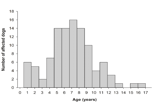
Dog age was not reported in 13 cases (11.60%). The reported age of dog cases in this study ranged from 6 months to 17 years (mean = 6.85 years; SE = 0.31). More than half of the dogs were between 5 and 9 years of age (n = 65; 58.03%). Age distribution breakdown is summarized in Figure 1.
Tumor histopathological diagnoses, relative frequency, and the respective distribution of dog breed, age, sex, and tumor anatomical site of the 118 canine cutaneous tumors diagnosed during the 2-year study period are shown in Tables 2-4. Of 118 cutaneous tumors, 68 (57.63%) were malignant and 50 (42.37%) were benign. The five most frequently diagnosed tumors were basal cell carcinoma (n = 10; 8.47%), papilloma (n = 9; 7.63%), infundibular keratinizing acanthoma (n = 8; 6.78%), liposarcoma (n= 8; 6.78%) and fibrosarcoma (n = 7; 5.93%). These tumors comprised 35.59% of the total cutaneous tumor cases. Of the 118 tumor samples 21 (17.79%) were localized on the head, 4 (3.38%) on the neck, 30 (25.42%) on the trunk, 6 (5.08%) on the genital organs, and 21 (17.79%) on the extremities. For 36 tumors (30.50%), the location on the skin was not reported.
Multinomial Logistic Regression Analysis
The results from the multiple logistic regression analyses are shown in Tables 5 and 6. There was no association between dog breed and cutaneous tumor development (x2(18) = 23.47, P = 0.1733; Table 5). Cutaneous tumor development was not associated with dog sex (x2(1) = 1.49, P = 0.2218) nor dog age (x2(13) = 16.50, P = 0.2231). However, it was associated with tumor anatomical site (x2(4) = 714.23, P = 0.0066; Table 6). Malignant cutaneous tumors in dog extremities had a significantly increased odds ratio compared to malignant cutaneous tumors in dog head (x2(1) = 4.25, P = 0.0393; Table 6).
Histopathological and Immunohistochemical Diagnoses of Tumor Samples
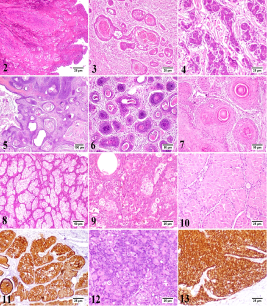
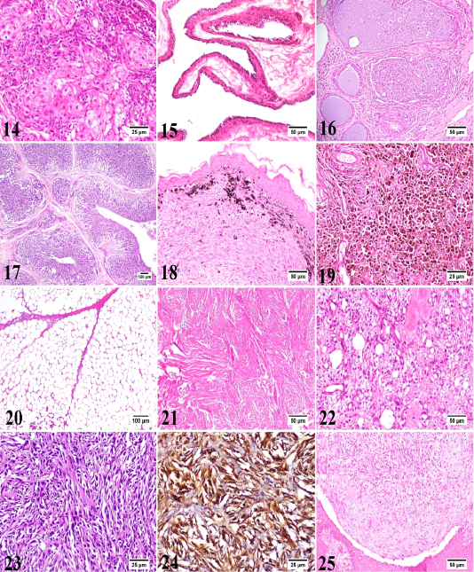
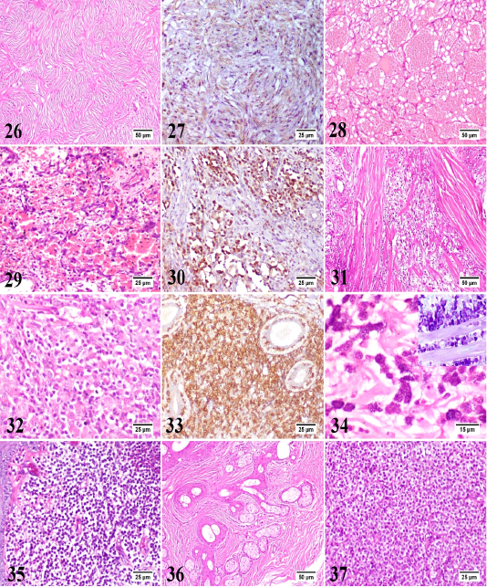
In the present study, 30 different tumor types have been diagnosed and classified based on their histopathological characteristics. Epidermal tumors consisted of papilloma, squamous cell carcinoma, and basal cell carcinoma (Figs. 2-4). Hair follicle tumors consisted of benign and malignant trichoepithelioma, and infundibular keratinizing acanthoma (Figs. 5-7). Sebaceous gland tumors consisted of sebaceous adenoma, sebaceous epithelioma, hepatoid gland adenoma (positive for cytokeratin [CK14+]), hepatoid gland adenocarcinoma (CK14+), and meibomian adenocarcinoma (Figs. 8-14). Apocrine gland tumors consisted of apocrine gland adenoma, ceruminous gland adenoma, and ceruminous gland adenocarcinoma (Figs. 15-17). Melanocytic tumors consisted of melanocytoma and malignant melanoma (Figs. 18 and 19). Benign soft tissue tumors consisted of lipoma and fibroma (Figs. 20 and 21). Soft tissue sarcomas consisted of liposarcoma, fibrosarcoma (positive for vimentin [V9+]), myxosarcoma, and malignant peripheral nerve sheath tumor (positive for glial fibrillary acidic protein [GFAP+]) (Figs. 22-27). Vascular tumors consisted of hemangioma and hemangiosarcoma (positive for vascular endothelial growth factor [VEGF+]) (Figs. 28-30). Rhabdomyosarcoma represented muscle tumors (Fig. 31). Round cell tumors consisted of lymphoma (positive for CD3+), mast cell tumor (inset: toluidine blue stain), and histiocytoma (Figs. 32-35). Hamartoma and transmissible venereal tumors are presented in Figs. 36 and 37.
The skin is the most common anatomical site for development of neoplasms due to its continuous exposure to risk factors (physical, chemical and environmental [e.g. ultraviolet light]. A wide variety of tumor types can be found in the skin, subcutaneous tissue, and adnexa ; ; . The frequency of various types of skin tumors in a particular geographic region should be recorded to collect information for potential investigations. Awareness of the frequency of various tumor types of skin tumors in a certain population enables clinicians to improve their diagnosis and to evaluate more appropriate treatment plans for their patients , and this is the key reason for performing epidemiological studies of this kind . The current study described the epidemiological trends of canine cutaneous tumors in Egypt and provided extensive information on the breed, age, sex, and tumor anatomical site of 118 canine skin biopsy and surgical excision cases submitted to the laboratory over the past 2 years. However, the relatively limited sample size compared to previous studies ; , this study is useful to elucidate frequently diagnosed tumor types, common dog breeds at risk, and anatomical sites of canine cutaneous tumors in Egypt.
In our study, the percentage of malignant tumors was higher than the percentage of benign tumors (57.63% vs. 42.37%), which is consistent with the previous studies ; ; The percentage of benign tumors may be underestimated, as cases have mostly been obtained from referral veterinary hospitals. Moreover, benign tumors have not been surgically resected or have not been regularly submitted for histological examination. More than 50% of the tumors obtained from dogs between 5 to 9 years of age could confirm an increased risk of developing canine skin tumors at an older age, as previously described ; . The percentage of tumor samples submitted from male dogs was higher than that of female dogs (52.67% vs 32.14%). These findings are consistent with most previous studies ; ; .
There were differences in the frequency of the five most common skin tumor types reported in this study (basal cell carcinoma, papilloma, infundibular keratinizing acanthoma, liposarcoma, and fibrosarcoma) when compared to previous literatures ; ; . This is undoubtedly due to variations in tumor samples, classification systems, and follow-up diagnostic recommendations. Moreover, the discrepancy in tumor frequency may be related to the population of dog breeds in the studied region and to environmental factors, in particular exposure to ultraviolet light, which is responsible for the development of certain skin tumors ;; .
Contrary to a previous study , the present study found that basal cell carcinoma was the most commonly diagnosed skin tumor types. Golden Retriever and German Shepherd were predisposed to basal cell carcinoma in our study, which needs further studies with a larger sample size to validate the hereditary factor in tumor development. In our study, basal cell carcinoma was often found on the head which was consistent with previous reports ; ;. Our findings revealed a higher predisposition for basal cell carcinoma in males compared to females, in contrast to a previous report .
The high incidence of cutaneous papilloma in dogs is well-documented . The second most common tumor type in the present study was cutaneous papillomatosis. In accordance with a previous report , young dogs below the age of 1 up to 5 years were affected. Our findings, in contrast to a previous report , suggest a higher predisposition for papilloma in male dogs compared to females. In our study, Baladi breed was predisposed to papillomatosis, although it has been reported that French Bulldog, Rhodesian Ridgeback, Whippet, Vizsla, and Bull Mastiff are at increased risk of developing cutaneous papillomatosis .
Being the third most common cutaneous neoplasm, infundibular keratinizing acanthoma was often found on the trunk and increased predisposition in mixed breeds. However, infundibular keratinizing acanthoma was predominantly found in Norwegian Elkhound as described in a previous literature . It was previously reported that dogs are the only species affected . No sex predilection was observed. In accordance with the literature , the age range of affected dogs in our study was 1-7 years suggesting that infundibular keratinizing acanthoma occurs in young and middle-aged dogs. Infundibular keratinizing acanthoma should be distinguished from well-differentiated squamous cell carcinoma. Infundibular keratinizing acanthoma develops as solitary or multiple cysts filled with keratin . On the other hand, well-differentiated squamous cell carcinoma occurs as an erosive, plaque-like lesion developed from islands and trabeculae of squamous cells into the dermis . Infundibular keratinizing acanthoma is a benign tumor, thus, microscopic characteristics such as high mitotic index and invasive behavior may also be useful in differentiating infundibular keratinizing acanthoma from well-differentiated squamous cell carcinoma ; ; .
In soft tissue sarcomas, liposarcomas and fibrosarcomas were the fourth and fifth most common encountered malignant neoplasms, respectively, in agreement with previous studies ; . Liposarcomas were most common in dogs and in our study, often found on the trunk. Labrador Retriever was predisposed to liposarcoma and this finding is supported by a previous study . Our findings, in contrast to a previous report , suggest a higher predisposition for liposarcoma in male dogs compared to females. Fibrosarcomas had a wide age range (2-12 years). No breed or tumor anatomic site predispositions were observed in fibrosarcomas which are in contrast to previous reports ; . Fibrosacromas were predisposed in female dogs than males.
In conclusion, the current study demonstrated the epidemiology of canine cutaneous tumors in Egypt. Basal cell carcinoma, papilloma, infundibular keratinizing acanthoma, liposarcoma, and fibrosarcoma were the most common tumor types. Golden Retriever, German Shepherd, Labrador Retriever, Mixed breed, and Rottweiler were predisposed to the development of cutaneous tumors. The results from this study will help veterinarians to plan their therapeutic approaches to improve the quality of life of their patients.
Acknowledgments
The authors offer their sincere appreciation for cases provided by different veterinary clinics in Egypt; The ARK veterinary clinic (Dr. M. Abdelaziz and Dr. Mina Roshdy), Pawsitive veterinary clinic (Dr. Marco Kelleny), Modern veterinary clinic (Dr. Aziz Sharaf Eldin and Dr. Moataz Shaaban), Country club veterinary clinic (Dr. Hatem Elsherbiny), and Dr. Nevein H. Hammad’s veterinary clinic.
Conflict of Interest
None.
authors contribution
Coceptualization, BH, AA, MM; Sample collection, AS; Diagnosis, MM, AA; Methodology, investigation and data curation, BH, AA, MM; Writing the original draft BH, HA; Review and editing, All authors; statistical analysis, HA. All authors read and approved the final manuscript.
References
• Araújo MR, Preis IS, Lavalle GE, Cassali GD, Ecco R (2012). Histomorphological and immunohistochemical characterization of 172 cutaneous round cell tumours in dogs %. J. Pesquisa Vet. Brasil. 32: 772-780. https://doi.org/10.1590/S0100-736X2012000800016[CrossRef]
• Aroni K, Mastoraki A, Kyriazi E, Ioannidis E, Patsouris E (2007). Silver-stained organizer regions and immunoglobulins in cutaneous keratoacanthomas and squamous cell carcinomas. Pathol. Res. Pract. 203: 659-665. https://doi.org/10.1016/j.prp.2007.05.015[CrossRef]
• Bongiovanni L, Romanucci M, Fant P, Lagadic M, Della Salda L (2008). Apoptosis and anti-apoptotic heat shock proteins in canine cutaneous infundibular keratinizing acanthomas and squamous cell carcinomas. Vet. Dermatol. 19: 271-279. https://doi.org/10.1111/j.1365-3164.2008.00687.x[CrossRef]
• Bostock DE (1986). Neoplasms of the skin and subcutaneous tissues in dogs and cats. Br. Vet. J. 142: 1-19. https://doi.org/10.1016/0007-1935(86)90002-3[CrossRef]
• Bronden LB, Eriksen T, Kristensen AT (2010). Mast cell tumours and other skin neoplasia in Danish dogs--data from the Danish Veterinary Cancer Registry. Acta Vet. Scand. 52: 6. https://doi.org/10.1186/1751-0147-52-6[CrossRef]
• Chikweto A, McNeil P, Bhaiyat MI, Stone D, Sharma RN (2011). Neoplastic and nonneoplastic cutaneous tumors of dogs in grenada, west indies. ISRN Vet. Sci. 416435. https://doi.org/10.5402/2011/416435[CrossRef]
• Dobson JM, Samuel S, Milstein H, Rogers K, Wood JL (2002). Canine neoplasia in the UK: estimates of incidence rates from a population of insured dogs. J. Small Anim. Pract. 43: 240-246. https://doi.org/10.1111/j.1748-5827.2002.tb00066.x[CrossRef]
• Fuertes L, Santonja C, Kutzner H, Requena L. (2013). Immunohistochemistry in dermatopathology: a review of the most commonly used antibodies (part II). Actas Dermosifiliogr. 104: 181-203. https://doi.org/10.1016/j.adengl.2013.02.001[CrossRef]
• Goldschmidt MH, Dunstan RW, Stannard AA, Von Tscharner C, Walder EJ, Yager JA (2007). Histological classification of epithelial and melanocytic tumors of the skin of domestic animals. In: WHO Histological Classification of Tumors of Domestic Species, S. FY, Ed, Vol. 3, Armed Forces Institute of Pathology, Washington, DC.
• Goldschmidt MH, Goldschmidt KH (2016). Epithelial and Melanocytic Tumors of the Skin. In: Tumors in Domestic Animals, D. J. Meuten, Ed, Wiley Blackwell, Ames, Iowa, pp. 88-141. https://doi.org/10.1002/9781119181200.ch4[CrossRef]
• Goldschmidt MH, Hendrick MJ (2002). Tumors of the Skin and Soft Tissues. In: Tumors in Domestic Animals, D. J. Meuten, Ed, Ames, Iowa, Iowa State University Press, pp. 45-117. https://doi.org/10.1002/9780470376928.ch2[CrossRef]
• Graf R, Pospischil A, Guscetti F, Meier D, Welle M, Dettwiler M (2018). Cutaneous Tumors in Swiss Dogs: Retrospective Data From the Swiss Canine Cancer Registry, 2008-2013. Vet. Pathol. 55: 809-820. https://doi.org/10.1177/0300985818789466[CrossRef]
• Gross TL, Ihrke PJ, Walder EJ (1992). Veterinary dermatopathology : a macroscopic and microscopic evaluation of canine and feline skin disease. St Louis (Mo.) : Mosby-Year Book.
• Gruntzig K, Graf R, Boo G, Guscetti F, Hassig M, Axhausen KW, Fabrikant S, Welle M, Meier D, Folkers G, Pospischil A (2016). Swiss Canine Cancer Registry 1955-2008: Occurrence of the Most Common Tumour Diagnoses and Influence of Age, Breed, Body Size, Sex and Neutering Status on Tumour Development. J. Comp. Pathol. 155: 156-170. https://doi.org/10.1016/j.jcpa.2016.05.011[CrossRef]
• Gruntzig K, Graf R, Hassig M, Welle M, Meier D, Lott G, Erni D, Schenker NS, Guscetti F, Boo G, Axhausen K, Fabrikant S, Folkers G, Pospischil A (2015). The Swiss Canine Cancer Registry: a retrospective study on the occurrence of tumours in dogs in Switzerland from 1955 to 2008. J. Comp. Pathol. 152: 161-171. https://doi.org/10.1016/j.jcpa.2015.02.005[CrossRef]
• Hargis AM, Ihrke PJ, Spangler WL, Stannard AA (1992). A retrospective clinicopathologic study of 212 dogs with cutaneous hemangiomas and hemangiosarcomas. Vet. Pathol. 29: 316-328. https://doi.org/10.1177/030098589202900406[CrossRef]
• Hendrick MJ (2016). Mesenchymal Tumors of the Skin and Soft Tissues. In: Tumors in Domestic Animals, D. J. Meuten, Ed, Wiley Blackwell, Ames, Iowa, pp. 142-175. https://doi.org/10.1002/9781119181200.ch5[CrossRef]
• Hueper WC (1963). Environmental Carcinogenesis in Man and Animals. Ann. NY Acad. Sci. 108: 963-1038. https://doi.org/10.1111/j.1749-6632.1963.tb13433.x[CrossRef]
• Kaldrymidou H, Leontides L, Koutinas AF, Saridomichelakis MN, Karayannopoulou M (2002). Prevalence, distribution and factors associated with the presence and the potential for malignancy of cutaneous neoplasms in 174 dogs admitted to a clinic in northern Greece. J. Vet. Med. A Physiol. Pathol. Clin. Med. 49: 87-91. https://doi.org/10.1046/j.1439-0442.2002.jv408.x[CrossRef]
• Kok MK, Chambers JK, Tsuboi M, Nishimura R, Tsujimoto H, Uchida K, Nakayama H (2019). Retrospective study of canine cutaneous tumors in Japan, 2008-2017. J. Vet. Med. Sci. 81: 1133-1143. https://doi.org/10.1292/jvms.19-0248[CrossRef]
• Kozaki M, Nakamura Y, Iguchi M, Kano R, Watanabe S, Fujiwara K, Hasegawa A (2001). Immunohistochemical analysis of cytokeratin expression in dog skin. J. Vet. Med. Sci. 63: 1-4. https://doi.org/10.1292/jvms.63.1[CrossRef]
• Merlo DF, Rossi L, Pellegrino C, Ceppi M, Cardellino U, Capurro C, Ratto A, Sambucco PL, Sestito V, Tanara G, Bocchini V (2008). Cancer incidence in pet dogs: findings of the Animal Tumor Registry of Genoa, Italy. J. Vet. Intern. Med. 22: 976-984. https://doi.org/10.1111/j.1939-1676.2008.0133.x[CrossRef]
• Moraes J, Moraes E, Baretta D, Zanetti A, Garrido E, Miyazato L, Sevarolli A, Moraes (2009). Cutaneous Tumors in Dogs - A Retrospective Study of Ten Years. Vet. Not. 15: 59.
• Nikula KJ, Benjamin SA, Angleton GM, Saunders WJ, Lee AC (1992). Ultraviolet Radiation, Solar Dermatosis, and Cutaneous Neoplasia in Beagle Dogs. Radiat. Res. 129: 11-18. https://doi.org/10.2307/3577898[CrossRef]
• Painter JT, Clayton NP, Herbert RA (2010). Useful immunohistochemical markers of tumor differentiation. Toxicol. Pathol. 38: 131-141. https://doi.org/10.1177/0192623309356449[CrossRef]
• Pakhrin B, Kang MS, Bae IH, Park MS, Jee H, You MH, Kim JH, Yoon BI, Choi YK, Kim DY (2007). Retrospective study of canine cutaneous tumors in Korea. J. Vet. Sci. 8: 229-236 https://doi.org/10.4142/jvs.2007.8.3.229.[CrossRef]
• Ramos-Vara JA, Kiupel M, Baszler T, Bliven L, Brodersen B, Chelack B, Czub S, Del Piero F, Dial S, Ehrhart EJ, Graham T, Manning L, Paulsen D, Valli VE, West K, American Association of Veterinary Laboratory Diagnosticians Subcommittee on Standardization of I (2008). Suggested guidelines for immunohistochemical techniques in veterinary diagnostic laboratories. J. Vet. Diagn. Invest. 20: 393-413. https://doi.org/10.1177/104063870802000401[CrossRef]
• Romanucci M, Bongiovanni L, Marruchella G, Marà M, Di Guardo G, Preziosi R, Della Salda L (2005). Heat shock proteins expression in canine intracutaneous cornifying epithelioma and squamous cell carcinoma. 16: 108-116. https://doi.org/10.1111/j.1365-3164.2005.00436.x[CrossRef]
• Rudolph R, Gray AP, Leipold HW (1977). Intracutaneous cornifying epithelioma (“keratoacanthoma”) of dogs and keratoacanthoma of man. Cornell. Vet. 67: 254-264.
• SAS (2013). C. SAS Institute Inc., North Carolina, USA., Ed, SAS Institute Inc., Cary, North Carolina, USA.
• Stannard AA, Pulley L T (1975). Intracutaneous cornifying epithelioma (keratoacanthoma) in the dog: a retrospective study of 25 cases. J. Am. Vet. Med. Assoc. 167: 385-388.
• Stockhaus C, Teske E, Rudolph R, Werner HG (2001). Assessment of cytological criteria for diagnosing basal cell tumours in the dog and cat. 42: 582-586. https://doi.org/10.1111/j.1748-5827.2001.tb06033.x[CrossRef]
• Strafuss AC (1976). Basal cell tumors in dogs. J. Am Vet. Med. Assoc. 169: 322-324.
• Strafuss AC (1985a). Skin Tumors. Vet. Clin. North America: Small Anim. Pract. 15: 473-492. https://doi.org/10.1016/S0195-5616(85)50051-0[CrossRef]
• Strafuss AC (1985b). Skin tumors. Vet. Clin. North Am. Small Anim. Pract. 15: 473-492. https://doi.org/10.1016/S0195-5616(85)50051-0[CrossRef]
• Tostes R, Branco A, Cestari F, Caleffo T, Viott A (2017). Retrospective study of canine cutaneous neoplasia. Archiv. Vet. Sci. 22: 71-80.
• Villamil JA, Henry CJ, Bryan JN, Ellersieck M, Schultz L, Tyler JW, Hahn AW (2011). Identification of the most common cutaneous neoplasms in dogs and evaluation of breed and age distributions for selected neoplasms. J. Am. Vet. Med. Assoc. 239: 960-965. https://doi.org/10.2460/javma.239.7.960[CrossRef]