Research Article · Volume 10, Issue 1, Pages 160–169, 20 December 2021 · Open Access
Assessment of Phytochemical, Antioxidant and Antibacterial Activity of Balanites Aegyptiaca and Curcuma Longa against Some Bacterial Pathogens Isolated from Dairy Cow Infected with Mastitis
The prevalence of subclinical and clinical mastitis was measured in different Egyptian governorates, and the most common bacteria causing mastitis was identified.
According to this study, the AE-BAF and EE-CLP can be used as an alternative to standard mastitis therapy because of their chemical composition, antioxidant capability, and efficiency against mastitis-associated bacteria.
AE-BAF exhibited potent antibacterial activities at 800 mg/ml and EE-CLP exhibited potent antibacterial at 20 and 50 mg/ml against the Gram-positive and Gram-negative bacteria isolated from clinical and subclinical mastitic cow.
However, more research into their applicability to mastitis-affected dairy cows is needed and further studies are needed to discover the definite fractions from C. longa and or B. aegyptiaca to explain the definite mechanism of action of the tested extracts as antibacterial.
Selected from the article's own text.
Abstract
Doaa Sedky1*, Amany M. Mohamed1, Rasha Fouad2, Manal H.M. Khafagi1, Elsayed A. Omer2, Mohamed K. Elbayoumy1, Mohammad M. Effat3, Hala A. A. Abou-Zeina1
1Department of Parasitology and Animal Diseases, Veterinary Research Institute, National Research Centre, 33 Bohouth St., Dokki, 12622 Giza, Egypt; 2Department of Medicinal and Aromatic Plants Research, National Research Centre, 33 Bohouth St., Dokki, 12622 Giza, Egypt; 3Department of Microbiology and Immunology, Veterinary Research Institute, National Research Centre, 33 Bohouth St., Dokki, 12622 Giza, Egypt.
Abstract | Bovine mastitis is very important disease threatening the dairy industry and animal wealth globally, resulting in great economic losses. The present work was aimed to assess the phytochemical components, antioxidant activity and the antibacterial effectiveness of Balanites aegyptiaca fruits (BAF) and Curcuma longa powder (CLP) extracts. A total of 287 quarter milk samples were collected from dairy cows raised in four different governorates of Egypt; 95 from clinical mastitis, 37 from subclinical mastitis and 155 from normal milk samples. The isolates from milk samples were identified and differentiated by microbiological cultures, colony morphology, hemolytic activity of the colony, Gram’s stain and biochemical tests. Antibacterial activity of the aqueous extract of BAF (AE-BAF) and ethanolic extract of CLP (EE-CLP) at different concentrations were investigated against Gram-positive and Gram-negative bacteria isolated from clinical and subclinical mastitic cows using agar well diffusion technique. The isolated bacteria from collected milk samples were Staphylococcus aureus (30.30%), Escherichia coli (28.03%), Streptococcus agalactiae (19.70%), Salmonella spp. (10.60%), Bacillus subtilis (6.06%), Klebsiella pneumoniae (3.03%) and Pseudomonas aeruginosa (2.27%). Preliminary phytochemical screening of AE-BAF indicated the presence of flavonoids, saponins, tannins, phenols, carbohydrate, cardiac glycoside, terpenoids and steroids. Except the cardiac glycoside, the EE-CLP contains the same components. The antioxidant efficacy of the tested plant extracts was evaluated by using free radical scavenging assay method. The AE-BAF and EE-CLP posses 86.6% and 85.9% free radical scavenging activity with 1.5 and 0.125 mg /ml concentrations, respectively. The AE-BAF possess significant antibacterial activity at 400 and 800 mg/ml against all isolates, except K. pneumoniae which was not suppressed at the 400 mg/ml concentration. The significant (P ≤ 0.05) antibacterial activities of EE-CLP were observed at 20 and 50 mg/ml concentrations. It could be concluded that AE-BAF and EE-CLP exhibited potent in-vitro antibacterial activities, thus justifying their application in treatment of clinical cases of bovine mastitis.
Bovine mastitis is the most common and commercially significant infectious disease in the world. Bovine mastitis, which affected both milk output and milk quality, resulted in increased treatment expenses, labour expenditures, and culling, resulting in significant economic losses. Mastitis caused due to a variety of agents including environmental and microbial factors. Mastitis in cattle is produced by a variety of pathogenic microorganisms. Staphylococcus aureus(S. aureus), Streptococcus agalactiae (S. agalactiae), Streptococcus uberis(S. uberis), Streptococcus dysgalactiae(S. dysgalactiae) and Escherichia coli(E. coli) are the most common pathogens accounting for the majority of cases being . The distribution of mastitis pathogen strains differs within individual animals in a herd, as well as herds, host species and countries . The frequent use of antibiotics intra-mammary and parenterally in the treatment of mastitis has several adverse effects, including antibiotic residues entering the human food chain and the transmission of antibiotic-resistant bacteria strains (Krömker and Leimbach, 2017; . Additionally, antibiotic-resistant resulting in low rate of intramammary cures .
Controlling udder health is essential for the dairy production and reducing food borne illness and giving healthy dairy food . Furthermore, the development and spread of antibiotics resistance bacteria as a result of mastitis treatment is a public health hazard to consumers, affecting both human and animal health . There is a considerable interest in developing new antimicrobial drugs from medicinal plants because of increasing evidence of multidrug-resistant bacteria .
Balanites aegyptiaca Delile (L.) (Family: Balanitaceae) is commonly known as the desert date, is a spiny tree that grows up to 10 meters tall and is found in Africa and South Asia. B. aegyptiaca grows in Western and Eastern deserts of Egypt and at the borders between Egypt and Sudan . B. aegyptiaca fruit is reported to have a wide variety of compounds with different biological and pharmacological effects such as anti-inflammatory, antioxidant, cytotoxic and antimicrobial activities .
Desert date is rich in antimicrobial compounds including phenolics as simple phenols, quinones, phenolic acids, flavonols, flavones, flavonoids, tannins, coumarins, terpenoids, essential oils and alkaloids ; . In African folk medicine the bark of B. aegyptiaca is commonly used for the treatment of wounds and skin diseases. The aqueous extract of B. aegyptiaca bark inhibited the growth of Pseudomonas aeruginosa (P. aeruginosa) and S. aureus isolated from wounds . The antifungal activity of saponin-rich extracts of fruit mesocarp against phytopathogenic fungi was investigated in vitro .
Curcuma longa L. (Turmeric) belongs to the Zingiberaceae family botanically. Turmeric has antibacterial, antifungal, antiviral, anti-aging, antimalarial, anticancer, anti-Alzheimer’s disease, antioxidant, and anti-inflammatory properties ; . The curcumin and the oil fraction inhibit the growth of many bacteria such as E. coli, Streptococcus, Salmonella Typhimurium, Staphylococcus, Yersinia enterocolitica, Bacillus subtilis and B. cereus (KaiKai et al., 2020).
The main objective of this study was to evaluate the prevalence of mastitis in dairy cattle and the principal bacterial pathogens that cause mastitis in four Egyptian governorates, and assess the antibacterial activities of B. aegyptiaca fruits and Curcuma longa powder as a model of medicinal plants against mastitis-associated pathogens. The purpose was also to assess the phytochemical contents and the in vitro antioxidant capabilities of the medicinal plant extracts studied.
MATERIALS AND METHODS
Ethical approval
The Ethical Committee for Medical Research (ECMR) at the National Research Centre (NRC) in Egypt approved this study and in accordance with local laws and regulations. Approval Protocol No.: 16229.
Collection of milk samples
After clinical examination of udder, milk samples were aseptically collected as previously described by Amer et al. (2018). California mastitis test (CMT) were applied for detection of clinical and subclinical mastitis . The quarter which gave positive result for CMT was considered as sub-clinically affected.
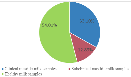
A total of 287 milk samples (95 from clinically mastitis, 37 from subclinical mastitis and 155 normal milk samples) were obtained from cows housed in private dairy farms located in four different provinces of Egypt at Kaluobia- Giza –Sharkia and EL Fayoum, 87, 95, 70 and 35 milk samples were collected, respectively.The distribution of the collected milk samples were illustrated in Table 1.
Bacteria isolation and identification from milk samples
Microbiological protocols for the diagnosis of udder infection published by the National Mastitis Council (1999), were used to isolate and identify bacterial strains from milk samples. A loopful of the milk sample was streaked on blood agar and then sub cultured on Eosin methylene blue (EMB), Mannitol salt agar, Edwards Agar, Salmonella-Shigella Agar, and MacConkey Agar. After that, plates were then incubated aerobically at 37ºC for 24 hours. Morphology of the suspected colonies were examined microscopically in Gram stained films. They were then subjected for biochemical tests for identification of pathogens including oxidase activity, acid production (lactose sucrose and glucose fermentation), indole production, catalase test, mannitol fermentation test, coagulase test, Voges–Proskauer and hydrogen sulfide production as formerly designated by Quinn (2011). In addition, each strain was confirmed the identification by using Analytical Profile Index API-20 tests (API, bio Meraux, France).
Table 1: Incidence of clinical, subclinical mastitic and healthy milk samples in four different provinces.
Provinces
clinical mastitic milk samples
Subclinical mastitic milk samples
Healthy milk samples
Total milk samples
Kaluobia
23 (26.44%)
21 (24.14%)
43 (49.43%)
87
Giza
31 (32.63%)
6 (6.32%)
58 (61.05%)
95
Sharkia
27 (38.57%)
8 (11.43%)
35 (50.00%)
70
El-Fayoum
14 (40%)
2 (5.71%)
19 (54.29%)
35
total
95 (33.10%)
37 (12.89%)
155(54.01%)
287
Preparation of plant extracts
Ethanolic extract of C. longa
C. longa dried rhizomes were purchased from a local market in Cairo, Egypt. It was extracted according to the techniques described by El-Newary et al. (2016). Briefly, dried rhizomes were coarsely powdered, then C. longa powder (1kg) were soaked in 4 L. 70% ethyl alcohol, and kept in tightly sealed vessels at room temperature for three weeks, stirring several times daily with a sterile glass rod. This mixture was filtered. The extraction of the residue was repeated 3-5 times in the same manner until a clear colorless supernatant extraction liquid was obtained. The extracted liquid was filtered and concentrated using rotary evaporator under reduced pressure at 50oC until the solvent was completely removed. The extract was stored at 4°C until used.
Aqueous extract of B. aegyptiaca
The fruits of B. aegyptiaca were obtained from Aswan, Egypt. The fresh fruits of B. aegyptiaca (1kg) were subjected to triple maceration then they were soaked in 4L distilled water and continued the next steps as shown previously in the EE-CLP.
Phytochemical screening
The AE-BAF and EE-CLP were analyzed for the presence of the phytoconstituents through preliminary phytochemical screening using modified and standard methods ; . The plant extracts were tested for the following phytochemical components: phenolics, cardiac glycosides, tannins, saponins, alkaloids, steroids, flavonoids, carbohydrates, proteins and terpenoids.
In vitro antioxidant activity
The antioxidant activities of AE-BAF and EE-CLP were assessed in terms of hydrogen-donating or radical-scavenging ability, using the stable radical DPPH (2, 20-Diphenyl-1-picrylhydrazyl) according to Brand-Williams et al. (1995). This activity is comparable with that of the standard drug ascorbic acid as strong antioxidant. The DPPH solution was prepared by dissolving 9.85 mg in 100 ml 70% methanol. Different concentrations of AE-BAF and EE-CLP were added to methanol 70 % and 335 µl DPPH. The mixtures were thoroughly shaken before being placed in the dark at room temperature for 30 minutes. If free radicals have been scavenged the purple colour of DPPH will degenerate to yellow, indicating free radical scavenging activity. A spectrophotometer was used to measure the decrease in absorbance at 517 nm. The Absorbance of the radical without sample was used as control (665 µl methanol 70 % and 335 µl DPPH). The amount of sample required to reduce the DPPH absorbance was determined.
The inhibition percentage of the DPPH radical by the extracts was calculated according to the following equation:
DPPH inhibition % = [(AB - AS)/AB] x 100
Where, AB=absorbance of control sample, and AS= absorbance of a tested sample at the end of the reaction.
Assay of the antibacterial effect of the plant extracts
Inoculums preparation
The identified isolated micro-organisms were cultured on a nutrient agar plate and incubated at 37°C for 24 hours. Discrete colonies were collected with sterile wire loop and inoculated into a 5ml sterile saline solution. To obtain 108 CFU/ml bacterial cultures, the test tubes were thoroughly shaken and the turbidity of the bacterial suspension was adjusted by comparing it with the 0.5 McFarland standards using spectrophotometer. Its absorbance was set to 580 um .
Agar well diffusion test
Muller Hinton agar was poured into sterile Petri dishes and was allowed to solidify. Using a sterile swap stick one milliliter of fresh bacterial culture was spread on the surface of the media. A sterile cork borer 6 mm in diameter was used to make wells in agar plates containing inoculums. Then, 100 μl of different concentrations of EE-CLP100, 50, 20 and 10 mg/ml then dispensed into the wells. The plates were placed in the refrigerator for 30 minutes to allow the extracts to full then diffuse into the agar. The plates were then incubated at 37°C for 18 hours. The zone of inhibition that appeared after the incubation period was recorded to detect antimicrobial activity . Gentamycin was used as positive control as described by NCCL (1999). The experiment was carried out in triplicate, and the results were expressed as means ± standard error of the three parallel measurements. This procedure was repeated again by using 800 and 400mg/ml of AE-BAF. Different concentrations of the extracts were prepared separately by dissolving the extract in sterile distilled water.
Statistical analysis
Using SPSS (version 17), data were statistically analyzed according to Snedecor and Cochran (1982). Data are represented as mean and standard error (mean ± SE). Using one-way analysis of variance (ANOVA) and Duncan’s multiple range test, differences between zones of inhibition (mm) induced by different concentrations of tested plant extracts and the antibiotic against bacteria, as well as between means in different bacteria species, were tested for significance. At the P ≤ 0.05 level of probability, differences were judged significant.
RESULTS
Bacteria isolation and identification from milk samples
The incidence of clinical, subclinical mastitis and healthy collected milk samples in all provinces is demonstrated in Figure 1 and Table 1. Out of 287 cow’s milk samples 132 isolates were identified and differentiated from 95 clinical and 37 subclinical masitic milk samples. Bacterial isolates were S. aureus (30.30%), E. coli(28.03%), S. agalactiae(19.70%), Salmonella species (10.60%), Bacillus subtilis (B. subtilis) (6.06%), Klebsiella pneumoniae (K. pneumoniae)(3.03%) and Pseudomonas aeruginosa (P. aeruginosa) (2.27%)as shown in Table 2.
Phytochemical screening
The results of preliminary phytochemical analysis of the different crude extracts of the medicinal plants in respect; AE-BAFand EE-CLP are presented in Table 3. The results show that all of the phytochemical compounds that were tested were present in the in AE-BAF, while EE-CLP contains all the same components, with the exception of cardiac glycosides which were not present.
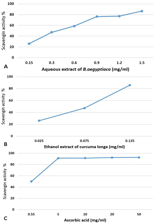
The obtained results indicated that the extracts of tested medicinal plants showed scavenging activity and illustrated in Figure 2A, B, C. It is seen that the AE-BAF possess high antioxidant activity at higher concentration (86.6% with 1.5mg/ml), whereas the EE-CLP possess good antioxidant activity (85.9% with 0.125mg/ml). The ascorbic acid possesses antioxidant activity at very high concentration where the percentage of scavenging activity was 92.73 % with 50mg/ml concentration.
Table 2: Percentage of isolated bacteria from clinical and subclinical mastitic cow’s milk in four provinces.
Provinces
Total CM and SCM
Isolated bacteria (n (%))
Gram -ve bacteria
Gram + ve bacteria
E.coli
K. pneumoniae
P. aeruginosa
Salmonella spp.
S. aureus
S. agalactiae
B. subtilis
Kaluobia
44
14(31.81%)
2(4.56%)
1(2.27%)
7(15.90%)
9(20.45%)
7(15.90%)
4(9.09%)
Giza
37
10(27.03%)
0(0%)
2(5.41%)
3(8.11%)
16(43.24%)
5(13.51%)
1(2.70%)
Sharkia
35
8(22.86%)
2(5.71%)
0(0%)
2(5.71%)
8(22.86%)
12(34.29%)
3(8.57%)
El-Fayoum
16
5(31.25%)
0(0%)
0(0%)
2(12.50%)
7(43.75%)
2(12.50%)
0(0%)
Total
132
37(28.03%)
4(3.03%)
3(2.27%)
14(10.60%)
40(30.30%)
26 (19.70%)
8(6.06%)
CM and SCM= clinical mastitic and subclinical mastitic milk samples.
Table 3: Phytochemical constituents of different crude extracts of B. aegyptiaca fruits and C. longa powder.
Test
Aqueous extract of B. aegyptiaca
Ethanol extract of C. longa
Phenolics
+
+
Alkaloids
+
+
Steroids
+
+
Cardiac Glycosides
+
-
Tannins
+
+
Saponins
+
+
Flavonoids
+
+
Proteins
+
+
Carbohydrates
+
+
Terpenoids
+
+
( + )= Detected, (-) = Not detected
Antibacterial activity of plant extracts
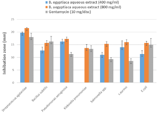
Evaluation of the antibacterial activity of AE-BAF against the bacteria isolated from clinical and subclinical mastitic cow’s milk was recorded in Table 4 and illustrated in Figure 3. The results revealed that AE-BAF at 400 and 800 mg/ml had the potential to limit growth of all bacteria except K. pneumoniae, which was not suppressed at the 400 mg/ml concentration. When AE-BAF was compared with the control antibiotic (Gentamycin), it showed considerable (P ≤ 0.05) antimicrobial activity againstP. aeruginosa, Salmonella species and S. aureus at 800 mg/ml concentration. Antibacterial activity of AE-BAF at 400 mg/ml concentration was found to be substantial (P ≤ 0.05) against P. aeruginosa and S. aureus when compared to the Gentamycin. There were no significant differences in antibacterial activity between the two concentrations of AE-BAF against all bacteria, with the exception of K. pneumoniae, Salmonella species and S. aures which were considerably (P ≤ 0.05) suppressed at the 800 mg/ml concentration. Among bacteria species, S. agalactiae and P. aeruginosa were the most significantly (P ≤ 0.05) inhibited bacterium species at 400 mg/ml, while S. agalactiae was the most inhibited bacteria species at 800 mg/ml, compared to other species.
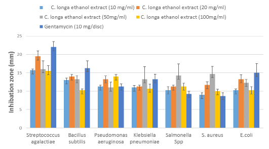
The antimicrobial properties of the EE-CLP against bacteria isolated from clinical and subclinical mastitic cow’s milk presented in Table 5 and Figure 4, where all the test organisms were inhibited with different concentrations of extract. The significant (P ≤ 0.05) antibacterial activity of EE-CLP was observed at 20 and 50 mg/ml concentrations against S. aureus compared to the Gentamycin. There were no significant differences in antimicrobial activity between various concentrations of EE-CLP and the control antibiotic against S. agalactiae, P. aeruginosa, K. pneumoniae, Salmonella species, and E. coli, except that 100mg/ml which had a significantly (P ≤ 0.05) lower effect against B. subtilis compared to concentration 20, 50 and the antibiotic. While concentration 50 had a significant (P ≤ 0.05) higher effect against S. aureus compared to the Gentamycin and, 10 and 100 mg/ml. At concentrations of 10, 20 and 100 mg/ml, S. agalactiae is the most affected (P ≤ 0.05) among bacterium species, whereas there was no significant difference between them at concentration 50mg/ml.
DIscussion
Mastitis is considered the most common serious health problem on dairy farms which lead to significant bio health hazard to human particularly in developing countries such as in Egypt . Clinical inspection and the California mastitis test were found to be effective diagnostic tools for detecting and distinguishing clinical and subclinical mastitis from apparently healthy cattle, which is consistent with previous findings .
According to clinical inspection and CMT obtained results, the prevalence of clinical and subclinical mastitic and normal milk samples in total collected samples were 33.10%, 12.89% and 54.01 %, respectively. These findings were nearly similar to those reported by , who recorded 34.5%, 24.7%, 40.8% prevalence of clinical mastitis, subclinical mastitis and healthy dairy cows respectively, at Beni-Suef, El-Fayoum, Behera and Monofia Governorates in Egypt. However, they disagree with , who reported that 56.3% of mastitis was subclinical and 13.3% was symptomatic. This variation in the percentage of clinical and subclinical mastitis could be attributed to differences in hygienic condition and management in rural areas .
Bacteria are the most important microorganisms that cause mastitis in dairy herds, and they can act as a contagious pathogen or an opportunistic pathogen . A total of 132 different bacterial isolates were isolated from 287 cow’s milk samples; they were identified by biochemical tests and found to belong to seven different species, as shown in Table 2.Das et al. (2010) and Alkhamaiseh et al. (2011) believe that bacterial culture is a good standard method for isolating causative bacteria.
The most predominant bacteria identified in milk samples from clinical mastitic and subclinical cows were S. aureus (30.30%), E. coli (28.03%) and S. agalactiae (19.70%). The highest prevalent of S. aureus, E. coli and S. agalactiae may occur through transmission by the contaminated Milker’s hands, teat-to-teat hygiene and cow-to-cow. These findings were in line with Das et al. (2010), who considered these microorganisms are the major etiological agents of clinical and subclinical mastitis worldwide. Because E. coli infects the udder via the teat canal, a higher incidence rate of E. coli could be attributed to poor hygienic conditions of the environment. S. aureus causes food poisoning and commonly found in milk from dairy animals that have mastitis from the environment .
Other species of bacterial isolates in this study, as Salmonella species, B. subtilis, K. pneumoniae and P. aeruginosa are minor causes of bovine mastitis, and this is in agreement with , who isolated also Salmonella species in low incidence (4.2%), K. pneumoniae (8.5%), and P. aerguinosa (4.2%), as well as Salih and Ahmed (2011), who isolated B. subtilis (9%) from cow’s milk. The distribution of mastitis pathogen strains differs within individual animals in a herd, among herds and among host species .
In the present results, phytochemical screening of the AE-BAF detected the presence of alkaloids, flavonoids, steroids, saponins, phenolic compounds, terpenoids and tannins. This was consistent with the work of Abdulhamid and Sani (2016), who detected the presence of the same phytochemicals in the AE-BAF. It was slightly similar to the observations of Henna et al. (2010), who reported the presence of the same phytochemicals in the AE-BAF except steroids and alkaloids were not detected in their study. This is in line with other phytochemical study of Wakawa et al. (2018), who demonstrated the same in the AE-BAF, except cardiac glycosides and alkaloids were not detected in their study.
Phytochemical screening of the EE-CLP indicated the presence of flavonoids, steroids, saponins, alkaloids, phenolic compounds, terpenoids and tannins, these findings are in agreement with Oghenejobo et al. (2017), who detected the same compounds in the EE-CLP. Also phytochemical screening of the C. longa powder confirmed the presence of carbohydrates, proteins and amino-acids, alkaloids, terpenoids and flavinoids but no tannins, glycosides and saponins . Contrary to the findings of Pawar et al. (2014), tannins and saponins were found in EE-CLP in this study. The chemical composition, toxicity, and bioactivity of the extracts may differ due to the solubility of the active component in various solvents , the chronological age of the plant, percentage humidity of the harvested material, and time of harvest, as well as the method of extraction .
The active phytochemical constituents indicated in these results as shown in Table 3 alone or in combination may be responsible for the observed antioxidant activity . Plant phenols, flavonoids, tannins and terpenoids are reported to possess potent antioxidant activity ; . Usman et al. (2020); Chothani and Vaghasiya (2011) reported that different parts of B. egyptiaca possess potent antioxidant activity. Miquel et al. (2002) reported that curcumin exhibit more potent antioxidant activity comparable of that to vitamin E.
The AE-BAF possess significant antibacterial activity at 400 and 800 mg/ml against the Gram-positive and Gram-negative bacteria isolated from clinical and subclinical mastitic cow as shown in Table 4 and Figure 3.The results obtained showed that the antibacterial activity of the extract significantly increased with increasing concentration, which is consistent with the findings . Furthermore, these results were supported by Jahan et al. (2013), who found that B. aegyptiaca have a broad spectrum activity againstvarious bacteria including S. aureus, S. pyogenes, E. coli, K. pneumoniae, P. aeruginosa, S. typhimurium, B. subtilis and resistance bacteria harboring bla genes. Also, Ibrahim (2016) reported that AE-BAF was more potent than petroleum ether extract of Phyllanthus reticulatus against S. aureus and E. coli. It has been reported that B. aegyptiaca extracts inhibited the in vitro growth of several strains of S. aureus and P. aeruginosa ; .
The presence of saponins and alkaloids in this plant may be responsible for its antibacterial effect, as some previous research have suggested . The presence of the phytochemical constituents in B. aegyptiaca extracts in this study was reported to be responsible for the plant extract’s antibacterial action ; .
Table 4: Mean zones of inhibition (mm) ± standard error by different concentrations of B. aegyptiaca aqueous extract and antibiotic against bacteria isolated from clinical and subclinical mastitic cow’s milk.
Test pathogens
B.aegyptiaca aqueous extract (400 mg/ml)
B. aegyptiaca aqueous extract (800 mg/ml)
Gentamycin
(10 mg/disc)
F value
P value
Streptococcus agalactiae
19.7±0.33Aa
21.5±0.29Aa
18.0±1.52Aa
3.637
NS
Bacillus subtilis
12.7±1.33BCa
15.7±0.89BCa
16.3±2.02Aa
1.717
NS
Pseudomonas aeruginosa
16.3±0.88ABa
17.3±0.67Ba
11.3±0.66BCb
18.600
0.003
Klebsiella pneumoniae
0.0±0.00Db
13.7±1.33Ca
13.3±1.33ABCa
51.281
<0.001
Salmonella Spp.
11.0±1.00Cb
15.3±0.88BCa
9.3±0.67Cb
12.950
0.007
S.aureus
14.0±2.52BCab
16.0±1.00BCa
8.6±0.89Cb
5.315
0.047
E.coli
11.3±1.33Ca
15.7±0.66BCa
15.0±2.51ABa
1.909
NS
F value
22.516
8.072
5.400
P value
<0.001
0.001
0.004
Different small letters at the same row indicateto significant differences between different concentrations of B. aegyptiacaaqueous extract and antibiotic,while different capital lettersat the same column indicate to significant differences between bacteria species at P ≤ 0.05, NS= Non-significant.
Table 5: Mean zones of inhibition (mm) ± standard error by different concentrations of C. longa ethanol extractand antibiotic against bacteria isolated from clinical and subclinical mastitic cow’s milk.
Test pathogens
C. longa ethanol extract(10 mg/ml)
C. longa ethanol extract(20 mg/ml)
C. longa ethanol extract(50mg/ml)
C. longa ethanol extract(100mg/ml)
Gentamycin (10 mg/disc)
F value
P value
Streptococcus agalactiae
15.7±0.33Aa
19.5±1.44Aa
16±2.30Aa
15.5±1.44Aa
22±1.52Aa
1.283
NS
Bacillus subtilis
13.0 ±0.57Bab
14.0±0.58Ba
13.3±0.88Aab
10.3±0.33Cb
16.3±2.02Aa
4.088
0.032
Pseudomonas aeruginosa
11.3±0.33Ca
13.3±0.88Ba
11.0±1.52Aa
14.0±0.57ABa
11.3±0.66BCa
2.333
NS
Klebsiella pneumoniae
11.0±0.58Ca
11.3±0.33Ba
13.3±3.38Aa
10.7±1.20Ca
13.3±1.33ABCa
0.559
NS
Salmonella Spp.
10.3±0.88CDa
11.3±0.33Ba
14.3±3.17Aa
11.3±0.88BCa
9.3±0.66Ca
1.432
NS
S.aureus
9.0±0.58Db
11.7±0.88Bab
14.7±2.02Aa
10.0±0.57Cb
8.7±0.88Cb
4.763
0.021
E.coli
10.3±0.33CDa
13.3±1.20Ba
12.3±0.88Aa
10.3±0.88Ca
15±2.51ABa
2.129
NS
F value
15.947
10.149
0.533
5.483
5.400
P value
<0.001
<0.001
NS
0.004
0.004
Different small letters at the same row indicate to significant differences between different concentrations of C. longa ethanol extract and antibiotic, while different capital letters at the same column indicate to significant differences between bacteria isolates at P ≤ 0.05, NS= Non significant.
This study evaluated the antibacterial effects of the crude EE-CLP, ethanolic extract of C. longa inhibits the growth of both Gram-positive and Gram-negative bacteria as shown in Table 5 and Figure 4. The significant antibacterial activities were observed with 20 and 50 mg/ml concentrations. These results are in agreement with those of Jha et al. (2013), who investigated the effects of turmeric and curcuminoids against S. aureus, B. subtilis and P. aeruginosa bacteria. They mentioned that the highest antimicrobial activity resulted from ethanol extract at 20 mg/ml. The C. longa rhizome extract was founded to be more potent antibacterial agent than the leaf extract , and when ethanol was utilized as the extraction solvent, higher quantities of curcumin and antimicrobial activity were produced .
The antibacterial activity of ethanolic extract was due to the presence of various types of terpenoids and glycosides like compounds and these compounds have been found to have strong antimicrobial activity against Gram positive and Gram negative bacteria ; . Curcuma aromatica extract (Wild turmeric) was reported to have antibacterial activity against B. lichaniformis, Micrococcus leutus, and S. Thyphorium . They also discovered that the antimicrobial action is due to the presence of high phenolic content. According to Wilson et al. (2005), B.subtilis was the most sensitive organism to C. longa extract curcuminoid and oil extracts.
CONCLUSIONS and Recommendations
The prevalence of subclinical and clinical mastitis was measured in different Egyptian governorates, and the most common bacteria causing mastitis was identified. According to this study, the AE-BAF and EE-CLP can be used as an alternative to standard mastitis therapy because of their chemical composition, antioxidant capability, and efficiency against mastitis-associated bacteria. AE-BAF exhibited potent antibacterial activities at 800 mg/ml and EE-CLP exhibited potent antibacterial at 20 and 50 mg/ml against the Gram-positive and Gram-negative bacteria isolated from clinical and subclinical mastitic cow. However, more research into their applicability to mastitis-affected dairy cows is neededand further studies are needed to discover the definite fractions from C. longa and or B. aegyptiaca to explain the definite mechanism of action of the tested extracts as antibacterial.
ACKNOWLEDGMENTS
This work was financially supported by National Research Centre as a part of the project No.11020303-the 11th Research plan, under supervision of Prof. Dr. Hala A.A. Abou- Zeina
Novelty Statement
The AE-BAF and EE-CLP exhibited potent antibacterial activities against the Gram -positive and Gram-negative bacteria isolated from clinical and subclinical mastitic cow, thus justifying their application in treatment of clinical cases of bovine mastitis.
AUTHOR’S CONTRIBUTION
All authors shared equally in designing, conducting the study and writing the manuscript.
Conflict of interest
The authors have declared no conflict of interest.
References
• Abdel-Farid IB, El-Sayed MA (2021). Phytochemical analysis of the desert date Balanites aegyptiaca Egypt. J. Bot., 61(1): 95-103.
• Abdulhamid A, Sani I (2016). Preliminary phytochemical screening and antimicrobial activity of aqueous and methanolic leave extracts of Balanites aegyptiaca (L.). Int. Res. J. Pharm. Biosci., pp. 1-7.
• Ahuja C, Kaur H, Sharma R (2015). Antibacterial activity of Terminalia Chebula fruit by agar well diffusion method. J. Chem. Eng. Sci. A., 1(2): 67–72. https://doi.org/10.15415/jce.2015.12006[CrossRef]
• Alkhamaiseh SI, Taher M, Ahmad F (2011). The phytochemical contents and antimicrobial activities of Malaysian Calophyllum rubiginosum. Am. J. Appl. Sci., 8: 201-205. https://doi.org/10.3844/ajassp.2011.201.205[CrossRef]
• Amer S, Galvez FLA, Fukuda Y, Tada C, Jimenez IL, Valle WFM, Nakai Y (2018). Prevalence and etiology of mastitis in dairy cattle in El Oro Province, Ecuador. Vet. Med. Sci., 80(6): 861–868. https://doi.org/10.1292/jvms.17-0504[CrossRef]
• Ammar AMA, Soumaya SAE, AbdElwahab AMO, Zeinab AEA (2016). Be detection of multidrug resistance genes in Pseudomonas Aeruginosa isolated from bovine mastitic milk. J. Dairy Vet. Anim. Res., 3(2): 43‒49. https://doi.org/10.15406/jdvar.2016.03.00071[CrossRef]
• Anani K, Adjrah Y, Ameyapoh Y, Karou SD, Agbonon A, De Souza C, Gbeassor M (2015). Effects of hydroethanolic extracts of Balanites aegyptiaca (L.) Delile (Balanitaceae) on some resistant pathogens bacteria isolated from wounds. J. Ethnophamacol. Moghadamtousi., 164: 16–21. https://doi.org/10.1016/j.jep.2015.01.051[CrossRef]
• Ara G, Durreshahwar KF, Imran M, Akbar A (2011). In vitro antibacterial activity of essential oils extracted from locally available medicinal plants. J. Chem. Soc. Pak., 33: 205-208.
• Boroumand N, Samarghandian S, Hashemy SI (2018). Immunomodulatory, anti-inflammatory, and antioxidant effects of curcumin. J. Herbmed. Pharmacol., 7: 211–219. https://doi.org/10.15171/jhp.2018.33[CrossRef]
• Brand-Williams W, Cuvelier ME, Berset C (1995). Use of a free radical method to evaluate antioxidant activity. LWT-Food Sci. Technol., 28(1): 25-30. https://doi.org/10.1016/S0023-6438(95)80008-5[CrossRef]
• Chapagain BP, Wiesman Z, Tsror L (2007). In vitro study of the antifungal activity of saponin-rich extracts against prevalent phytopathogenic fungi. Ind. Crop Prod., 26: 109–115. https://doi.org/10.1016/j.indcrop.2007.02.005[CrossRef]
• Chhetri HP, Yogol NS, Sherchan J (2008). Phytochemical and antimicrobial evaluations of some medicinal plants of Nepal. Kathmandu Univ. J. Sci. Eng. Technol., 1(5): 49-54. https://doi.org/10.3126/kuset.v4i1.2883[CrossRef]
• Chothani DL, Vaghasiya HU (2011). A review on Balanites aegyptiaca Del (desert date): Phytochemical constituents, traditional uses and pharmacological activity. Pharmacogn. Rev., 5(9): 55–62. https://doi.org/10.4103/0973-7847.79100[CrossRef]
• Cowan MM (1999). Plant products as antimicrobial agents. Clin. Microbiol. Rev., 12: 564–582. https://doi.org/10.1128/CMR.12.4.564[CrossRef]
• Das K, Tiwari SKR, Shrivastav KD (2010). Techniques for evaluation of medicinal plant products as antimicrobial agent: Current methods and future trends. J. Med. Plants Res., 4: 104-111.
• Dingwell RT, Kelton DF, Leslie KE (2003). Management of the dry cow in control of peripartum disease and mastitis. Vet. Clin. N. Am. Food Anim., 19(1): 235–265. https://doi.org/10.1016/S0749-0720(02)00072-5[CrossRef]
• Doughari JH, Pukuma MS, De N (2007). Antibacterial effects of Balanites aegyptiaca L. Drel. and Moringa oleifera Lam. on Salmonella typhi. Afr. J. Biotechnol., 6(19): 2212-2215. https://doi.org/10.5897/AJB2007.000-2346[CrossRef]
• Ekpo MA, Etim, PC (2009). Antimicrobial activity of ethanolic and aqueous extracts of Sidaacuta on microorganisms from skin infections. J. Med. Plants Res., 3(9): 621-624.
• El-Newary SA, Ismail RF, Shaffie N, Hendawy SF, Omer E (2016). Hepatoprotective, therapeutic and in vivo anti-oxidant activities of Tagetes lucida leaves alcoholic extract against paracetamol-induced hepatotoxicity rats. Int. J. Pharm. Tech. Res., 9(12): 327-341.
• Ezemokwe GC, Aguiyi JC, Chollom FP (2020). The antibacterial activity of aqueous and ethanolic leaf extracts of Balanites aegyptiaca (L.) Del plant on some selected clinical human pathogens. J. Adv. Microbiol., 20(10): 51-66. https://doi.org/10.9734/jamb/2020/v20i1030290[CrossRef]
• Felix MT (1982). Medical microbiology. Churchill livingstone (Publishers): London, UK.
• Fusco V, Chieffi D, Fanelli F, Logrieco AF, Cho GS, Kabisch J, Böhnlein C, Franz CMAP (2020). Microbial quality and safety of milk and milk products in the 21st century. Comp. Rev. Food Sci. Food Saf., 19: 2013–2049. https://doi.org/10.1111/1541-4337.12568[CrossRef]
• Gianneechini R, Concha C, Rivero R, Delucci I, Moreno LJ (2002). Occurrence of clinical and sub-clinical mastitis in dairy herds in the West Littoral Region in Uruguay. Acta Vet. Scand., 43: 221-230. https://doi.org/10.1186/1751-0147-43-221[CrossRef]
• Henna JS, Adamu AK, Lortsuan DN, Olonitola OS (2010). Phytochemical screening and antibacterial effect of the aqueous and methanolic extracts of roots of Balanites aegyptiaca (Del) on some bacteria species on some bacteria species. Sci. World J., 5(2): 59-62. https://doi.org/10.4314/swj.v5i2.61518[CrossRef]
• Ibrahim EE (2016). Evaluation of antimicrobial activity of Balanites aegyptiaca and Phyllanthus reticulates Leaves. Pharm. Glob., 7(1): 1-4.
• Jahan N, Khatoonn RM, Shahzad A, Shahid M (2013). Antimicrobial activity of medicinal plant Balanites aegyptiaca Del. and it’s in vitro raised calli against resistant organisms especially those harbouring Bla genes. J. Med. Plants Res., 7:1692-1698. https://doi.org/10.5897/AJPS2013.1058[CrossRef]
• Jha H, Barapatre A, Prajapati M, Aadil KR, Senapati S (2013). Antimicrobial activity of rhizome of selected curcuma variety. Int. J. Life Sci. Bt Pharm. Res., 2(3): 183-189.
• Kai Kai, Wanling BI, Yukun BO, Yingwang YE, Danfeng Z (2020). Curcumin-A review of its antibacterial effect. Biomed. J. Sci. Tech. Res., 26(1): 19585-19587.
• Khatua S, Roy T, Acharya K (2013). Antioxidant and free radical scavenging capacity of phenolic extract from Russula laurocerasi. Asian J. Pharm. Clin. Res., 6(4): 156-160.
• Kotb EEZ, EL Sawah AM, Kortam LE, Abd El Fattah OA, Barghooth WM (2021). Evaluation of using different adjuvants used for preparation of Staphylococcus aureus mastitis vaccine on the immune response. J. Appl. Vet. Sci., 6(1): 9–17. https://doi.org/10.21608/javs.2021.140065[CrossRef]
• Kovacevic Z, Radinovic M, Cabarkapa I, Kladar N, Bozin B (2021). Natural agents against bovine mastitis Pathogens. Antibiotics, 10(2): 205-221. https://doi.org/10.3390/antibiotics10020205[CrossRef]
• Kromker V, Leimbach S (2017). Mastitis treatment- Reduction in antibiotic usage in dairy cows. Reprod. Dom. Anim., 52: 21–29. https://doi.org/10.1111/rda.13032[CrossRef]
• Martinez-Correa HA, Paula JT, Kayano ACA, Queiroga CL, Magalhaes, PM, Costa, FTM, Cabral, FA (2017). Composition and antimalarial activity of extracts of Curcuma longa L. Obtained by a combination of extraction processes using supercritical CO2, ethanol and water as solvents. J. Supercrit. Fluids, 119(1): 122-129. https://doi.org/10.1016/j.supflu.2016.08.017[CrossRef]
• Maryam Z, Farrukh A, Iqbal A (2009). The in vitro antioxidant activity and total phenolic content of four Indian medicinal plants. Int. J. Pharm. Pharm. Sci., 1: 88-95.
• Miquel J, Bernd A, Sempere JM, Diaz-Alperi J, Ramirez A (2002). The curcuma antioxidants: Pharmacological effects and prospects for future clinical use a review. Arch. Gerontol. Geriatr., 34: 37-46. https://doi.org/10.1016/S0167-4943(01)00194-7[CrossRef]
• Mir AQ, Bansal BK, Gupta DK (2014). Subclinical mastitis in machine milked dairy farms in Punjab: prevalence, distribution of bacteria and current antibiogram. Vet. World, 7: 291-294. https://doi.org/10.14202/vetworld.2014.291-294[CrossRef]
• Moghadamtousi SZ, Kadir HA, Hassandarvish P, Tajik H, Abubakar S, Zandi K (2014). A review on antibacterial, antiviral, and antifungal activity of curcumin. Biomed. Res. Int., 2014: 186864. https://doi.org/10.1155/2014/186864[CrossRef]
• Mohandes SSE, Gamal IM, Abou-Zeina HA, Elbayoumy MK (2021). Some studies on phenotypic and genotypic characters of small colony variants staphylococcus aureus isolated from dairy cows infected with mastitis in Egypt. Adv. Anim. Vet. Sci., 9(5): 637-647. https://doi.org/10.17582/journal.aavs/2021/9.5.637.647[Full text][CrossRef]
• Mostafa AA, Al-Askar AA, Almaary KS, Dawoud TM, Sholkamy EN, Bakri MM (2018). Antimicrobial activity of some plant extracts against bacterial strains causing food poisoning diseases. Saudi J. Biol. Sci., 25(2): 361-366. https://doi.org/10.1016/j.sjbs.2017.02.004[CrossRef]
• Murthy HN, Yadav GG, Dewir YH, Ibrahim A (2021). Phytochemicals and biological activity of Desert Date (Balanites aegyptiaca L.) Delile. Plants, 10(32). https://doi.org/10.3390/plants10010032[CrossRef]
• Mutwali IFA, Abdelgadir S (2016). Phytochemical screening and biological activity of Balanites aegyptiaca Stem Bark. J. Chem. Pharm. Res., 8(4): 489-498.
• National Mastitis Council (1999). Laboratory handbook on bovine mastitis. NMC Inc., Madison, W.I.
• NCCL (1999). Performance standards for antimicrobial susceptibility testing. Supplement M100-S9. National Committee for Clinical Laboratory Standards, Wayne, PA, USA.
• Obasi NL, Egbuonu ACC, Ukoha PO, Ejikeme PM (2010). Comparative phytochemical and antimicrobial screening of some solvent extracts of Samanea saman pods. Afr. J. Pure Appl. Chem., 4(9): 206-212.
• Oghenejobo M, Opajobi OA, Bethel OUS, Uzoegbu U (2017). Antibacterial evaluation, phytochemical screening and ascorbic acid assay of turmeric (Curcuma longa). MOJ Bioequiv. Availab., 4(2): 232‒239. https://doi.org/10.15406/mojbb.2017.04.00063[CrossRef]
• Okigbo R N, Anuagasi CL, Amadi JE, Ukpabi UJ (2009). Potential inhibitory effects of some African tuberous plant extracts on Escherichia coli, Staphylococcus aureus and Candida albicans. Int. J. Integr. Biol., 6(2): 91-98.
• Oliver SP, Jayarao BM, Almeida RA (2005). Food borne pathogens in milk and the dairy farm environment: Food safety and public health implications. Foodborne Pathog. Dis., 2(2):115–129. https://doi.org/10.1089/fpd.2005.2.115[CrossRef]
• Paloi S, Acharya K (2014). Evaluation of antioxidative activity and chemical composition of ethanolic extract from Amanita vaginata (Bull.) Lam.: An in vitro study. Asian J. Pharm. Clin. Res., 7(2): 88-92.
• Pawar H, Karde M, Mundle N, Jadhav P, Mehra K (2014). Phytochemical evaluation and curcumin content determination of turmeric Rhizomes collected from Bhandara district of Maharashtra (India). Med. Chem., 4(8): 588-591. https://doi.org/10.4172/2161-0444.1000198[CrossRef]
• Pyörälä S (2002). New strategies to prevent mastitis. Reprod. Dom. Anim., 37(4): 211–216. https://doi.org/10.1046/j.1439-0531.2002.00378.x[CrossRef]
• Quinn PJ (2011). Veterinary microbiology and microbial Disease. (2nd Ed.). Wiley-Blackwell, J. Wiley and Sons Ltd Publication, UK.
• Reyher KK, Dohoo R, Scholl DT, Keefe GP (2012). Evaluation of minor pathogen intramammary infection, susceptibility parameters, and somatic cell counts on the development of new intramammary infections with major mastitis pathogens. J. Dairy Sci., 95(7): 3766–3780. https://doi.org/10.3168/jds.2011-5148[CrossRef]
• Salih MRR, Ahmed MFA (2011). Treatment trial of bovine bacterial mastitis in Khartoum state Sudan. World’s Vet. J., 1(1): 20-24.
• Sayed RH, Salamas S, Soliman RT (2014). Bacteriological evaluation of present situation of mastitis in dairy cows. Glob. Vet., 13(5): 690-695.
• Sedky D, Ghazy AA, Soliman KA (2020a). Incidence and control of infectious bacteria in bovine milk. J. Microbiol. Biotech. Food Sci., 10(3): 398-404. https://doi.org/10.15414/jmbfs.2020.10.3.398-404[CrossRef]
• Sedky D, Ghazy AA, Soliman NA, Shaapan RM (2020b). Comparative diagnosis of infectious bacteria in bovine milk. J. Anim. Health Prod., 8(4): 171-182. https://doi.org/10.17582/journal.jahp/2020/8.4.171.182[Full text][CrossRef]
• Singh N, Gupta S, Rathore V (2017). Comparative antimicrobial study of ethanolic extract of leaf and rhizome of Curcuma longa Linn. Pharmacogn. J., 9(2): 208-212. https://doi.org/10.5530/pj.2017.2.35[CrossRef]
• Snedecor GW, Cochran WG (1982). Statistical methods, 8th edition, Iowa State University USA.
• Tarazona-Manrique LE, Villate-Hernández JR, Andrade-Becerra RJ (2019). pathogen or contagious pathogen Bacterial and fungal infectious etiology causing mastitis in dairy cows in the highlands of Boyacá (Colombia). Rev. Med. Vet. Zoot., 66(3): 208-218. https://doi.org/10.15446/rfmvz.v66n3.84258[CrossRef]
• Tiwari P, Kumar B, Kaur M, Kaur G, Kaur H (2011). Phytochemical screening and extraction: A review. Int. Pharm. Sci., 1(1): 98–106.
• Usman A, Mohammed Y, Muhammed HO, Usman N L, Zakari AH (2020). Phytochemical screening and antioxidant activity of Balanites aegyptiaca root bark extracts: Influence of solvent. Commun. Phys. Sci., 5(2): 156-164.
• Wakawa AI, Sambo AB, Yusuf S (2018). Phytochemistry and proximate composition of root, stem bark, leaf and fruit of desert date, Balanites aegyptiaca. J. Phytopharmacol., 7(6): 464-470. https://doi.org/10.31254/phyto.2018.7602[CrossRef]
• Wilson B, Abraham G, Manju VS, Mathew M, Vimala B, Sundaresan S, Nambisan B (2005). Antimicrobial activity of Curcuma zedoaria and Curcuma malabarica tuber. J. Ethnopharmacol., 99(1): 147-153. https://doi.org/10.1016/j.jep.2005.02.004[CrossRef]
• Yadav JP, Panghal M (2010). Balanites aegyptiaca (L.) Del. A review of its traditional Uses, phytochemistry and pharmacological properties. Int. J. Green Pharm., 4: 140-146. https://doi.org/10.4103/0973-8258.69158[CrossRef]
• Zeedan GSG, Abdalhamed AM, Abdeen E, Ottai ME, Abdel-Shafy S (2014). Evaluation of antibacterial effect of some Sinai medicinal plant extracts on bacteria isolated from bovine mastitis. Vet. World, 7(11): 991-998. https://doi.org/10.14202/vetworld.2014.991-998[CrossRef]
Assessment of Phytochemical, Antioxidant and Antibacterial Activity of Balanites Aegyptiaca and Curcuma Longa against Some Bacterial Pathogens Isolated from Dairy Cow Infected with Mastitis · ResearchersLinks