From the collected 12 cases, 11 cases were diagnosed as malignant carcinomas and only one case was benign fibroepithelial hyperplasia. Tubulo-papillary carcinoma represented the most frequently observed histological pattern of all examined cases, in which the epithelial neoplastic cells showed the criteria of malignancy, including cellular and nuclear pleomorphism, hyperchromasia and frequent atypical mitosis. These proliferating cells projected toward the ductal lumina forming inward projections supported with fibrous connective tissue stroma (Fig. A.1). Micropapillary invasive carcinoma (Fig. A.2) was also noticed, in which a population of neoplastic cells were forming intraductal aggregates or small papillae without supporting fibrovascular stroma.
Some cases were bearing the criteria of non-invasive ductal carcinoma in situ (DCIS) in which the neoplastic cells
Table 1: Pathological features and animal data for mammary gland samples
were proliferating in the ductal lumen with no tendency for invasion or basement membrane destruction (Fig. A.3 and A.4). Cystic-papillary carcinoma (Fig. A.5) was also detected, in which the tubular lumen was greatly ectatic with presence of multiple papillary projections with some eosinophilic debris in some instances.
Tubular carcinoma (Fig. A.6 and A.7) as a simple type of tumor was quite common as well, the neoplastic cells were arranged in tubular or gland-like structure of one or two cells thickness. Some of the examined cases showed cribriform pattern (Fig. A.8 and A.9) that characterized by the sieve-like appearance of neoplastic cells as they arranged with central narrow aperture.
Comedocarcinoma (Fig. A.10 and A.11) was described as an aggregate of neoplastic cells with central area of necrosis. The necrotic foci were consisted of homogenous eosinophilic material, cell debris, degenerating neutrophils and macrophages.
Solid carcinoma (Fig. A.12) was recorded; sheets of proliferating neoplastic cells were seen without tendency for tubular or luminal formation. Solid carcinoma pattern was
usually accompanied by another pattern of the previously described carcinomas and was associated with marked local and vascular invasion.
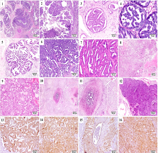
Figure (A): Mammary gland of cat , H&E stained, showing (1) papillary carcinoma, (2) Micropapillary carcinoma, (3) Ductal carcinoma in situ, (4) higher magnification, frequent mitosis of intraductal neoplastic cells, (5) cystic papillary carcinoma, (6) Tubular carcinoma, (7) higher magnification, tubular carcinoma, (8) cribriform carcinoma, (9) higher magnification, cribriform carcinoma, (10) comedocarcinoma, (11) higher magnification, comedocarcinoma with central necrotic area, (12) Solid carcinoma, (13) – (16) Positive Immunostaining of CK 8/18 in the different histologic patterns of carcinoma.
All previously described carcinoma patterns were CK8/18+ positive by immunostaining (Fig. A.13-A.16).
Some of the examined cases were accompanied by desmoplastic stromal reaction, in which the proliferating fibrous tissue stroma were separating an islands of tumor cells (Fig. B.1). Vascular invasion was observed in many cases that was represented by the presence of intravascular aggregates of neoplastic cells (Fig. B.2).
A case of proliferating pleomorphic myoepithelial cells was detected (Fig. B.3) these cells were strong vimentin positive by immunostaining (Fig. B.4).
In the current study three types of rare carcinomas were spotted, the first one was glycogen-rich or clear cell carcinoma (Fig. B.5) in which the neoplastic cells were arranged predominately in solid form with their characteristic clear vacuolated cytoplasm. The second case was that of mucinous carcinoma (Fig. B.6); which was characterized by the presence of blue stained mucus within the tumor cells. The last case was lipid-rich carcinoma (Fig. B.7) characterized by microvacuolated cytoplasm of neoplastic cells with presence of variably sized, intracytoplasmic, distinct vacuoles pushing the nucleus to the periphery of the cells.
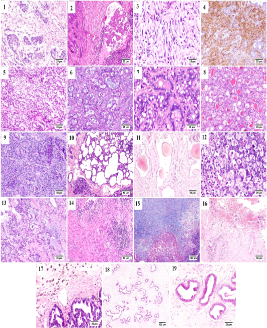
Figure (B): Mammary gland of cat, showing (1) Desmoplasia, (2) vascular invasion by anaplastic cells, (3) Pleomorphic population of myoepithelial cells, (4) Vimentin positive cells (immunostaining), (5) Glycogen-rich carcinoma, (6) mucinous carcinoma, (7) lipid-rich carcinoma, (8) lobular hyperplasia, (9) lobular hyperplasia with atypia, (10) cystically dilated ductules, (11) Duct ectasia with corpora amylacea, (12) Apoptosis with karyorrhexis, (13) Myxomatous change, (14) Stromal fibroplasia with lymphocytic infiltration, (15) proliferating fibrous tissue stained blue with MTC stain, (16) Extensive hemorrhage, (17) hemosiderin-laden macrophages, (18) fibroepithelial hyperplasia and (19) higher magnification, proliferating ductal epithelial tissue with surrounding myoepithelial cells and fibrous stroma.
Generally, in all the diagnosed cases of carcinoma, the non-affected lobules also suffered from some changes including; lobular hyperplasia (Fig. B.8) was detected in some cases in which the proliferation of non-neoplastic cells resulting in increased number of acini and ductules per lobule. Lobular hyperplasia with atypia (Fig. B.9) was noticed as well, in which the proliferating cells bearing some resemblance to malignant cells as pleomorphism, anisokaryosis and frequent mitosis. Cystic dilatation of the ductules was frequently detected (Fig. B.10) as well as duct ectasia with presence of several corpora amylacea (Fig. B.11).
Within the proliferating tumor cells, massive necrosis, apoptosis and karyorrhexis (Fig. B.12) and presence of extended areas of myxomatous change (Fig. B.13) were frequently seen.
Stromal inflammatory and scirrhous reactions (Fig. B.14 and B.15) were quite common. Some carcinomas were accompanied by extensive stromal hemorrhages (Fig. B.16) with presence of increased numbers of hemosiderin laden macrophages (Fig. B.17). The benign case of fibroadenomatous hyperplasia was characterized histologically by the presence of hyperplastic ducts lined by one to three layers of proliferating cells surrounded by multiple layers of myoepithelial cells and finally embedded within a loose fibrillar stroma (Fig. B.18 and B.19).
The benign case of fibroadenomatous hyperplasia was characterized histologically by the presence of hyperplastic ducts lined by one to three layers of proliferating cells surrounded by multiple layers of myoepithelial cells and finally embedded within a loose fibrillar stroma (Fig. B.18 and B.19).
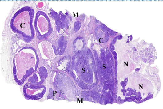
Figure (C): WSI of feline mammary gland tumor, resized, illustrating (C) Comedocarcinoma, (P) Tubulopapillary carcinoma, (S) Solid carcinoma, (N) necrosis and (M) surgical margins.

