
Uncovering intraspecific genetic differentiation is important for evolutionary and phylogenetic investigations of marine fish species. Compared with partial mitochondrial sequences, the mitochondrial genome (mitogenome) is more informative, providing higher resolution for determining evolutionary patterns of species. The yellow goosefish (Lophius litulon) is a commercially important fish species in China, Korea and Japan, yet the genetic investigations of this species are lacking till now. In this study, we assembled and annotated a mitogenome of the yellow goosefish collected from the Taizhou coastal waters, China. The complete mitogenome is 16,468 bp in length, containing typical features with 13 protein-coding genes, 22 tRNAs, 2 rRNAs and one noncoding control region. Comparative analysis revealed mitogenome size variation in the L. litulon, which was mainly attributed to a 40-bp insertion fragment in the control region. Further BLAST analysis against the GenBank database indicated the observed insertion sequence matched with the control region sequence of L. piscatorius, showing the highest identity of 98.44%. Considering the publically available mitogenome sequence (KJ020931) was published ten years ago, our result suggested a recent mitochondrial introgression event between these two species. This should be the first time to detect mitochondrial introgression in goosefish species. Phylogenetic analysis revealed phylogenetic inconsistency in investigated goosefishes in family Lophiidae, which should be resolved by using more powerful approaches. The genetic data and information reported in this study will aid the mitochondrial evolution and differentiation of goosefishes and anglerfishes.
Short Communication · Volume 57, Issue 4, 6 July 2025 · Open Access
Comparative Analysis Reveals Recent Mitochondrial Introgression and Genome Size Variation in the Yellow Goosefish Lophius litulon
· · ·
Affiliations & Notes ⌄
- 1Fishery College, Zhejiang Ocean University, 316022 Zhoushan, China Jing Luo and Yaoren Zhang contributed equally for this study. * Corresponding author: kevin890223@163.com
Article Info ⌄
- Journal:
- Pakistan Journal of Zoology
- Published:
- 6 July 2025

From the article
- Compared with the published mitogenome (KJ020931), an unaligned fragment (a 40-bp insertion) in the control region was observed.
- The observed insertion fragment should be the major contributor to mitogenome size variation of the yellow goosefish.
- Considering that the publicly available mitogenome (KJ020931) was published ten years ago, and the sampling localities of these two mitogenomes were neighboring-sympatric, our result suggested a recent mitochondrial introgression event between L. litulon and L. piscatorius.
- For the first time, the mitochondrial introgression was observed in the goosefish species.
Selected from the article's own text.
Abstract
Materials and methods
A specimen of the yellow goosefish was sampled from the coastal waters of Taizhou, China (28.65N, 121.94E) in November, 2023. The examined specimen was preserved at Fisheries Ecology and Biodiversity Laboratory in Zhejiang Ocean University under specimen accession ZJOU-09517. The specimen was morphologically identified, then a piece of muscle tissue was collected and stored in 95% ethanol at -80 °C.
Total genomic DNA was extracted using a standard phenol-chloroform method. A 350-bp paired-end genome resequencing library was constructed and sequenced using an Illumina NovaSeq6000 platform (Illumina, Inc., San Diego, CA, USA). The library construction and sequencing were performed at Novogene Co., Ltd. (Beijing, China).
After quality control of raw sequencing data, the retained clean data were used for mitogenome assembly. The MEANGS software was employed to de novo assemble the mitogenome sequence based on genome sequencing data. The assembled sequence was then annotated using the online Mitochondrial Genome Database of Fish server .
Together with the publically available mitogenome sequence (KJ020931), the mitogenome sequences were aligned and compared using the Unipro UGENE v44.0 software . Unaligned fragments were blasted against the GenBank database using the online NCBI BLAST software . Phylogenetic analysis was conducted using the mitogenome sequences of seven goosefishes and the anglerfish (Haplophryne mollis; outgroup). The nucleotide and amino acid sequences of protein-coding genes (excluding stop codons) were concatenated manually and aligned using the MUSCLE algorithm implemented in the UGENE software. Maximum likelihood (ML) algorithm was run using MEGA v 7.0 software for 1000 replicates to construct phylogenetic topologies.
Results and discussion
The newly assembled mitogenome sequence in this study was a circularized DNA molecule with a length of 16,468 bp (Fig. 1). This mitogenome was deposited in the GenBank with accession number PP187730. The size of the newly assembled mitogenome was 37-bp larger than that of the published mitogenome in the GenBank database (KJ020931). Nucleotide frequencies of the newly assembled sequence were A: 28.64%, T: 25.53%, G: 16.80% and C: 29.02% with a slight bias to AT content (54.18%). The mitogenome possessed typical gene content, including two rRNAs (12S and 16s rRNAs), 22 tRNAs, 13 protein-coding genes (PCGs) and one noncoding control region. A total of 28 genes (2 rRNAs, 12 PCGs, and 14 tRNAs) were encoded on the heavy strand, while the remaining 9 genes (1 PCG and 8 tRNAs) were located on the light strand (Fig. 1).
Compared with the published mitogenome (KJ020931), an unaligned fragment (a 40-bp insertion) in the control region was observed. The observed insertion fragment should be the major contributor to mitogenome size variation of the yellow goosefish. We then investigated the origin of this insertion fragment using the BLAST program. Unexpectedly, the blast results against the GenBank database indicated that the insertion fragment matched with L. piscatorius, showing the highest identity of 98.44% (Fig. 2). Additionally, the MEANGS assembly for L. litulon was further confirmed through Polymerase Chain Reaction amplification and Sanger sequencing, indicating the accuracy of the MEANGS assembly. Considering that the publicly available mitogenome (KJ020931) was published ten years ago, and the sampling localities of these two mitogenomes were neighboring-sympatric, our result suggested a recent mitochondrial introgression event between L. litulon and L. piscatorius. Similarly, mitochondrial introgression events between closely related species have been detected in several taxa including Nasonia spp. , Crotaphytus spp. , freshwater fish species (i.e., , and among others. For the first time, the mitochondrial introgression was observed in the goosefish species. However, the origin and underlying mechanisms of
mtDNA introgression between these two goosefish species remain puzzled and further studies are needed to resolve these questions.
Phylogenetic analyses based on concatenated nucleotide and amino acid sequences of the PCGs revealed our assembled sequence was first clustered with L. litulon (KJ020931), confirming the accuracy of species identification (Fig. 3). Besides, species in genus Lophius formed a monophyletic group, and then grouped with species in genus Lophiodes, suggesting a close relationship between these two genera. Our topology was inconsistent with the results in Miya et al. (2010), in which genus Lophius was first clustered with Lophiomus, and then grouped with Lophiodes. The phylogenetic inconsistency of family Lophiidae might be due to the different genetic datasets. Therefore, more genomic data are warranted for further comprehensive phylogenetic inferences of goosefishes and anglerfishes.
Declarations
Acknowledgments
We sincerely thank Prof. Pengfei Li for helping in sample collection and species identification.
Funding
Data availability statement
The sequencing data in this study have been deposited in Sequence Read Archive (SRA) database under accession number PRJNA1034949. The complete mitogenome sequence assembled in this study has been deposited in GenBank database under accession number PP187730.
Statement of conflict of interest
The authors have declared no conflict of interest.
Figures
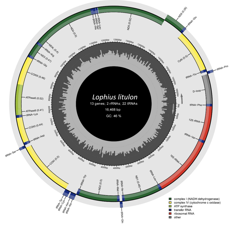
Organization and characterization of the yellow goosefish mitogenome.
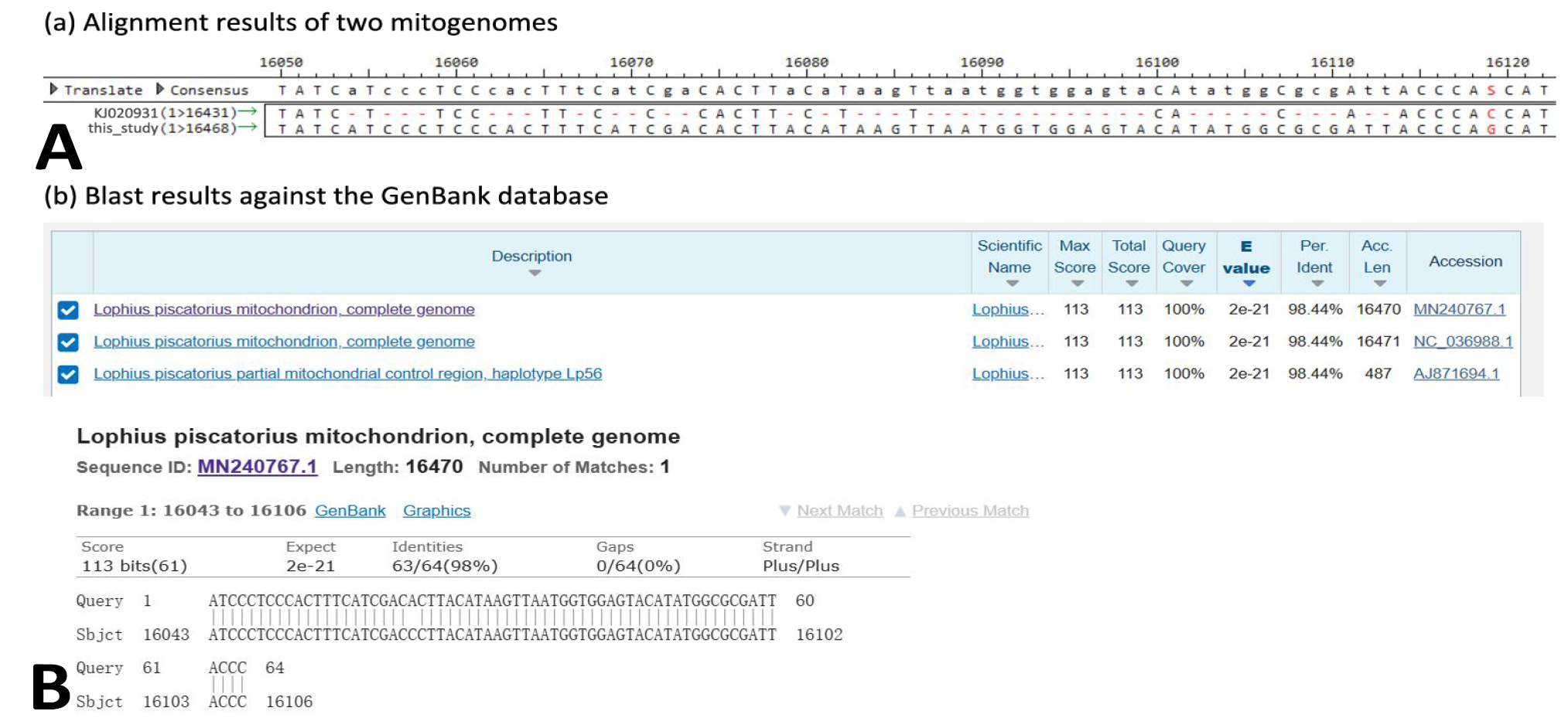
Alignment results showing the unaligned sequence in the control region (A) and subsequent BLAST results against the GenBank database (B).
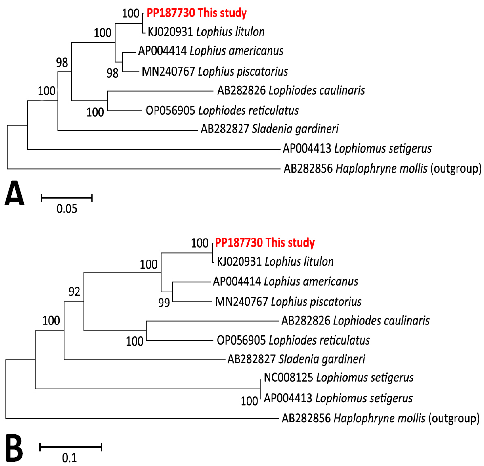
Phylogenetic maximum-likelihood topologies of investigated goosefish in family Lophiidae based on concatenated amino acid sequences (A) and nucleotide sequences (B) of 13 protein-coding genes, with the anglerfish Haplophryne mollis as the outgroup.
References
- Avise, J.C. and Saunders, N.C., 1984. Genetics, 108: 237-255. https://doi.org/10.1093/genetics/108.1.237[CrossRef]
- Iwasaki, W., Fukunaga, T., Isagozawa, R., Yamada, K., Maeda, Y., Satoh, T.P., Sado, T., Mabuchi, K., Takeshima, H., Miya, M. and Nishida, M., 2013. Mol. Biol. Evol., 30: 2531–2540. https://doi.org/10.1093/molbev/mst141[CrossRef]
- Johnson, M., Zaretskaya, I., Raytselis, Y., Merezhuk, Y., McGinnis, S. and Madden, T.L., 2008. Nucl. Acids Res., 36: W5–W9. https://doi.org/10.1093/nar/gkn201[CrossRef]
- Kumar, S., Stecher, G. and Tamura, K., 2016. Mol. Biol. Evol., 33: 1870–1874. https://doi.org/10.1093/molbev/msw054[CrossRef]
- Kwan, Y.S., Ko, M.H., Jeon, Y.S., Kim, H.J. and Won, Y.J. 2019. Ecol. Evol., 9: 1244-1254. https://doi.org/10.1002/ece3.4830[CrossRef]
- Lin, Z.J., Wang, X., Wang, J., Tan, Y., Tang, X., Werren, J.H., Zhang, D.P. and Wang, X., 2021. Int. J. Biol. Macromol., 166: 1246-1257. https://doi.org/10.1016/j.ijbiomac.2020.11.007[CrossRef]
- McGuire, J.A., Linkem, C.W., Koo, M.S., Hutchison, D.W., Lappin, A.K., Orange, D.I., Lemos-Espinal, J., Riddle, B.R. and Jaeger, J.R., 2007. Evolution, 61: 2879-2897. https://doi.org/10.1111/j.1558-5646.2007.00239.x[CrossRef]
- Miya, M., Pietsch, T.W., Orr, J.W., Arnold, R.J., Satoh, T.P., Shedlock, A.M., Ho, H., Shimazaki, M., Yabe, M. and Nishida, M., 2010. BMC Evol. Biol., 10: 1-27. https://doi.org/10.1186/1471-2148-10-58[CrossRef]
- Nevado, B., Fazalova, V., Backeljau, T., Hanssens, M. and Verheyen, E. 2011. Mol. Biol. Evol., 28: 2253-2267. https://doi.org/10.1093/molbev/msr043[CrossRef]
- Okonechnikov, K., Golosova, O., Fursov, M. and Ugene Team., 2012. Bioinformatics, 28: 1166-1167. https://doi.org/10.1093/bioinformatics/bts091[CrossRef]
- Šlechtová, V., Bohlen, J. and Perdices, A., 2008. Mol. Phylogenet. Evol., 47: 812-831. https://doi.org/10.1016/j.ympev.2007.12.018[CrossRef]
- Song, M.H., Yan, C. and Li, J.T., 2022. Brief. Bioinform., 23: bbab538. https://doi.org/10.1093/bib/bbab538[CrossRef]
- Wallis, G.P., Cameron-Christie, S.R., Kennedy, H.L., Palmer, G., Sanders, T.R. and Winter, D.J., 2017. Interspecific hybridization causes long-term Mol. Ecol., 26: 3116-3127. https://doi.org/10.1111/mec.14096[CrossRef]
- Wei, T., Wang, R., Shi, G. and Xu, T., 2016. Mitochondr. DNA Part A, 27: 286-288. https://doi.org/10.3109/19401736.2014.892080[CrossRef]
- Yoneda, M., Tokimura, M., Fujita, H., Takeshita, N., Takeshita, K., Matsuyama, M. and Matsuura, S., 1997. Fish. Sci., 63: 887-892. https://doi.org/10.2331/fishsci.63.887[CrossRef]
Services
Article usage statistics
Loading…