
Enzootic bovine leukosis (EBL) disturbs the immune system, increasing infection incidence. Clinical staging, along with hematological analysis is crucial for disease diagnosis and its progression. Our objective was to determine the clinical staging among the different age groups of BLV seropositive dairy cattle. Blood samples (n= 72) were collected from three age groups of American Friesian dairy cattle and 24 samples were collected from each group. The seroconversion against BLV was determined by using competitive ELISA, and the complete blood count was determined by using a Hematological analyzer. Hematological parameters were measured in all seropositive cattle and divided into three clinical stages based on lymphocyte and white blood cell counts. This study revealed a 37.5% seroprevalence of BLV in American Frisian dairy herd. During the progression of the disease, there was a significant change in white blood cells, lymphocytes, and monocytes. Lymphopenia developed in a later stage of the disease, with lower mean values of lymphocytes and white blood cells. The effect of age on disease occurrence and progression was significant, as evidenced by the higher seroprevalence in older animals. In conclusion, overall hematological alterations and clinical stages provide a clear visualization of the progression and management of the disease.
Research Article · Volume 58, Issue 6, 24 September 2026 · Open Access
Clinical Staging and Hematological Evaluation of Enzootic Bovine Leukosis in American Friesian Cattle
· · · · ·
Affiliations & Notes ⌄
- 1Department of Veterinary Medicine, University of Veterinary and Animal Sciences, Lahore, 54000, Pakistan 2University Diagnostic Laboratory, University of Veterinary and Animal Sciences, Lahore, 54000, Pakistan 3Department of Animal Nutrition, University of Veterinary and Animal Sciences, Pattoki, 55300, Pakistan 4School of Food and Biological Engineering, Jiangsu University, Zhenjiang, 212013, China * Corresponding author: zahid.iqbal@uvas.edu.pk
Article Info ⌄
- Journal:
- Pakistan Journal of Zoology
- Published:
- 24 September 2026

From the article
- In seropositive cattle, an earlier stage of disease was characterized as asymptomatic (AS) (n=10), during which all parameters were observed within the normal range.
- During disease progression, disruptions were observed in various parameters; however, statistically significant alterations were found only in white blood cell, lymphocyte,.
- Clinical staging is essential in evaluating the progression of disease in seropositive animals, as these animals may have different clinical presentations.
- After disease diagnosis, clinical staging provides a clear visualization of disease extent and progression, which helps to develop effective disease control strategies.
Selected from the article's own text.
Abstract
INTRODUCTION
Enzootic bovine leukosis (EBL) caused by the bovine leukemia virus (BLV) belongs to the Retroviridae family, Orthoretrovirinae subfamily and Deltaretrovirus genus which includes human T-cell leukemia virus types I and II, feline immunodeficiency virus, feline leukemia virus, and simian T-cell leukemia virus ; . BLV infection disrupts the cattle immune system, leading to poor resistance to infections and vaccinations ; . BLV-infected animals experience production loss, longevity, and trading restrictions, causing significant economic loss to the global cattle industry . The BLV-infected cattle’s apparent prevalence 46.5% of US dairy herds, 84% of Argentina dairy herds, 40.9% of dairy cattle in Japan, and other countries, impacting the cattle industry remains a complex epidemiological issue ; ; . Diagnostic testing for bovine leukosis has evolved with PCR and ELISA now commonly used, replacing the previously standard AGID (agar gel immuno-diffusion) test as the first serological method recommended by the OIE ; . ELISA is more efficient, sensitive, and accessible method than AGID . BLV is epidemiologically associated with malignancy in lymphocytes and a susceptibility to lymphoproliferative disorders. Based on disease progression, EBL is classified into aleukemic, persistent lymphocytosis (PL), and malignant stages ; . Clinical staging includes determining normal and abnormal lymphocytic and leukocyte counts indicating the occurrence of proliferative disorders. BLV positive animals remained infected throughout their lives, with 30% of animals developing persistent lymphocytosis, and more likely to develop B cell tumors . Hematological keys determined animals at risk for EBL, about half of tumor developing cattle exhibit persistent lymphocytosis, a subclinical infection stage . BLV infected animals develop persistent leukocytes, lymphocytes, and neoplastic proliferation, which mostly influence later stages of age. About 1-5% of infected animals develop malignancy conditions involving B lymphocytes in spleen, abomasum, and mammary glands. This significantly reduces productive life and carcass value ; .
Clinical staging is crucial for understanding disease progression, prognosis, and treatment . The European community leukosis key is a haematology technique used to detect BLV infection in dairy cattle by identifying persistent lymphocytosis through lymphocyte count and age . Neoplastic disease often resulting abnormalities in blood (hematopoietic system), is challenging to diagnose because it has a latent period during the asymptomatic stage. However, disruptions in normal hematological parameters indicate disease severity and its progression. Hematological analysis is a reliable method for determining EBL stages, but there is limited information on specific changes. The study aimed to determine changes in haematology cellular distribution in BLV infected cattle, excluding the uninfected cattle, across EBL clinical stages.
Materials and methods
American Friesian dairy cattle (n=72) were selected on a random basis from different age groups at the dairy farm located in Punjab with a milking herd size of about 700. The milking herd was categorized into three different age groups A (<4 years), B (<6 years), and C (>6years), and 24 animals were randomly selected from each group. Blood samples were collected using the 6 ml EDTA-K3 containing tube (Vacuum tube, Bio Vac, USA) via a jugular vein. Samples were transported in an ice box for further processing to the University Diagnostic Lab, UVAS at 4˚C and processed within 48 h. A 3 ml EDTA-mixed whole blood sample aliquot was set aside for hematology analysis. Serum was separated from the remaining EDTA-mixed blood by centrifugation at 1500 rpm for 5 min at 10˚C and stored in 1.5 ml Eppendorf tubes at -20 oC. Seroconversion against BLV has been determined by using a competitive ELISA to detect anti-gP51 antibodies (ID Screen® BLV Competition, USA) as per instructions provided by the manufacturer. The EDTA mixed whole blood aliquot of all seropositive samples was further evaluated to determine the complete blood count by using an automatic junior jet vet hematological analyzer (Abacus Junior Vet). Heamotological parameters (haemoglobin, red blood cell, mean corpuscular haemoglobin, mean corpuscular haemoglobin concentration, mean corpuscular volume, white blood cell, lymphocyte, granulocyte, monocyte, and platelet count) of all seropositive animals were measured, and divided into 3 clinical stages (asymptomatic, persistent lymphocytosis, lymphopenia) based on lymphocyte and white blood cell count.
To assess the BLV seropositivity, a binomial test with a mixed effect model was conducted by using GLIMMIX in SAS software (SAS 9.4 version; SAS Institute Cary, NC) with a logit link, as Elisa seropositivity status was binary (0 = negative and 1 = positive). We have performed descriptive statistics to summarise the distribution of each hematological parameter within the infectious status of the animal and analysis of variance (ANOVA) to assess the overall significant changes in the hematological parameters across the clinical stages.
Results and discussion
In the current investigation, overall seroprevalence of BLV was found 37.5% (27/72) in American Frisian dairy cattle. Table I presents the mean and standard deviation of haemotological parameters in seropositive animals. In seropositive cattle, an earlier stage of disease was characterized as asymptomatic (AS) (n=10), during which all parameters were observed within the normal range. However, the mean values of granulocyte and monocyte count (6.55×10⁹/L and 1.35×10⁹/L) were observed higher than the reference values (0.6-4.0×10⁹/L and 0.0-0.8×10⁹/L). In the second stage of disease, referred to as persistent lymphocytosis (PL) (n=12), the mean values of white blood cell (WBC), lymphocyte (LYM), granulocyte (GRAN), and monocyte (MONO) count increased, while haemoglobin (HB), red blood cell (RBC), mean corpuscular haemoglobin (MCH), mean corpuscular haemoglobin concentration (MCHC), mean corpuscular volume (MCV), and platelet count were all within normal range values. It was observed that lymphopenia typically emerged in later stages of the disease (n=5), characterized by lower mean values of WBC, LYM, and RBC count. During disease progression, disruptions were observed in various parameters; however, statistically significant alterations were found only in white blood cell, lymphocyte,
Table I. Hematological values (mean ± standard error) of BLV seropositive American Friesian cattle (n=27) across the clinical stages.
|
Hematological parameters3 |
Clinical stages1 |
P-value2 |
||
|
AS |
PL |
L |
||
|
Haemoglobin (g/dl) |
9.94 ± 1.46 |
10.11 ± 1.71 |
9.18 ± 1.73 |
0.56 |
|
Red blood cells (g/dl) |
6.63 ± 1.00 |
6.80 ± 1.38 |
5.85 ± 1.38 |
0.37 |
|
Mean corpuscular haemoglobin (pg) |
15.12 ± 0.42 |
14.97 ± 0.72 |
15.96 ± 1.53 |
0.09 |
|
Mean corpuscular haemoglobin concentration (g/dl) |
36.85 ± 0.32 |
34.87 ± 6.07 |
37.60 ± 1.47 |
0.39 |
|
Mean corpuscular volume (fl) |
40.84 ± 1.06 |
40.37 ± 2.64 |
42.28 ± 3.91 |
0.29 |
|
White blood cells (× 109/L) |
11.21 ± 4.66 |
25.25 ± 14.53 |
5.78 ± 1.23 |
<0.01 |
|
Lymphocytes (× 109/L) |
3.70 ± 1.04 |
18.40 ± 13.24 |
1.92 ± 0.27 |
<0.01 |
|
Granulocytes (× 109/L) |
6.55 ± 3.60 |
6.76 ± 3.83 |
3.38 ± 0.95 |
0.17 |
|
Monocytes (× 109/L) |
1.35 ± 0.35 |
1.75 ± 0.66 |
0.48 ± 0.08 |
<0.01 |
|
Platelets (× 109/L) |
225.80 ± 72.54 |
217.75 ± 77.14 |
180.60 ± 51.64 |
0.51 |
1AS, asymptomatic; PL, persistent lymphocytosis; L, lymphopenia; 2P value in the column represents the significant change in hematological parameters across the infection status (P <0.01). 3pg, pictogram; fl, femtoliter; L, liter.
and monocyte count (P <0.01) as shown in Table I. There was a statistically significant relationship between the animal age and occurrence of BLV because both were directly linked (P <0.01). It was observed that Seropositivity and progression of disease were higher in group C (> 6 years) as compared to other groups (Fig. 1).
Our objective was to evaluate the clinical staging among the BLV seropositive dairy cattle, and the impact of BLV infection on hematological parameters. BLV infection status in seropositive dairy cattle was determined by analyzing the alterations in blood cell counts. Clinical staging was classified into 3 categories based on the BLV infection status in seropositive cattle.
This study found a noteworthy association between the BLV infection and notable significant alterations in white blood cell, lymphocyte, and monocyte count (P <0.01) as the disease progressed in seropositive cattle. This suggests a higher level of infection severity. These hematological parameters play a crucial role in disease progression. This finding corroborates previous research by Williams et al. (1988); Nikolay et al. (2013); Chen et al. (2020), which also demonstrated significant associations between BLV infection and change in WBC, lymphocyte, and monocyte counts. While some studies, such as Petropavlovskiy et al. (2019) highlighted the significant impact by revealing increased leukocyte and lymphocyte numbers, but decreased monocytes.
During the asymptomatic stage (AS) of BLV infection, seropositive cattle exhibited significantly elevated WBCs, reaching the upper limit of the normal range (11.2 ×10⁹\L compared to the 4-12×109\L normal range). Moreover, the mean values of granulocytes and monocytes were observed to be higher than the normal range. This deviation suggests that BLV infection can significantly alter the host’s hematological profile. During this condition, the virus remained in latency, and cell-mediated immunity (CMI) suppresses disease progression. CMI is crucial for controlling BLV replication. However, its decline may accelerate infection . The alteration in WBC counts observed in this study may serve as an indicator of asymptomatic infection and provide insights into the host immune system.
The study outcomes revealed that BLV infection tends to remain latent during the asymptomatic stage but may progress to persistent lymphocystosis (PL), which is characterized by a significant increase in mean of WBC, LYM, GRAN, and MONO counts. This elevation in WBC and LYM counts may indicate BLV infection and can be used to assess infection activation status . B lymphocytes are the preferentially target of BLV, with B-cell expansion and its impairment mainly impaired the host immune system, especially in PL according to Libera et al. (2012). Meanwhile, Chen et al. (2020) reported an increase in T and B lymphocytes. PL involves an elevated lymphocyte count exceeding 8000/μL that remains despite the absence of visible signs, as it consistently triggers the immune response ; . Evidence suggests that mean values of monocyte count were significantly elevated during the both AS and PL stages of disease. However, this finding, regarding the role of monocytes in BLV infection was mixed. While some studies, such as Heeney et al. (1992) have reported evidence of BLV infection in monocytes, suggesting a broader impact on the immune system. Mirsky et al. (1996) found no evidence of BLV provirus in monocytes, suggesting that they may not be a significant target of virus.
One critical transition was the shift from persistent lymphocytosis stage to lymphopenia stage of disease. This stage was marked by the decrease in the mean values of WBCs and LYM count. Lymphopenia is a concerning development, accompanied by lymphocyte exhaustion. T-cell exhaustion was driven by the increased expression of immunosuppressive factors which induced immune exhaustion, specifically T-cell, and diminishing cell proliferation ; ; . This study revealed a significant correlation between animal age and BLV occurrence in older animals showing higher BLV infection seroprevalence. According to previous studies, age has a significant influence on the progression of infection and may increase the risk of BLV infection (Barros Filho et al., 2010; ; . The study emphasizes the significance of monitoring blood parameters as a key indicator of BLV infection severity, and provides an understanding of disease progression.
Conclusions
Clinical staging is essential in evaluating the progression of disease in seropositive animals, as these animals may have different clinical presentations. During the asymptomatic stage, the virus latency period may not affect animal health. However, as the disease progresses in animals, it weakens the host immune system and becomes dormant. After disease diagnosis, clinical staging provides a clear visualization of disease extent and progression, which helps to develop effective disease control strategies.
Declarations
Funding
This article was supported by a project of the Higher Education Commission of Pakistan (Project grant no. 16873) entitled “In depth genetic diversity and molecular epidemiology of newly emerging Bovine Leukemia Virus (BLV) in dairy animals of Punjab and development of strategies to control disease in Pakistani herd.
Ethical statement
All the experimental procedures involving animals were approved by the Institutional Guidelines of the Ethical Review Committee (No. DR 325) of the University of Veterinary and Animal Sciences, Lahore.
Statement of conflict of interest
The authors have declared no conflict of interest.
Figures
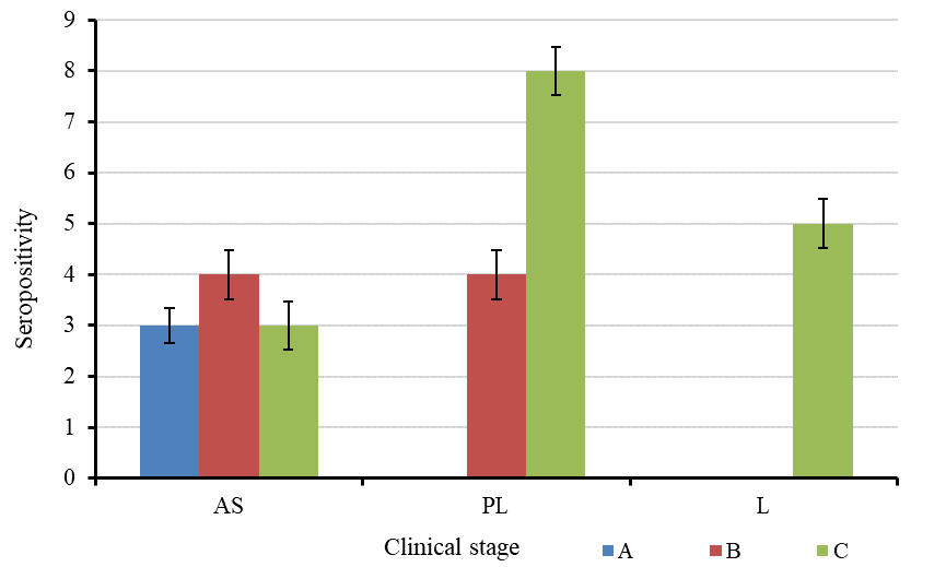
Progression of disease significantly influenced by age. Age groups A (<4 years), B (<6 years), and C (>6 years) had 3 different clinical stages. AS, asymptomatic; PL, persistent lymphocytosis; L, lymphopenia in BLV ELISA positive American Friesian cattle based on lymphocyte count. Error bars indicate standard deviation in age groups according to stages.
References
- Akagami, M., Oya, S., Kashima, Y., Seki, S., Ouchi, Y. and Hayama, Y., 2019. A hematologic key for bovine leukemia virus screening in Japanese black cattle. J. Vet. Diagn. Invest., 31: 568-571. https://doi.org/10.1177/1040638719848449[CrossRef]
- Barber, D.., Wherry, E.J., Masopust, D., Zhu, B., Allison, J.P., Sharpe, A.H., Freeman, G.J. and Ahmed, R., 2006. Restoring function in exhausted CD8 T cells during chronic viral infection. Nature, 439: 682-687. https://doi.org/10.1038/nature04444[CrossRef]
- Barros-Filho, I.R.d., Guimarães, A.K., Sponchiado, D., Krüger, E.R., Wammes, E.V., Ollhoff, R.D., Dornbusch, P.T. and Biondo, A.W., 2010. SoroprevalÊncia De Anticorpos Para O VÍrus Da Leucose EnzoÓtica Em Bovinos Criados Na RegiÃo Metropolitana De Curitiba, ParanÁ. Arq. Inst. Biol., 77: 511-515. https://doi.org/10.1590/1808-1657v77p5112010[CrossRef]
- Benitez, O.J., LaDronka, R.M., Norby, B., Grooms, D.L. and Bartlett, P.C., 2022. The effect of bovine leukemia virus on dairy cow longevity. JDS Commun., 3: 185-188. https://doi.org/10.3168/jdsc.2021-0187[CrossRef]
- Chen, Y.C., Chang, C.C., Hsu, W.L. and Chuang, S.T., 2020. Dairy cattle with bovine leukaemia virus RNA show significantly increased leukocyte counts. Vet. J., 257: 105449. https://doi.org/10.1016/j.tvjl.2020.105449[CrossRef]
- Emanuelson, U., Scherling, K. and Pettersson, H., 1992. Relationships between herd bovine leukemia virus infection status and reproduction, disease incidence, and productivity in Swedish dairy herds. Prev. Vet. Med., 12: 121-131. https://doi.org/10.1016/0167-5877(92)90075-Q[CrossRef]
- Erskine, R.J., Bartlett, P.C., Sabo, K.M. and Sordillo, L.M., 2011. Bovine Leukemia Virus Infection in Dairy Cattle: Effect on Serological Response to Immunization against J5 Escherichia coli Bacterin. Vet. Med. Int., 2011: 915747. https://doi.org/10.4061/2011/915747[CrossRef]
- Fatima, A., Iqbal, M. Z., Fatima, N., Riaz, S., Ahmad, H. and Ur Rehman, M., 2025. Prevalence and genotypic distribution of bovine leukemia virus across asian regions: Insights into economic significance and clinical staging. Bio Commun., 1: 29-39. https://doi.org/10.71320/bcs.0008[CrossRef]
- Frossard, J.P., 2022. Retroviridae. Vet. Microbiol., pp. 698-727. https://doi.org/10.1002/9781119650836.ch65[CrossRef]
- Gillet, N., Florins, A., Boxus, M., Burteau, C., Nigro, A., Vandermeers, F., Balon, H., Bouzar, A.B., Defoiche, J., Burny, A., Reichert, M., Kettmann, R. and Willems, L., 2007. Mechanisms of leukemogenesis induced by bovine leukemia virus: prospects for novel anti-retroviral therapies in human. Retrovirology, 4: 18. https://doi.org/10.1186/1742-4690-4-18[CrossRef]
- González, T., Licursi, M. and Bonzo, E., 2007. Enzootic bovine leukosis: performance of an indirect ELISA applied in serological diagnosis. Braz. J. Microbiol., 38: 1-5. https://doi.org/10.1590/S1517-83822007000100001[CrossRef]
- Heeney, J.L., Valli, P.J., Jacobs, R.M. and Valli, V.E., 1992. Evidence for bovine leukemia virus infection of peripheral blood monocytes and limited antigen expression in bovine lymphoid tissue. Lab. Invest., 66: 608-617. https://www.ncbi.nlm.nih.gov/pubmed/1315405
- Hoff-Jorgensen, R., 1989. An international comparison of different laboratory tests for the diagnosis of bovine leukosis: suggestions for international standardization. Vet. Immunol. Immunopathol., 22: 293-297. https://doi.org/10.1016/0165-2427(89)90016-0[CrossRef]
- Kabeya, H., Ohashi, K. and Onuma, M., 2001. Host immune responses in the course of bovine leukemia virus infection. J. Vet. med. Sci., 63: 703-708. https://doi.org/10.1292/jvms.63.703[CrossRef]
- Khudhair, Y.I., Hasso, S.A., Yaseen, N.Y. and Al-Shammari, A.M., 2016. Serological and molecular detection of bovine leukemia virus in cattle in Iraq. Emerg. Microb. Infect., 5: e56. https://doi.org/10.1038/emi.2016.60[CrossRef]
- LaDronka, R.M., Ainsworth, S., Wilkins, M.J., Norby, B., Byrem, T.M. and Bartlett, P.C., 2018. Prevalence of bovine leukemia virus antibodies in US Dairy Cattle. Vet. Med. Int., 2018: 5831278. https://doi.org/10.1155/2018/5831278[CrossRef]
- Libera, A.M.M.P.D., Blagitz, M.G., Batista, C.F., Latorre, A.O., Stricagnolo, C.R. and Souza, F.N.D., 2012. Quantification of B cells and T lymphocyte subsets in bovine leukemia virus infected dairy cows. Semin. Ciênc. Agrár., 33: 1487-1494. https://doi.org/10.5433/1679-0359.2012v33n4p1487[CrossRef]
- Mammerickx, M., Lorenz, R.J., Straub, O.C., Donnelly, W.J.C., Flensburg, J.C., Gentile, G., Markson, L.M., Ressang, A.A. and Taylor, S.M., 2010. Bovine Hematology1. Zent. Vet. Reihe B, 25: 257-267. https://doi.org/10.1111/j.1439-0450.1978.tb00001.x[CrossRef]
- Mirsky, M.L., Olmstead, C.A., Da, Y. and Lewin, H.A., 1996. The prevalence of proviral bovine leukemia virus in peripheral blood mononuclear cells at two subclinical stages of infection. J. Virol., 70: 2178-2183. https://doi.org/10.1128/jvi.70.4.2178-2183.1996[CrossRef]
- Morovati, H., Shirvani, E., Noaman, V., Lotfi, M., Kamalzadeh, M., Hatami, A., Bahreyari, M., Shahramyar, Z., Morovati, M.H., Azimi, M. and Sakhaei, D., 2012. Seroprevalence of bovine leukemia virus (BLV) infection in dairy cattle in Isfahan Province, Iran. Trop. Anim. Hlth. Prod., 44: 1127-1129. https://doi.org/10.1007/s11250-011-0062-4[CrossRef]
- Murakami, K., Kobayashi, S., Konishi, M., Kameyama, K. and Tsutsui, T., 2013. Nationwide survey of bovine leukemia virus infection among dairy and beef breeding cattle in Japan from 2009-2011. J. Vet. med. Sci., 75: 1123-1126. https://doi.org/10.1292/jvms.12-0374[CrossRef]
- Nakada, S., Fujimoto, Y., Kohara, J., Adachi, Y. and Makita, K., 2022. Estimation of economic loss by carcass weight reduction of Japanese dairy cows due to infection with bovine leukemia virus. Prev. Vet. Med., 198: 105528.https://doi.org/10.1016/j.prevetmed.2021.105528[CrossRef]
- Nikolay, S., Dimitrinka, Z., Ivanka, S., Nikolina, R. and Teodora, M., 2013. Investigation of some haematological and blood biochemical parameters in cattle spontaneously infected with bovine leukosis virus. Maced. Vet. Rev., 36: 107-110.
- Okagawa, T., Konnai, S., Ikebuchi, R., Suzuki, S., Shirai, T., Sunden, Y., Onuma, M., Murata, S. and Ohashi, K., 2012. Increased bovine Tim-3 and its ligand expressions during bovine leukemia virus infection. Vet. Res., 43: 45. https://doi.org/10.1186/1297-9716-43-45[CrossRef]
- Okagawa, T., Konnai, S., Nishimori, A., Maekawa, N., Goto, S., Ikebuchi, R., Kohara, J., Suzuki, Y., Yamada, S., Kato, Y., Murata, S. and Ohashi, K., 2018. Cooperation of PD-1 and LAG-3 in the exhaustion of CD4(+) and CD8(+) T cells during bovine leukemia virus infection. Vet. Res., 49: 50. https://doi.org/10.1186/s13567-018-0543-9[CrossRef]
- Petropavlovskiy, M., Vereschyak, N., Bezborodova, N. and Oparina, O., 2019. Immuno-biological evaluation of individual genetic variants of bovine leukemia virus in the conditions of the Ural region. International Scientific and Practical Conference Digital Agriculture-Development Strategy (ISPC, 2019). https://doi.org/10.2991/ispc-19.2019.84[CrossRef]
- Radostits, O.M., Gay, C., Hinchcliff, K.W. and Constable, P.D., 2006. Veterinary medicine e-book: A textbook of the diseases of cattle, horses, sheep, pigs and goats. Elsevier Health Sciences.
- Selim, A., Marawan, M.A., Ali, A.F., Manaa, E. and AbouelGhaut, H.A., 2020. Seroprevalence of bovine leukemia virus in cattle, buffalo, and camel in Egypt. Trop. Anim. Hlth. Prod., 52: 1207-1210. https://doi.org/10.1007/s11250-019-02105-8[CrossRef]
- Simard, C., Richardson, S., Dixon, P., Belanger, C. and Maxwell, P., 2000. Enzyme-linked immunosorbent assay for the diagnosis of bovine leukosis: comparison with the agar gel immunodiffusion test approved by the Canadian Food Inspection Agency. Can. J. Vet. Res., 64: 101-106. https://doi.org/10.1023/A:1019028229377[CrossRef]
- Trono, K.G., Perez-Filgueira, D.M., Duffy, S., Borca, M.V. and Carrillo, C., 2001. Seroprevalence of bovine leukemia virus in dairy cattle in Argentina: Comparison of sensitivity and specificity of different detection methods. Vet. Microbiol., 83: 235-248. https://doi.org/10.1016/S0378-1135(01)00420-5[CrossRef]
- Velu, V., Titanji, K., Zhu, B., Husain, S., Pladevega, A., Lai, L., Vanderford, T.H., Chennareddi, L., Silvestri, G., Freeman, G.J., Ahmed, R. and Amara, R.R., 2009. Enhancing SIV-specific immunity in vivo by PD-1 blockade. Nature, 458: 206-210. https://doi.org/10.1038/nature07662[CrossRef]
- Williams, D.L., Amborski, G.F. and Davis, W.C., 1988. Enumeration of T and B lymphocytes in bovine leukemia virus-infected cattle, using monoclonal antibodies. Am. J. Vet. Res., 49: 1098-1103. https://www.ncbi.nlm.nih.gov/pubmed/2844108
Services
Article usage statistics
Loading…