
In this study, expression pattern-and function of growth arrest specific gene 6 (Gas6) during porcine oocyte maturation and early embryonic development were systemically investigated. The expression of Gas6 in the process of follicular development was studied by immunohistochemical staining, immunofluorescence, QRT-PCR and western blot. Immunohistochemical staining results showed that Gas6 was expressed in the oocytes, especially the nucleus, from the primary to preovulatory stages. The further immunofluorescence results showed that Gas6 was expressed thoughout the in vitro matured porcine oocytes, and the staining signal decreased in the first 12 h but then increased during the process of oocyte maturation. QRT-PCR analysis revealed that Gas6 mRNA was continuously expressed during the process of oocyte maturation, disappeared after parthenogenetic activation, re-appeared and elevated in the blastocysts stage. The following western blot analysis revealed a similar expression pattern with QRT-PCR and immunofluorescence. In conclusion, the expression pattern of Gas6 gene is closely related to oocytes maturation and embryo development. Gas6 may play a crucial role in the process of oocytes maturation and improve the development potential of early embryos by promoting the cytoplasmic maturation.
Research Article · Volume 54, Issue 1, 18 December 2021 · Open Access
Growth Arrest Specific Gene 6 Improved in vitro Maturation of Oocytes and the Development Potential of Porcine Embryo after Parthenogenetic Activation
· · · · · · ·
Affiliations & Notes ⌄
- 1State Key Laboratory for Conservation and Utilization of Subtropical Agro-Bioresources, Guangxi University, Nanning, Guangxi 530005, China. , * Corresponding author: ardsshi@gxu.edu.cn
- 2qyliu-gene@gxu.edu.cn
Article Info ⌄
- Journal:
- Pakistan Journal of Zoology
- Published:
- 18 December 2021

From the article
- The expression of Gas6 was usually decreased in the first 12 h maturation of oocytes and recovered subsequently and was closely related to porcine oocytes maturation and embryo development.
- While tracking the embryonic development potential of the oocytes, it was found that the addition of Gas6 protein significantly increased the cleavage rate, blastocyst rate and blastocyst cell number.
- We report the expression of Gas6 in the process of follicular development and in vitro maturation porcine of oocyte and analysed the gene regulation mode of Gas6 in the process of oocyte maturation and embryonic development.
- This work provides a reference for further exploration of the regulatory mechanism of Gas6 in the process of oocyte maturation and early embryonic development.
Selected from the article's own text.
Abstract
INTRODUCTION
Oocyte growth and maturation are long-lasting and necessary processes for fertilization and embryonic development ; . Oocyte maturation involves in nuclear and cytoplasmic maturation, and amount of maternal mRNA, protein, and regulatory molecules were stored in oocytes to support completion of meiosis, fertilization, and early embryogenesis ; . This process is mainly controlled by post-transcriptional regulatory mechanisms, such as RNA polyadenylation, localization, sorting, and masking, as well as protein phosphorylation . Therefore, functional analysis of certain gene(s) in the oogenesismay provide important information on the molecular regulatory mechanisms of oocyte nuclear and cytoplasmic maturation .
The growth arrest specific (Gas) gene was first identified in cells under stressors such as nutrient deprivation or exposure to genotoxic agents . Growth arrest-special gene 6 (Gas6) was previously isolated as a gene product inducible expressed in fibroblasts in response to serum starvation . It has a structure similar to that of protein S, a vitamin K-dependent plasma protein with anticoagulant activityat 43% amino acid identity . Numerous studies have indicated that Gas6-mediated signalling is implicated in cell survival, growth arrest, proliferation, differentiation, and other cell type-specific functions ; ; . Clinically, Gas6 plays an important role in haematosis and thombosis . In mice, Gas6 is expressed in the gonads and supports primordial germ cell growth and survival in cultureand the regulation of spermatogenesis and apoptosis is also controlled by Gas6 ; . Previous studies haverevealed that Gas6 is expressed in germinal vesicle (GV) oocytes, 8-cell embryo and the blastocyst ; . The absence of Gas6 is associated with a deficit in embryonic erythoid precursor cells and Gas6–/– adult mice showed a platelet dysfunction .
Cytoplasmic maturation, a poorly understood physiological process, supports the fertilization of the oocyte and early embryonic development . Previous studies found that Gas6 downregulation impaired cytoplasmic maturation and pronuclear formation of oocytes in mice independent of maturation promoting factor (MPF) activity . This suggests that Gas6 may have crucial function on oocytes maturation and early embryonic development. To date, efforts to elucidate the function of Gas6 and receptor signalling have focused mainly on thombosis and spermatogenesis, while few studies on oocytes maturation and embryos development. Thus, the objective of the present study was to explore the role and expression pattern of Gas6 in porcine oocytes and preimplantation embryos.
MATERIALS AND METHODS
Reagents
Chemicals and reagents were obtained from Sigma Chemical Co. (St. Louis, MO), unless noted otherwise.
Immunohistochemical (IHC) studies
Porcine ovaries were obtained from a local abattoir. The ovaries of cyclic sows aged 5-8 months were selected for this study. We selected 10 ovaries from different cyclic sowsand fixed them overnight in 4% (w/v) paraformaldehyde in phosphate-buffered saline (PBS; 0.01 M, pH 7.6; Sigma, Poole) and transferred to 70% (v/v) ethanol until processing. The fixed tissue was embedded in paraffin wax after dehydration.
Sections (7 μm thickness) were dewaxed and rehydrated in decreasing concentrations of alcohol (95, 90, 80 and 70%, respectively, and distilled water), followed by two washes of 5 min each in PBS. Antigen retrieval was performed by microwaving the sections in citrate buffer (0.01 M, pH 6.0) at 100°C for 10 min, and the sections were kept in the buffer for 60 min until cool. After washing in PBS, the sections were incubated in 3% hydrogen peroxide in water for 15 min. After washing in PBS, a combined avidin-biotin block was performed according to the manufacturer’s instructions (Proteintech, Wuhan). In brief, avidin was added to the blocking buffer, which consisted of 5% (w/v) bovine serum albumin (BSA; Sigma) in PBS. The slides were incubated for 30 min and then washed in PBS. Biotin was added to the blocking buffer and the slides were treated for 15 min and washed in PBS. Rabbit polyclonal antibodies directed against Gas6 (ab136249, 1:250 dilutions; Abcam) were used in blocking buffer diluted at 1:250. Negative controls were obtained by replacing the primary antibody with pre-immune serum. Slides with serial sections were incubated with antibodies in a humidified chamber at 37°C overnight. Slides were washed twice for 5 min in 0.1% (v/v) Tween 20 in PBS (PBST) and incubated with goat anti-rabbit biotinylated antibody (Proteintech, Wuhan) diluted 1:300 in blocking buffer for 1 h at room temperature. The sections were washed twice in PBST for 5 min, incubated in ABC-HP (Proteintech, Wuhan) for 1 h at room temperature and washed again in PBST. Bound antibodies were visualized by incubation in HP-TAB (brown stain; Tiangen Biotech, Beijing); the sections were counterstained with haematoxylin (Beyotime, Shanghai), dehydrated though increasing concentrations of alcohol and mounted with cover slips. The slides were examined using an Olympus Provis microscope (Olympus Optical Co., London) and the images were captured with a Kodak DCS420 camera (Eastman Kodak, New York, USA) and assembled in Photoshop CS6 software (Adobe, San Jose, USA). Approximately 100 follicles from each ovary were examined in sections. Staining density and IOD (integrated optical density) was analysed by image-pro plus 6.0 software (Media Cybernetics, Washington, USA). As we focused on the function of Gas6 in oocytes maturation, only staining in oocytes was analysed.
Cell culture media (CCM)
The base media for in vitro maturation (IVM) of oocytes in this study was medium 199 (M-199) (Invitrogen, Grand Island, NY) and Porcine Zygote Medium (PZM)-3 for embryo culture. M-199 was supplemented with 10% (v/v) porcine follicular fluid, 0.6 mM cysteine, 0.91 mM pyruvate, 10 ng/ml epidermal growth factor, 75 mg/ml kanamycin, and 1 mg/ml insulin (PM). The in vitro culture medium for embryo development was PZM-3, which was modified by adding 2.77 mM myo-inositol, 0.34 mM tri-sodium citrate, and 10 μM β-mercaptoethanol .
Oocyte collection and IVM
Porcine ovaries were obtained from a local abattoir and transported to the laboratory at 29-31°C. Cumulus-oocyte complexes (COCs) were aspirated from superficial follicles (3-6mm in diameter) in the ovaries using an 18-gauge needle and a 10-ml syringe. Only COCs with uniform cytoplasm and at least three layers of compact cumulus cells were selected for maturation and further studies. COCs were placed into Petri dishes containing 150μL of IVM medium with 10 IU/ml equine chorionic gonadotropin (eCG) and 10 IU/ml human chorionic gonadotropin (hCG) (Intervet International BV, Boxmeer, Holland). The COCs were cultured at 39 °Cin 5% CO2 at maximum humidity. After 22 h in the maturation culture, the COCs were washed three times in fresh hormone-free IVM medium, and then cultured in hormone-free IVM medium for an additional 20 hs for parthenogenetic activation (PA). The oocytes in vitro matured to GVBD (1-12 h), MI (12-24 h), post-MI (24-36 h) and MII (36-42 h) were collected to check the expression of Gas6 mRNA and protein based on the previous study in our lab .
After 42 h of IVM, cumulus-cell-free oocytes were incubated for 15 min in PBS containing 2 μg/mL Hoechst 33342. Following incubation, the oocytes were washed twice with fresh PBS medium. An epifluorescence microscope (TE300; Nikon, Tokyo, Japan) was used to identify the polocyte.
Parthenogenetic activation (PA)
Parthenogenetic activation was performed according to the previous study . In brief, after IVM, oocytes were denuded of cumulus cells by repeated pipetting with 0.1% hyaluronidase in cell culture medium (CCM) on a warm stage at 39℃. Denuded oocytes were washed 3 times in CCM. Cumulus free oocytes showing first polar body were selected and activatedusing a cell fusion generator with thee pulses of 80 V/ mm direct current (DC) for 50 msec.
Post-activation treatment and embryo culture
Following electrical activation, the PA embryos were washed three times in fresh in vitro culture medium (PZM-3), and 35 μL in vitro culture droplets were transferred under mineral oil and cultured at 39°C in 5% CO2 at maximum humidity for 7 days. Cleavage and blastocyst formation were evaluated on day 2 and 7, with the day of PA designated as day 0. The total blastocyst cell count was obtained using Hoechst 33342 staining under an epifluorescence microscope. After incubation, oocytes were washed twice with fresh PBS medium, aliquoted into 10 μL droplets, and observed for fluorescence using an epifluorescence microscope (TE-300; Nikon) with a UV filter (370 nm). Fluorescent images were recorded and saved in TIFF format.
Immunofluorescence staining
Denuded oocytes and COCs were placed in 4% paraformaldehyde and fixed for 40 min at room temperature. Fixed oocytes were washed three times in PBS for 5 min each and were blocked with 1% BSA for 1 h and incubated with rabbit monoclonal anti-Gas6 antibody (ab136249, 1:250 dilutions; Abcam) at 4 °C overnight. Oocytes incubated with PBS containing 0.05% BSA were used as negative control. After washing, oocytes were incubated with FITC-conjugated anti-rabbit IgG (SA00003-3, 1:200, Proteintech, Wuhan) for 1 h at room temperature, and DNA was counterstained with propidium iodide. All of the experiments were repeated more than three times.
The occurrence of Gas6 immunofluorescence in pig oocytes was determined by confocal scanning laser microscopy. The analysis of oocyte immunofluorescence by scanning laser confocal microscopy enabled the precise detection of unique distributions of Gas6 because individual cells could be examined in sections and all portions of the oocytes could be studied in isolation. Oocytes were scanned at the same laser intensity and photomultiplier settings as far as possible. However, in some instances, such as the COCs and GCs, scans were performed at lower signal amplification in order to view cell-specific immunofluorescence in finer detail. For some images, such as the denuded oocytes, higher settings were used to accentuate the fluorescent signal in order to emphasize the differential distribution. Fluorescence unit was calculated and analysed with image-pro plus 6.0 software (Media Cybernetics, Washington, USA).
cDNA synthesis
Total RNA was extracted from COCs, oocytes and embryos (5 oocytes or embryos per tube, 3 repeats) using the RNeasy Micro Kit (Qiagen, Valencia, CA, USA) according to the manufacturer’s instructions and residual genomic DNA was removed by using RNase Free DNase (Qiagen) treatment for 15 min . Messenger RNA from GV stage COCs, GVBD stage COCs, MI stage COCs, post-MI stage COCs, MII stage COCs, denuded germinal vesicle stage oocytes, denuded MII stage oocytes (as confirmed by the presence of one polar body), 2-cell stage embryos, 4-cell stage embryos, 8-cell stage embryos and blastocysts were reverse transcribed to cDNA using the SuperScript® II Reverse Transcriptase kit (Invitrogen, Life Technologies), according to the manufacturer’s instructions. In brief, the reverse transcription system contains a 20-μL mixture as follows: 8 μL of mRNA, 1 μL of DNase Ⅰ, 1.3 μL of DNase I Buffer,1 μL of EDTA, l μL of dNTP, 2 μL of random primer,4 μL of 5×FS Buffer,0.5 μL of RNase Inhibitor,2 μL of DTT, and 0.3 μL of FS RT (reverse transcriptase).
Gene expression analysis by QRT-PCR
Preceding QRT-PCR amplification, primers for Gas6 (GenBank: KC526197.1) and 18S were designed using Oligo 6.0 software (Table I). Primers were synthesized by Sangon Biotech (Shanghai) Co., Ltd. Gene transcripts were quantified using SYBR Green supermix (4913914001, Roche) and fluorescence data were acquired using a fluorescence ration PCR instrument (ABI 7500, Applied Biosystems, Foster City, CA, USA). In each run, 1 μL of cDNA was used as the template for the amplification reaction and 1 μL of ddH2O was used as negative control. The sample was added to 19 μL of reaction mixture, containing 8 μL of ddH2O, 10 μL of Faststart Universal SYBR Green Master (ROX), and 0.5 uM aliquots of both forward and reverse primers. The thermal cycling profile started with a 3-min dwell temperature of 50°C, followed by 40 cycles of 30 sec at 94°C, 30 secs at the primer specific annealing temperature (60°C), 30 secs at 72°C, and a final step during which fluorescence was acquired. After 40 cycles, the program continued with a post-dwell of 1 min at 94°C. Finally, a melt curve was generated by temperature increments of 0.1°C starting from 60 to 100°C, with fluorescence acquisition after each step. Each sample was repeated 4 times, and the relative gene expression was calculated using the 2-△△Ct method with 18s as the reference gene and the expression ratio was calculated against Gas6 expression in the GV oocyte.
Table I. Primer sequences used in this study.
| Gene | Primer sequences (5’ to 3’) | Product(bp) |
| Gas6 | F: CTACTCCTGCCTGTGTGACG | 188 |
| R: GGATGTCCTCACAGGTGCTC | ||
| 18S | F: GATGGGCGGCGGAAAATTG | 107 |
| R: TCCTCAACACCACATGAGCA |
Western blot analysis
Protein extracts (150 oocytes or 150 embryos per lane, 3 repeats) were separated using 10% SDS-PAGE and transferred onto a 0.2μm nitrocellulose membrane (Pall, New York, US). The membrane was blocked for 1 h in TBST containing 5% non-fat dry milk. The blocked membranes were then incubated with rabbit polyclonal anti-gas6 antibody (ab136249, 1:1000, Abcam) in TBST. After incubation, membranes were incubated with horseradish-peroxidase-conjugated anti-rabbit IgG (SA00003-3, 1:2000, Proteintech, Wuhan) in TBST for 1 h at room temperature. After each step, the membranes were washed several times with TBST, and bound antibody was detected using an enhanced chemiluminescence detection system (Pierce ECL Western Blotting Substrate, Thermo).
Statistical analysis
All of the data we obtained in this study were analyzed using one-way ANOVA followed by Duncan’s Multiple Comparison Tests as implemented in the SPSS statistical software package (IBM, Chicago, USA).
Results
Immunohistochemistry (IHC)
The expression of Gas6 in porcine ovarian tissue was firstly studied by immunohistochemical staining. Approximately 100 follicles from each ovary were examined in sections. Strong immunostaining for Gas6 was observed in the oocytes, especially the nucleus, from the primary to preovulatory stages, with no staining in the negative control (Fig. 1A). Staining was also observed in zonapellucida and most granulosa layers of follicles, except the primordial follicles (Fig. 1A). The staining density (only staining in oocytes was analysed) was statistical analysed by IPP 6.0 software and results showed that the staining of Gas6 lessened from primary follicles to secondary follicles, while gradually enhanced following the growth of the follicles (Fig. 1B).
Immunofluorescence staining
The expression of Gas6 in in vitro matured oocytes was analysed by using Immunofluorescence staining. Immunofluorescence images of the oocytes stained with anti-Gas6 antibodies are presented in Figure 2. Analysis of oocytes stained with anti-Gas6 antibody demonstrated that Gas6 was expressed persistently throughout the in vitro maturing of the oocytes. No expression was observed in the negative control. Specifically, comparing the immunofluorescence staining of the denuded oocytes and COCs showed that Gas6 localized in the nucleus of the oocytes andcumulus cells (Fig. 2A, B), which is consistent with the immunohistochemical localization results in follicles. Further analysis of the relative fluorescent signal demonstrated the expression pattern of the Gas6 protein in the COCs and denuded oocytes, which decreased to the bottom in the first 12 h, and then gradually increasing following the maturation of the oocytes (Fig. 2C).
Gas6 mRNA expression pattern in oocytes and embryos
The expression pattern of Gas6 mRNA during oocytes maturation and preimplantation embryo development was determined by QRT-PCR. In each reaction, the absence of nonspecific amplification was confirmed by checking the melting curve of the Gas6 and 18s gene with a single peak. During oocyte maturation, the expression of Gas6 decreased gradually in the first 12 h, while increased 5-folds in the following 12 h and remained in the MII stage (Fig. 3A), which is in close agreement with the immunofluorescence results above. In the pre-implanted embryos, the Gas6 transcript was rapidly decreased to an undetectable level from MII oocytes to 2-cell and 4-cell stage, while recovered at 8-cell stage embryos and increased in the following development (Fig. 3B).
Western blot analysis
To confirm the expression of Gas6 protein, we further performed western blot analysis for samples of denuded oocytes, COCs and early embryos. A high level of expression of Gas6 was observed in COCs matured for 42 h compared to the cumulus-cell-free oocytes and pre-maturation samples. No specific hybridization bands were found in the protein samples of embryos until the blastocyst stage. These data suggest that the expression of Gas6 was constitutively throughout oocyte maturation and silenced in the early development of embryo, which is consistent with the results of QRT-PCR. The peak expression also appeared at the blastocyst stage (Fig. 4).
DISCUSSION
First time we reported that the Gas6 is continuously expressed in the process of porcine follicular development and oocyte maturation. The expression of Gas6 was usually decreased in the first 12 h maturation of oocytes and recovered subsequently and was closely related to porcine oocytes maturation and embryo development.
Gas6 is an important participant in cell survival, growth arrest, proliferation and differentiation, and maternal effect gene that is essential for cytoplasmic maturation of oocytes and early embryogenesis. It was suggested that Gas6 contributes in many aspects for gonad development , and numerous studies have indicated that the Gas6 gene is involved in the regulation of early embryonic development ; . Cytoplasmic maturation is critical for embryonic development and is directly related to the normal activation of zygote genome, especially for the genome reprogramming of nuclear transfer embryonic. Multiple studies have focused on genes that affect the cytoplasmic maturation, and Gas6 has been discussed as a candidate gene. Gas6 is expressed in germinal ridge and primordial germ cells and the developing ovary of mice . When it comes to porcine, we found that Gas6 is continuously expressed during oocyte maturation both in the follicle and IVM medium, and the expression were mainly located in the nucleus, cumulus cells and zona pellucida. Further analysis of the express pattern indicated that the expression was reduced before the GVBD stage (in the first 12 h), while increased again in the following maturation and maintained to the MII stage. The expression of Gas6 is closely related and plays important role in process of oocytes maturation. Supplement of Gas6 protein in the process of oocytes maturation did not significantly affect the polar body rate at MII stage, indicating that Gas6 is not essential for the progression of nuclear maturation in porcine oocytes. While tracking the embryonic development potential of the oocytes, it was found that the addition of Gas6 protein significantly increased the cleavage rate, blastocyst rate and blastocyst cell number. Therefore, we believe that Gas6 improves the embryonic development potential of the oocytes by promoting the cytoplasmic maturation during the in vitro maturation.
The expression of Gas6 was located in the nucleus of the oocytes, while regulated the cytoplasm maturation of the oocytes becomes an interesting problem. Analysis suggested that, the cytoplasmic maturation begins mainly after GVBD, at which time the typical components of the nucleus, such as nuclear membrane and nucleolus dissolved, breaking the physical separation between the nucleus and the cytoplasm. Meanwhile, most of the organelles are located in the middle of the oocytes around the nucleus and facilitated the interaction between the nucleus and the cytoplasm. It is well known that oocyte maturation is regulated mainly by the activities of MPF and MAPK ; . Previous studies have demonstrated that downregulation of Gas6 expression dramatically decreased the activities of MPF. Further study showed that oocytes rescued of the MPF activity with Gas6-silenced still failed to undergo PN formation despite sperm penetration into the cytoplasm, indicating that Gas6 is directly involved in the regulation of cytoplasmic maturation, independent of MPF activity . This study systematically investigated the role of Gas6 in the cytoplasmic maturation of oocytes but did not explain how Gas6 regulates cytoplasmic maturation of oocytes independent of MPF activity. Combined with our findings, we speculate that Gas6 acts directly or indirectly as a downstream receptor of MPF which can activate the activity of MPF. The silence of Gas6 may reduce the downstream receptor and block the MPF pathway, which made the active MPF out of working, and the superfluous MPF activity was degraded by degradation of the cyclin B1 and increasing of the phosphorylation of Tyr15 in p34cdc2. Therefore, the supplement of Gas6 protein in the oocytes may improve the downstream receptor of MPF and promote the MPF pathway, and result in better cytoplasmic maturation of the oocytes.
conclusion
We report the expression of Gas6 in the process of follicular development and in vitro maturation porcine of oocyte and analysed the gene regulation mode of Gas6 in the process of oocyte maturation and embryonic development. It is generally accepted that a complex equilibrium of interactions links oocyte maturation and the culture media, which plays an active role by focusing growth factors, proteases, and protease inhibitors close to reproduction. We speculate that Gas6 may be one of the key factors involved in the regulation of downstream receptors of MPF and may directly or indirectly affect the MPF pathway. This work provides a reference for further exploration of the regulatory mechanism of Gas6 in the process of oocyte maturation and early embryonic development.
Funding sources
This work was supported by the National Natural Science Foundation of China (No. 31260552) and the National High Technology Research and Development Program of China (No.2011AA100607).
Conflict of interest
The authors have declared no conflict of interest.
Figures
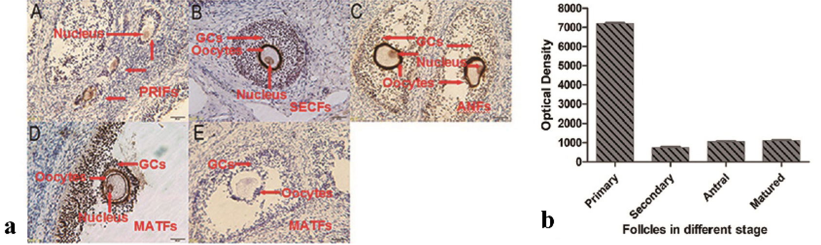
Immunohistochemistry analysis of Gas6 in the ovaries of porcine. a) Gas6 protein were staining brown by immunostaining using anti-Gas6 antibody (A, B, C and D), pre-immune serum instead of anti-Gas6 antibody was used as negative control (E). PRIFs, primary follicles; SECFs, secondary follicles; ANFs, antral follicles; MATFs, mature follicles. Scale bars=100 μm. b) The optical density of oocytes in different stage was analyzed by image-pro plus 6.0 software. The results are presented as mean ± SEM. GCs, granule cells.
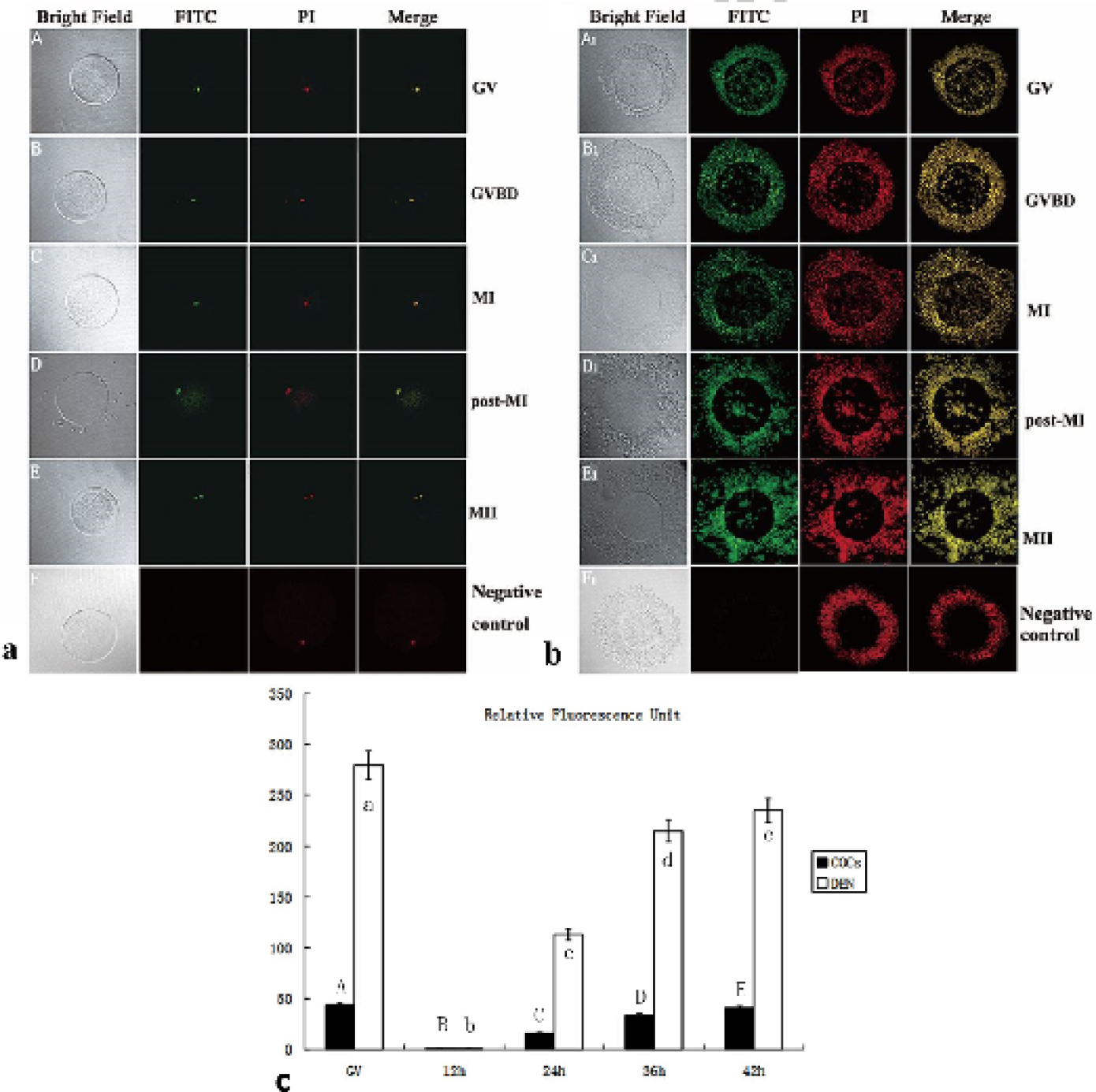
Immunofluorescence analysis of the Gas6 in oocytes in vitro maturing. a and b, Identification of Gas6 in oocytes and COCs. The green staining indicates a FITC signal and the red stain indicates counterstaining with propidium iodide (PI). C, Relative fluorescence unit during the oocyte maturation was analyzed by image-pro plus 6.0 software. COCs, Cumulus-oocyte complexes. GV, germinal vesicle; GVBD, germinal vesicle broken down; MI, metaphase I; MII, metaphase II; COCs, cumulus-oocyte complexes; DEN, denuded oocytes. The results are presented as mean ± SEM.
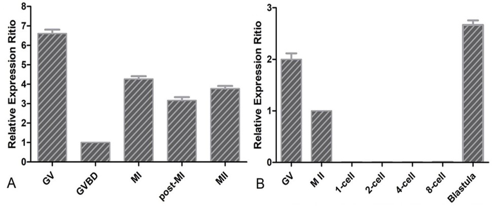
QRT-PCR analysis of Gas6 in the process of oocytes maturation (A) and embryo development (B). The relative gene expression was calculated using the 2-△△Ct method with 18S as the reference gene, and the expression ratio was calculated against Gas6 expression in the GV oocyte. Experiments were repeated 4 times, and data were presented as mean ± SEM.
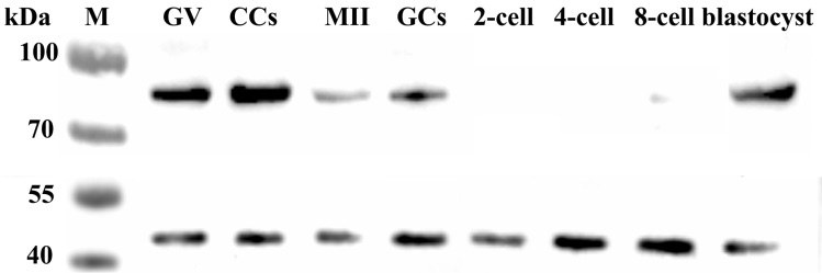
Western blot analysis of Gas6 expressed in the oocytes and early embryos. GV, Germinal vesicle; CCS, Cumulus oocyte complexes; MII, Metaphase II stage oocytes; GCS, Granulosa cells.
References
- Angelillo Scherrer, A., Burnier, L., Lambrechts, D., Fish, R.J., Tjwa, M., Plaisance, S.E.P., Sugamele, R., DeMol, M., Martinez-Soria, E., Maxwell,P.H., Lemke, G., Goff, S.P. Matsushima, G.K., Earp, H.S., Chanson, M., Collen, D., Izui, S., Schapira, M., Conway, E.M. and Carmeliet, P., 2008. Role of Gas6 in erythopoiesis and anemia in mice. J. clin. Invest., 118: 583-596. https://doi.org/10.1172/JCI30375[CrossRef]
- Coticchio, G., Dal Canto, M., Renzini, M.M., Guglielmo, M.C., Brambillasca, F., Turchi, D., Novara, P.V. and Fadini, R., 2015. Oocyte maturation: Gamete-somatic cells interactions, meiotic resumption, cytoskeletal dynamics and cytoplasmic reorganization. Hum. Reprod. Update, dmv011. https://doi.org/10.1093/humupd/dmv011[CrossRef]
- Eppig, J.J. and Wigglesworth, K., 2000. Development of mouse and rat oocytes in chimeric reaggregated ovaries after interspecific exchange of somatic and germ cell components. Biol. Reprod., 63: 1014-1023. https://doi.org/10.1095/biolreprod63.4.1014[CrossRef]
- Fair, T., 2009. Mammalian oocyte development: checkpoints for competence. Reprod., Fert. Dev., 22: 13-20. https://doi.org/10.1071/RD09216[CrossRef]
- Farghaly, T., Khalifa, E., Mostafa, S., Hussein, M., Bedaiwy, M. and Ahmady, A., 2015. The effect of temporary meiotic attenuation on the in vitro maturation outcome of bovine oocytes. In Vitro Cell. Dev. Biol. Anim., 51: 662-671. https://doi.org/10.1007/s11626-015-9878-y[CrossRef]
- Fleming, J.V., Fontanier, N., Harries, D.N., Rees, W.D., 1997. The growth arrest genes gas5, gas6 and CHOP-10 (gadd153) are expressed in the mouse preimplantation embryo. Mol. Reprod. Dev., 48: 310-316. https://doi.org/10.1002/(SICI)1098-2795(199711)48:3<310::AID-MRD2>3.0.CO;2-U[CrossRef]
- Gandolfi, T.A. and Gandolfi, F., 2001. The maternal legacy to the embryo: cytoplasmic components and their effects on early development. Theriogenology, 55: 1255-1276. https://doi.org/10.1016/S0093-691X(01)00481-2[CrossRef]
- Goruppi, S., Ruaro, E., Schneider, C., 1996. Gas6, the ligand of Axl tyrosine kinase receptor, has mitogenic and survival activities for serum starved NIH3T3 fibroblasts. Oncogene, 12: 471-480.
- Hafizi, S. and Dahlbäck, B., 2006. Gas6 and protein S. FEBS J. 273: 5231-5244. https://doi.org/10.1111/j.1742-4658.2006.05529.x[CrossRef]
- Keoung-Hwa, K.K.K., Eun-Young K.E.K. and Kyung-Ah, L.K.L., 2008. Role of Gas6 in the mouse oocyte maturation and early embryogenesis. Reprod. Dev. Biol., (Supplement), 32: 133-133.
- Kim, K.H., Kim, E.Y., Kim, Y., Kim, E., Lee, H.S., Yoon, S.Y. and Lee, K.A., 2011. Gas6 downregulation impaired cytoplasmic maturation and pronuclear formation independent to the MPF activity. PLoS One, 6: e23304. https://doi.org/10.1371/journal.pone.0023304[CrossRef]
- Kotani, T. and Yamashita, M., 2002. Discrimination of the roles of MPF and MAP kinase in morphological changes that occur during oocyte maturation. Dev. Biol., 252: 271-286. https://doi.org/10.1006/dbio.2002.0853[CrossRef]
- Labrecque, R., Sirard, M., 2011. Gene expression analysis of bovine blastocysts produced by parthenogenic activation or fertilisation. Reprod., Fert. Dev., 23: 591-602. https://doi.org/10.1071/RD10243[CrossRef]
- Lee, W., Jang, S., Lee, S., Park, J., Jeon, R., Subbarao, R.B., Bharti, D., Shin, J.K., Park, B.W. and Rho, G.N., 2015. Selection of reference genes for quantitative real-time polymerase chain reaction in porcine embryos. Reprod., Fert. Dev., 29: 357-367 http://dx.doi.org/10.1071/RD14393[CrossRef]
- Li, R. and Albert, D.F., 2013. The road to maturation: somatic cell interaction and self-organization of the mammalian oocyte. Nat. Rev. Mol. Cell. Biol., 14: 141-152. https://doi.org/10.1038/nrm3531[CrossRef]
- Li, R., Chen, J., Hammonds, G., Phillips, H., Armanini, M., Wood, P., Bunge, R., Godowski, P.J., Sliwkowski, M.X. and Mather, J.P., 1996. Identification of Gas6 as a growth factor for human Schwann cells. J. Neurosci., 16: 2012-2019. https://doi.org/10.1523/JNEUROSCI.16-06-02012.1996[CrossRef]
- Liu, S., Cui, K., Li, H.L., Sun, J.M., Lu, X.R., Shen, K.Y., Liu, Q.U. and Shi, D.S., 2015. Comparison of chemical, electrical, and combined activation methods for in vitro matured porcine oocytes. In Vitro Cell. Dev. Biol. Anim., 51: 103-112. https://doi.org/10.1007/s11626-014-9819-1[CrossRef]
- Manfioletti, G., Brancolini, C., Avanzi, G., Schneider, C., 1993. The protein encoded by a growth arrest-specific gene (gas6) is a new member of the vitamin K-dependent proteins related to protein S, a negative coregulator in the blood coagulation cascade. Mol. Cell. Biol., 13: 4976-4985. https://doi.org/10.1128/MCB.13.8.4976[CrossRef]
- Matsubara, N., Takahashi, Y., Nishina, Y., Mukouyama, Y., Yanagisawa, M., Watanabe, T., Nakano, T., Nomura, K., Arita, H., Nishimune, Y., Obinata, M. and Matsui, Y., 1996. A receptor tyrosine kinase, Sky, and its ligand Gas 6 are expressed in gonads and support primordial germ cell growth or survival in culture. Dev. Biol., 180: 499-510. https://doi.org/10.1006/dbio.1996.0323[CrossRef]
- Nagata, K., Ohashi, K., Nakano, T., Arita, H., Zong, C., Hanafusa, H. and Mizuno, K., 1996. Identification of the product of growth arrest-specific gene 6 as a common ligand for Axl, Sky, and Mer receptor tyrosine kinases. J. biol. Chem., 271: 30022-30027. https://doi.org/10.1074/jbc.271.47.30022[CrossRef]
- Nakano, T., Higashino, K., Kikuchi, N., Kishino, J., Nomura, K., Fujita, H., Ohara, O. and Arita, H., 1995. Vascular smooth muscle cell-derived, Gla-containing growth-potentiating factor for Ca (2+)-mobilizing growth factors. J. biol. Chem., 270: 5702-5705. https://doi.org/10.1074/jbc.270.11.5702[CrossRef]
- Palmer, A. and Nebreda, A.R., 2000. The activation of MAP kinase and p34cdc2/cyclin B during the meiotic maturation of Xenopus oocytes. Prog. Cell Cycle Res., 4: 131-143. https://doi.org/10.1007/978-1-4615-4253-7_12[CrossRef]
- Shuai, L., 2015. Study on porcine transgene of CI-MGT and ICSI-MGT. pp.166.
- Stenhoff, J., Dahlbäck, B. and Hafizi, S., 2004. Vitamin K-dependent Gas6 activates ERK kinase and stimulates growth of cardiac fibroblasts. Biochem. biophs. Res. Commun., 319: 871-878. https://doi.org/10.1016/j.bbrc.2004.05.070[CrossRef]
- Xiong, W., Chen, Y., Wang, H., Wu, H., Lu, Q. and Han, D., 2008. Gas6 and the Tyro 3 receptor tyrosine kinase subfamily regulate the phagocytic function of Sertoli cells. Reproduction, 135: 77-87. https://doi.org/10.1530/REP-07-0287[CrossRef]
- Yoon, S.J., Chung, H.M., Cha, K.Y., Kim, N.H. and Lee, K.A., 2005. Identification of differential gene expression in germinal vesicle vs. metaphase II mouse oocytes by using annealing control primers. Fertil. Steril., 83: 1293-1296. https://doi.org/10.1016/j.fertnstert.2004.09.037[CrossRef]
- Yoshioka, K., Suzuki, C., Tanaka, A., Anas, I.M., Iwamura, S., 2002. Birth of piglets derived from porcine zygotes cultured in a chemically defined medium. Biol. Reprod., 66: 112-119. https://doi.org/10.1095/biolreprod66.1.112[CrossRef]
- You, J., Lee, J., Hyun, S.H. and Lee, E., 2012. L-carnitine treatment during oocyte maturation improves in vitro development of cloned pig embryos by influencing intracellular glutathione synthesis and embryonic gene expression. Theriogenology, 78: 235-243. https://doi.org/10.1016/j.theriogenology.2012.02.027[CrossRef]
Services
Article usage statistics
Loading…