
Here, we aimed to determine the protective effects of tea leaf extract (TLE) (Camellia sinensis) against cypermethrin (CMN)-induced toxicity in gill and liver of rainbow trout (Oncorhynchus mykiss). CMN exposure led to increase in aspartate aminotransferase (AST), serum alanine aminotransferase (ALT) and malondialdehyde (MDA) levels, and reduction in superoxide dismutase (SOD), catalase (CAT) activities, glutathione (GSH), total immunoglobulin (T. Ig), and white blood cell (WBC) levels. Moreover, CMN exposure led to degeneration, steatosis, and necrosis in liver hepatocytes as well as hyperemia and inflammation in the liver and caused degeneration, desquamation, necrosis and adhesion in the gill epithelium. Additionally, expression levels of 8-hydroxy-2-deoxyguanosine (8-OHdG) and Caspase 3 were severe in the liver and gill tissues. However, both 50 and 100 mg doses of TLE had protective effects against CMN-induced toxicity in all the above parameters. As a result, we have shown that 100 mg of tea leaf extract is more effective in preventing harmful effects on blood biochemistry (AST and ALT), oxidative stress, immunity, apoptosis, histopathology and DNA damage caused by CMN.
Research Article · Volume 56, Issue 2, 16 February 2024 · Open Access
Protective Effects of Tea Leaf Extract (Camellia sinensis) Against Cypermethrin-Induced Toxicity in Gill and Liver of Rainbow Trout (Oncorhynchus mykiss)
· ·
Affiliations & Notes ⌄
- 1Vocational School of Health Services, Agri Ibrahim Cecen University, TR-04100 Agri, Turkey 2Department of Biochemistry, Faculty of Veterinary Medicine, Atatürk University, TR-25240 Erzurum, Turkey. 3Department of Pathology, Faculty of Veterinary Medicine, Atatürk University, TR-25240 Erzurum, Turkey * Corresponding author: tkaratas025@gmail.com
Article Info ⌄
- Journal:
- Pakistan Journal of Zoology
- Published:
- 16 February 2024

From the article
- Our results confirm that the natural antioxidants in tea leaf extract support the immune system against CMN toxicity and overcome the formation of necrosis in liver and gill tissues.
- However, 100 mg dose TLE against CMN toxicity prevented necrosis, hyperemia and inflammation in liver tissue as well as necrosis in gill tissue and significantly reduced degeneration and steatosis in the liver and desquamation and adhesion in the gill tissue of group 4.
- The severe of 8-OHdG expression caused by CMN in liver and gill tissue was significantly decreased in the groups 3 and 4.
- It shows that increased apoptosis in liver and gill tissues may be associated with oxidative stress.
Selected from the article's own text.
Abstract
INTRODUCTION
Pesticides have widely been used to increase agricultural productivity and to control undesired pests and diseases for centuries a). Although there are many legal regulations regarding pesticides, it has caused an important rise in pesticide use worldwide because of its wide spectrum and low cost ; . Pesticides contain highly toxic substances, and their direct or indirect contamination into water resources through agricultural, industrial, and domestic runoff poses a major risk to aquatic organisms including fish ; ; . For this reason, fish are considered one of the most important organisms in toxicity studies. In recent years, it has been determined that pyrethroid pesticides are more toxic and used more than organophosphates due to their high sensitivity in living things a). One of the widely used pesticides among pyrenoids is cypermethrin (CMN). CMN is one of the photostable synthetic pyrethroids and is the active ingredient of commonly used insecticides . CMN is highly toxic to fish, and the 96 h LC50 value for rainbow trout is between 0.5-8.2 µg/L , 1983; . The daily Kow for CMN is 6.06 and its solubility in water is 0.01 mg/L and the half-life in water is more than 50 days ; .
The tea plant belongs to the genus Camellia of the Theaceae family. Tea leaves (Camellia sinensis) contain flavan-3-ols (catechins), flavonol glycosides and phenolic acids ; ; . About 75% of the polyphenols in the tea leaf are flavanols, and 60-70% of the flavanols are epigallocatechin-3-gallate . Aslo, the flavanols are known to scavenge free radicals, have powerful antioxidant properties, inhibit hydrolytic and oxidative enzymes (phospholipase A2, cytochrome oxygenase, lipoxygenase), and anti-inflammatory activities ; ; . However, there is no study on the protective efficacy of tea leaf extract (TLE) on rainbow trout. This study was carried out to determine the protective efficiency of TLE on blood biochemistry (total protein, albumin, AST and ALT), oxidative stress, immunity, antioxidant enzymes, histopathology, apoptosis, and DNA damage against the toxicity induced by CMN in gill and liver tissues of rainbow trout (Oncorhynchus mykiss).
MATERIALS AND METHODS
Experimental animals
After purchasing healthy rainbow trout, they were kept in the stock pond for 15 days for adaptation. During this time, the fish were fed commercial trout feed (Abalıoğlu, Turkey), . After the adaptation period, 10 fish for each tank were stocked in tanks with a flow of 0.5 L and a water volume of 380 L (temperature 11.3°C; dissolved oxygen 7.95; and pH 6.8) a).
Preparation of tea leaf extract and diets
Fresh tea leaves were gathered from Rize, Turkey, dried in the room temperature, and pulverized using a grinder. Then, the mixture containing ethanol (300 ml) and sample (100 g) was prepared. This mixture was kept in 96% ethanol solution for 24 h at room temperature and filtered. Ethanol was removed under vacuum. Then, 50 and 100mg doses of the prepared TLE were added to commercial trout feed ; .
Experimental design and toxicity
Rainbow trout with weight of 40 g at the beginning of the experiment were randomly divided into 4 groups, each of 10 fish. Until the end of the trial, two groups (group 1 and 2) were fed with commercial trout feed, group 3 fed with 50 mg TLE and group 4 was fed with 100 mg dose of TLE added to commercial trout feed. No deaths were observed in the groups throughout the trial. Then, groups 2, 3 and 4 were exposed to 10 % of the LC50 value of CMN (CAS Number 52315-07-8, ≥98%, Molecular Weight 416.30) obtained from Sigma–Aldrich (Germany) for 21 days.
Biochemical and immunological analysis
Blood from the fish tail vein was drawn into vacuum-sealed gel serum tubes, where it was allowed to coagulate for seven minutes. Then, the coagulated blood was centrifuged at 3000 rpm for 10 min. and separated into serum . Serum metabolites such as total protein, albumin, aspartate aminotransferase (AST), serum alanine aminotransferase (ALT) (Kit no: 3183734190) were measured by a Cobas 6000 autoanalyzer , 2020). White blood cell (WBC) was measured using the sysmex XN9500 modular system. T. Ig level in fish was determined as described by Siwicki and Anderson (1993).
Malondialdehyde (MDA), and antioxidant enzyme analysis
After removing the gill and liver tissues, they dissected using Tissue Lyser II. The tissues were diluted with 1.15% potassium chloride to obtain 1:10 (w/v) homogenate for MDA, GSH and protein analysis. The homogenates were centrifuged at 3500 rpm for 15 min for MDA and protein analysis, and at 11000 rpm for 20 min for glutathione (GSH) analysis . Protein concentration in supernatants (liver and gill tissues) was measured spectrophotometrically at 650 nm according to the Lowry method using standard bovine serum albumin . MDA was determined at 532 nm as described by Placer et al. (1966). GSH was determined at 412 nm as described by Stahr (1977). The superoxide dismutase (SOD) activities were determined at 560 nm as described by Sun et al. (1988). The catalase (CAT) activities was determined at 405 nm as described by Goth (1991).
Histopathological examination
After the gill and liver tissues were removed, they were kept in 10% formalin for 48 h. Tissues taken from formalin were cut in 4 μm thickness and embedded in paraffin blocks. Then, sections were stained with Hematoxylin-Eosin (HE) and observed under microscope (Bar: 50µm) , 2021).
Immunohistochemical examination
The histological sections passed through xylol and alcohol series for immunoperoxidase analysis were kept in 3% H2O2 for 10 min. for endogenous peroxidase inactivation after washing with PBS. To determine the antigen in the tissues, they were treated with antigen retrieval solution for 2x5 min at 500 watts in the oven. As shown in the immunohistochemistry kit process (AbcamHRP/ DAB Detection IHC kit), tissues were incubated with 8-OHdG and Caspase 3 Antibody (Catalog no: sc66036, sc-56053, dilution 1/50; Santa Cruz, USA) for 30 min at 37˚C for detection of DNA and apoptotic cell damage. The chromogen was 3-3’ Diaminobenzidine. The sections were showed according to their immune positivity .
Statistical analysis
SPSS 20.0 program was used for all of the statistical analyses of this study. ANOVA (one way analysis of variance) test was used to determine the statistical differences of the results obtained from the study, Duncan test was used in multiple comparisons and p<0.05 was considered significant. Kruskal-Wallis (nonparametric) test was used for semi-objective findings in histopathology and Mann Whitney U test was used for pairwise comparisons.
RESULTS
Protective effects of TLE on liver functions
The changes in liver functions of the groups exposed to CMN toxicity after being fed with 50 and 100 mg doses of tea leaf extract are given in Table I. Serum total protein, and albumin levels were decreased in the group 2 exposed to CMN (p <0.05). In contrast, total protein and albumin levels in groups 3 and 4 were increased compared to group 2 (p <0.05). ALT and AST activities, known as liver enzymes, were increased in the group 2 (p <0.05). However, these enzymes were decreased in groups 3 and 4 (p <0.05). There was no statistically significant difference between the liver functions of groups 3 and 4 (Table I).
Protective effects of TLE on oxidative stress and antioxidant enzymes
CMN exposure led to a significant increase in MDA levels and a significant decrease in SOD, CAT activities and GSH levels in the liver and gill tissues of group 2 compared to group 1 (p<0.05). However, while MDA levels
Table I. Effects of tea leaf extract on blood biochemistry against Cypermethrin (CMN) toxicity in rainbow trout.
| Blood biochemistry | Group 1 | Group 2 | Group 3 | Group 4 |
| T. Protein (mg/dL) | 4.15±0.03 a | 3.79±0.01 c | 3.90±0.05 b | 4.02±0.04 ab |
| Albumin (mg/dL) | 2.01±0.03 a | 1.77±0.03 c | 1.84±0.04 b | 1.93±0.02 ab |
| ALT (IU/L) | 15.4±0.78 a | 24.6±0.60 c | 18.8±0.40 b | 16.5±0.54 ab |
| AST (IU/L) | 640.8±6.25a | 721.3±5.35 c | 671.0±5.86 b | 654.0±3.74ab |
The results were given as mean and standard deviation. Different letters indicate differences between groups. Group 1: Control; Group 2: CMN exposure; Group 3: CMN exposure after feeding with 50 mg of tea leaf extract; Group 4: CMN exposure after feeding with 100 mg of tea leaf extract.
ALT, alanine aminotransferase; AST, aspartate aminotransferase.
Table II. Effects on oxidative stress biomarkers of tea leaf extract (TLE) against CMN toxicity in rainbow trout.
| Stress parameters | Group 1 | Group 2 | Group 3 | Group 4 |
| Liver tissue | ||||
| MDA (mmol/g tissue) | 49.8±0.65 d | 77.9±1.67 a | 59.4±0.23 bc | 54.7±0.53 dc |
| SOD (EU/g protein) | 50.6±0.59 a | 38.9±1.28 c | 43.5±2.02 b | 47.5±2.44 ab |
| CAT (kU/g doku) | 51.7±2.94 a | 35.6±0.23 d | 44.3±0.65 b | 48.6±1.47 ab |
| GSH (mmol/g tissue) | 1.99±0.05 a | 1.64±0.07 d | 1.82±0.02 bc | 1.90±0.04 ab |
| Gill tissue | ||||
| MDA (mmol/g tissue) | 74.2±1.25 d | 94.5±0.93 a | 82.1±0.30 bc | 77.4±0.78 dc |
| SOD (EU/g protein) | 52.3±1.48 a | 41.6±1.0 c | 46.1±1.73 b | 50.1±1.0 ab |
| CAT (kU/g doku) | 31.1±0.57 a | 22.6±0.47 c | 26.4±0.75 b | 29.5±0.79 ab |
| GSH (mmol/g tissue) | 2.02±0.02 a | 1.65±0.06 d | 1.73±0.03 bc | 1.90±0.08 ab |
The results were given as mean and standard deviation. Different letters indicate differences between groups. For details of groups, see Table I.
CAT, catalase; GSH, glutathione; MDA, malondialdehyde; SOD, superoxide dismutase.
Table III. Effects of tea leaf extract on immunity against CMN toxicity in rainbow trout.
| Blood biochemistry | Group 1 | Group 2 | Group 3 | Group 4 |
| T. Ig (mg/ml) | 3.02±0.03a | 2.80±0.08c | 2.87±0.07b | 2.95±0.02ab |
| WBC (104mm-3) | 62.6±1.50a | 51.3±2.11d | 56.3±0.54bc | 60.4±1.27ab |
The results were given as mean and standard deviation. Different letters indicate differences between groups. For details of groups, see Table I.
T. Ig, total immunoglobulin; WBC, white blood cells
decreased in the liver and gill tissues of the groups 3 and 4, SOD, CAT activities and GSH levels increased (p<0.05). Moreover, group 4 was more effective in reducing the toxicity effects caused by CMN compared to group 3 (Table II).
Protective effects of TLE on immunity
T. Ig and WBC were reduced in the group 2 exposed to CMN (p <0.05). In contrast, T. Ig and WBC levels in groups 3 and 4 were higher than those of group 2 treated with CMN (p <0.05). There was no statistically significant difference between the liver functions of groups 3 and 4 (Table III).
Table IV. Scoring histopathological findings in liver and gill tissues.
| Histopathological findings | Group 1 | Group 2 | Group 3 | Group 4 |
| Liver | ||||
| Degeneration in hepatocytes | - | +++ | ++ | + |
| Necrosis in hepatocytes | - | +++ | + | - |
| Steatosis in hepatocytes | - | +++ | ++ | + |
| Inflammation | - | +++ | + | - |
| Hyperemia | - | +++ | + | - |
| Gill | ||||
| Degeneration in the gill epithelium | - | +++ | +++ | ++ |
| Desquamation in the gill epithelium | - | +++ | +++ | + |
| Necrosis in the gill epithelium | - | +++ | + | - |
| Adhesion in the gill epithelium | - | +++ | ++ | + |
None (−), Mild (+), Moderate (++), and Severe (+++). For details of groups, see Table I.
Protective effects of TLE on liver and gill histopathology
The liver and gill tissues of the groups 1 had normal histological appearance (Figs. 1A, 2A). CMN exposure caused severe degeneration, steatosis, inflammation, necrosis, hyperemia, enlargement of sinusoids and moderate mononuclear cell infiltration in the liver tissue (Fig. 1B), and severe degeneration, desquamation, necrosis of the lamellar epithelium, as well as severe adhesion in the lamella due to cell infiltration in the interlamellar spaces and dilatation in the vessels in the gill tissue of group 2 (Fig. 2B). While 50 mg dose of TLE caused moderate degeneration, moderate steatosis, mild inflammation and mild necrosis in the liver hepatocytes (Fig. 1C) and severe degeneration, severe desquamation, moderate adhesion, and mild necrosis in the gill lamellar epithelium of group 3 (Fig. 2C), 100 mg dose TLE caused mild degeneration and steatosis in liver hepatocytes (Fig. 2D), and mild adhesion and proliferation in gill interlamellar epithelium of group 4 (Fig. 2D) (Table IV).
Effects on caspase 3 and 8-OHdG of TLE against CMN toxicity in rainbow trout
8-OHdG and caspase 3 expressions in the liver (Figs. 3A, 5A) and gill (Figs. 4A, 6A) tissues of groups 1 were negative. Expressions of 8-OHdG and Caspase 3 of group 2 were severe in liver hepatocytes (intracytoplasmic localization) (Figs. 3B, 5B) and gill lamellar epithelium (Figs. 4B, 6B). The 8-OHdG and Caspase 3 expressions were moderate in liver hepatocytes (intracytoplasmic) (Figs. 3C, 5C) and gill lamellar epithelium (Figs. 4C, 6C) of group 3 and mild in liver (Figs. 3D-5-D) and gill (Figs. 4D, 6D) tissues of group 4 (Table V).
Table V. Scoring immunohistochemical findings in liver and gill tissues.
| Immunohistochemical findings | Group 1 | Group 2 | Group 3 | Group 4 |
| Liver | ||||
| 8 OHdG | - | +++ | ++ | + |
| Caspase 3 | - | +++ | ++ | + |
| Gill | ||||
| 8 OHdG | - | +++ | ++ | + |
| Caspase 3 | - | +++ | ++ | + |
None (−), Mild (+), Moderate (++), and Severe (+++). For details of groups, see Table I.
DISCUSSION
In recent years, studies have focused on the safety, dosage and action mechanisms of different plants in the protection of living things against pesticides . One of these plants is tea. There are many different types of tea, such as green, black and white, which is an important nutritional component. Tea, which contains active phytochemicals (polyphenols), has an active role in protection against pesticides and diseases due to its rich antioxidant source . The main polyphenol of tea is epigallocatechin-3-gallate (EGCG; 59%) and it has therapeutic effects in protection against pesticides and diseases by regulating biochemical, physiological and molecular processes .
AST and ALT have high sensitivity to hepatotoxicity and histopathological changes, including amino acid metabolism , and are used to evaluate the level of liver tissue damage such as cell inflammation and necrosis caused by pesticides ; , b). CMN exposure led to increase in serum ALT and AST activities in group 2. The increase in these enzymes may be the most important cause of liver dysfunction and damage. However, there were significant decreases in serum AST and ALT activities in groups 3 and 4. This may be an indication of the protective effect of tea leaf extract on the liver. Our results were similar to those of studies on animals such as the African catfish and Wistar rat . Moreover, there are significant decreases in serum protein and albumin levels of group 2 exposed to CMN. These reductions may be related to decreased oxygen availability, inhibit energy production, suppress oxidative metabolism, as well as hypoxia due to lactic acid accumulation . Similar results were observed in common carp exposed to bifenthrin , brown trout exposed to deltamethrin and rainbow trout exposed to tetrachlorobiphenyl and diazinon ; ; .
Reactive oxygen species (ROS) could lead to the cellular and molecular changes such as lipid, protein, DNA and antioxidant, damage the membranes by making them permeable, and different physiological instabilities lead to necrosis and thus apoptosis ; ; ; . Reduced SOD, CAT and GSH and increased MDA in liver and gill tissue of group 2 may be due to excessive free radical production . Histopathological results support these findings. However, the reduce in MDA levels and increment in CAT and GSH levels in groups 3 and 4 may be a result of suppressing the formation of reactive oxygen molecules and free radicals, as well as preventing oxidative stress. Moreover, the increase in SOD activities may be associated with the detoxification of superoxide radicals (O2-) . The increase in both of these antioxidants, even under stress, may be an indication that tea leaf extract supports the defense system. Similar results have been observed in African catfish exposed to 4-NP and winstar rat exposed to doxorubicin . Moreover, there was a significant decrease in T. Ig and WBC levels of group 2 treated with CMN compared to group 1. Decreased WBC and T. Ig may be the result of chronic and/or persistent immunosuppression of prolonged CMN exposure . Moreover, the reduction in protein content may be partially associated with the decrease in WBC levels which are the main source of protein production such as immunoglobulin, lysozyme, complementary factors, and bactericidal peptides (Misra et al., 2006a, b; . However, there was an increase in T. Ig and WBC levels of groups 3 and 4. Hasanpour et al. (2017) reported that green tea supports the immune system such as T. Ig, lysozyme, and ACH50. Our results confirm that the natural antioxidants in tea leaf extract support the immune system against CMN toxicity and overcome the formation of necrosis in liver and gill tissues. Histopathologically, severe degeneration, desquamation, necrosis and adhesion in the gill epithelium and severe steatosis, degeneration, necrosis, inflammation and hyperemia in the liver tissue of group 2 exposed to CMN were observed. One of the deadly effects of pyrethroids such as cypermethrin and deltamethrin on fish is gill damage. Therefore, pyrethroids have a high absorption rate by gill even at low levels due to their high lipophilicity ; ; ; . The liver is considered to be responsible for detoxification of toxic substances in living things. Karatas et al. (2019a) showed that hyperemia may be a result of increased inflammation in liver tissue. Colakoglu and Donmez (2012) determined that impaired lipid biosynthesis or lipid transport is the most important cause of steatosis and degeneration in the liver. Salim et al. (2011) and Karatas et al. (2019a) showed that toxic substances such as aflatoxins and deltamethrin cause hepatocyte necrosis in sinusoids due to the expansion and swelling of hepatocytes. Different researchers determined different lesions in fish such as Rohu exposed to CMN , Rainbow trout exposed to CMN , Common carp exposed to CMN (Dobsíková et al., 2006), Clarias gariepinus exposed to CMN , Zebrafish exposed to CMN , and Nile tilapia exposed to CMN . However, 100 mg dose TLE against CMN toxicity prevented necrosis, hyperemia and inflammation in liver tissue as well as necrosis in gill tissue and significantly reduced degeneration and steatosis in the liver and desquamation and adhesion in the gill tissue of group 4. Sayed and Soliman (2018) determined that green tea extract reduced the hepatotoxic effects of 4-NP in catfish.
8-OHdG is one of the markers used to determine the levels of DNA damage caused by ROS ; ; . Hydroxyl radicals formed due to oxidative stress are an indicator of hydrogenation of nucleic acid leading to 8-OHdG ; . The 8-OHdG expression level was severe in both liver and gill tissues of the group 2 exposed to CMN. The increase in the 8-OHdG expression levels in liver and gill tissues may be due to oxidative stress and superoxide anion (O2) production ; ; , b). Previous studies showed that pyrethroids increase 8-OHdG expression levels ; , b). The severe of 8-OHdG expression caused by CMN in liver and gill tissue was significantly decreased in the groups 3 and 4. This suggests that the powerful antioxidant agents found in tea leaf play an important role in reducing DNA damage caused by oxidative stress.
Apoptosis has a vital effect on the survival of multicellular organisms by getting rid of damaged or infected cells that cannot perform their normal functions ; ; . Caspase 3 is an effector protein that plays an important role in the mitochondrial and the death receptor pathways ; . Caspase-3 expression levels were severe in gill and liver cells of group 2 exposed to CMN. It shows that increased apoptosis in liver and gill tissues may be associated with oxidative stress. Topal et al. (2014) reported that chlorpyrifos increases gill and liver cell apoptosis in rainbow trout and can activate caspase-3. The severity of caspase 3 in groups 3 and 4 was lower than that in group 2. Reduced apoptosis in liver and gill tissues may be the result of suppressing oxidative stress and increasing antioxidant defense system of flavonoids found in tea extract . In addition, Spencer et al. (2001) determined that 3′-O-methyl epicatechin found in tea reduces caspase-3 activity by inhibiting H2O2-induced cell death.
Consequently, TLE had positive effects on all processes such as suppression of oxidative stress, strengthening of immune and antioxidant defense system, regulation of the effects of inflammatory signaling pathways and anti-apoptotic. Especially, 100 mg dose of tea leaf extract was determined to be more effective in preventing liver and gill tissue damage caused by CMN. Further studies are needed to determine the effects of TLE on different tissues against different pesticides in rainbow trout.
ACKNOWLEDGEMENTS
The authors thank Ataturk University, Faculty of Aquaculture, Inland Water Fish Research and Application Center. A part of this work was presented at the 2lst National Fisheries Symposium.
Funding
This work was supported by the Scientific Research Project Coordination Unit of Agri Ibrahim Cecen University (Project number: SHMYO.20.001).
IRB approval
The experimental protocol was approved by ethics committee of Agri Ibrahim Cecen University.
Ethical statement
This study was performed within the ethical rules determined by Agri Ibrahim Cecen University (Writing and decision number: 47825/207).
Statement of conflict of interest
The authors have declared no conflict of interest.
Figures
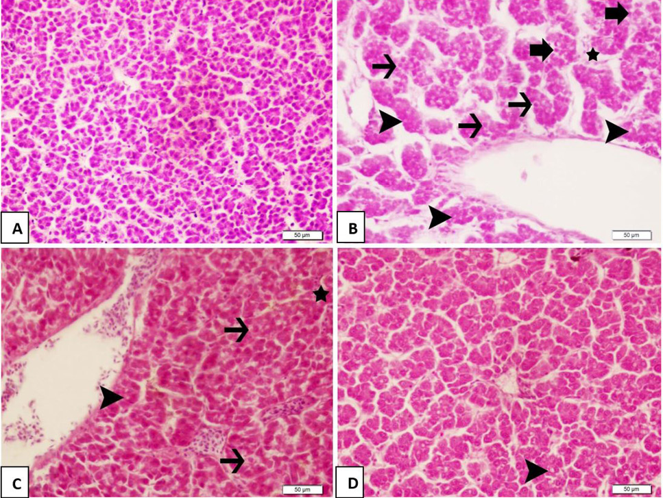
Effect of tea leaf extract on histological structure of fish liver induced with cypermethrin. A, normal histological structure of liver. B shows severe degeneration (thin arrow), severe steatosis (arrowhead), and severe necrosis (thick arrow) in hepatocytes, as well as severe inflammation (star) after treatment with cypermethrin. C shows effect of 50 mg of TLE on CMN exposure, modarate degeneration, modarate steatosis and mild necrosis in hepatocytes, mild hyperemia (empty star) and inflammation (star) is visible. D shows effect of 100 mg of TLE on CMN exposure, mild steatosis in hepatocytes (arrowhead) is visible. H and E, Bar: 50 µm.
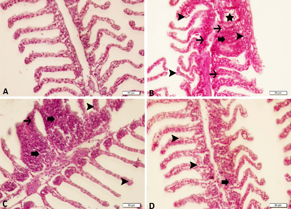
Effect of tea leaf extract on histological structure of fish gill induced with Cypermethrin. A normal histological structure of gill. B shows severe adhesion (thick arrow), severe necrosis (thin arrow), severe inflammation (star), and severe degeneration (arrowhead) in gill lamellar epithelium after treatment with cypermethrin. C shows effect of 50 mg of TLE on CMN exposure, severe degeneration (arrowhead), mild necrosis (thin arrow), and moderate adhesion (thick arrow) in gill lamellar epithelium is visible. D shows effect of 50 mg of TLE on CMN exposure, moderate degeneration (arrowhead) and mild adhesion (thick arrow) in gill lamellar epithelium is visible, H and E, Bar: 50 µm.
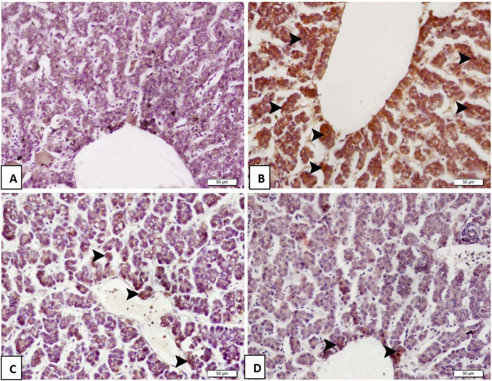
Effect of tea leaf extract on 8-OHdG expression in liver hepatocytes. A shows negative 8-OHdG expression in hepatocytes. B shows severe 8-OHdG expression in hepatocytes (arrowhead) of fish after exposure to CMN. C shows moderate 8-OHdG expression in hepatocytes of 50 mg of TLE on CMN exposure. D shows mild 8-OHdG expression in hepatocytes of 100 mg of TLE on CMN exposure H and E, Bar: 50 µm.
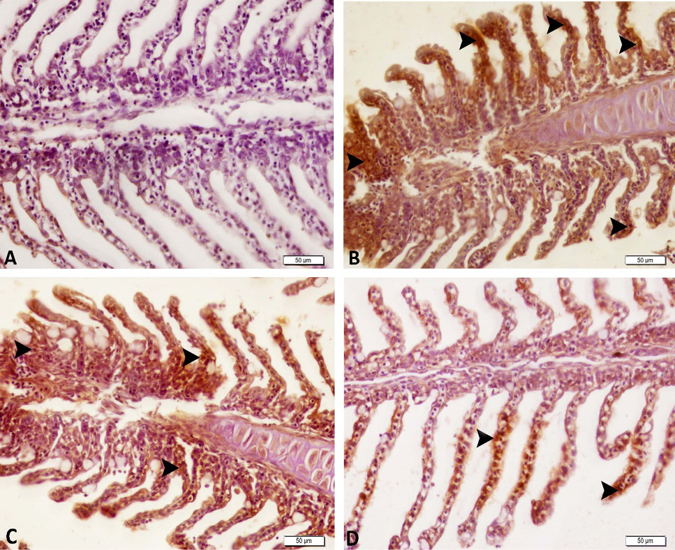
Effect of tea leaf extract on 8-OHdG expression in gill epithelium. A shows negative 8-OHdG expression in the gill epithelium. B shows severe 8-OHdG expression in the gill epithelium (arrowhead) of CMN exposure. C shows moderate 8-OHdG expression in the gill epithelium (arrowhead) of 50 mg of TLE on CMN exposure. D shows mild 8-OHdG expression in the gill epithelium (arrowhead) of 100 mg of TLE on CMN exposure, H and E, Bar: 50 µm.
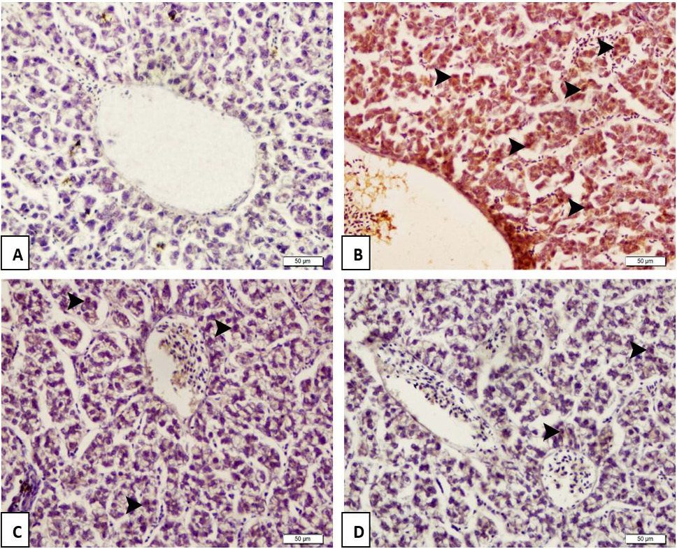
Effect of tea leaf extract on Caspase 3 expression in liver hepatocytes. A shows negative Caspase 3 expression in hepatocytes of control fish. B shows severe Caspase 3 expression in hepatocytes (arrowhead) of fish exposed to CMN. C shows effect of moderate Caspase 3 expression in hepatocytes (arrowhead) of TLE (50mg) pretreated fish after exposure to CMN. D shows, mild Caspase 3 expression in hepatocytes (arrowhead) of TLE (100mg) pretreated fish after exposure to CMN, H and E, Bar: 50 µm.
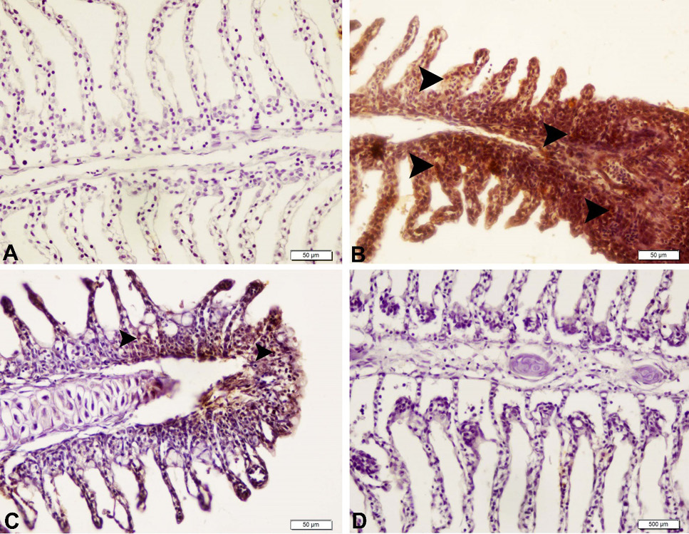
Effect of tea leaf extract on Caspase 3 expression in gill epithelium. A shows negative Caspase 3 expression in epithelium of control fish. B shows severe Caspase 3 expression in the gill epithelium (arrowhead) of fish exposed to CMN. C shows effect of moderate Caspase 3 expression in the gill epithelium (arrowhead) of TLE (50mg) pretreated fish after exposure to CMN. D shows mild Caspase 3 expression in the gill epithelium (arrowhead) of TLE (100mg) pretreated fish after exposure to CMN, H and E, Bar: 50 µm.
References
- Ahmed, O.M., Abdul-Hamid, M.M., El-Bakry, A.M., Mohamed, H.M. and Abdel Rahman, E., 2019. Camellia sinensis and epicatechin abate doxorubicin-induced hepatotoxicity in male Wistar rats via their modulatory effects on oxidative stress, inflammation, and apoptosis. J. appl. Pharm. Sci., 9: 30-44. https://doi.org/10.7324/JAPS.2019.90405[CrossRef]
- Anjana, V.V.S., Tinu, S.K., Geetha, C.S., Lissy, K.K. and Mohanan, P.V., 2013. Effect of fibrin glue on antioxidant defense mechanism, oxidative DNA damage and chromosomal aberrations. Toxicol. Mech. Methods, 23: 500-508. https://doi.org/10.3109/15376516.2013.785625[CrossRef]
- Arslan, H., Altun, S. and Özdemir, S., 2017. Acute toxication of deltamethrin results in activation of iNOS, 8-OHdG and up-regulation of caspase 3, iNOS gene expression in common carp (Cyprinus carpio L.). Aquat. Toxicol., 187: 90-99. https://doi.org/10.1016/j.aquatox.2017.03.014[CrossRef]
- Banaee, M., Sureda, A., Mirvaghefi, AR. and Ahmadi, K., 2011. Effects of diazinon on biochemical parameters of blood in rainbow trout (Oncorhynchus mykiss). Pestic. Biochem. Physiol., 99: 1-6. https://doi.org/10.1016/j.pestbp.2010.09.001[CrossRef]
- Ben Slima, A., Chtourou, Y., Barkallah, M., Fetoui, H., Boudawara, T. and Gdoura, R., 2017. Endocrine disrupting potential and reproductive dysfunction in male mice exposed to deltamethrin. Hum. exp. Toxicol., 36: 218-226. https://doi.org/10.1177/0960327116646617[CrossRef]
- Bradbury, S.P. and Coast, J.R., 1989. Comparative toxicology of the pyrethroid insecticides. Environ. Contam. Toxicol., 108: 1341–1377.
- Cadet, J., Douki, T., Gasparutto, D. and Ravanat, J.L., 2003. Oxidative damage to DNA: Formation, measurement and biochemical features. Mutat. Res., 531: 5-23. https://doi.org/10.1016/j.mrfmmm.2003.09.001[CrossRef]
- Cadet, J.L., 2016. Epigenetics of stress, addiction, and resilience: Therapeutic implications. Mol. Neurobiol., 53: 545-560. https://doi.org/10.1007/s12035-014-9040-y[CrossRef]
- Clifford, M.N., Copeland, E.L., Bloxsidge, J.P. and Mitchell, L.A., 2000. Hippuric acid as a major excretion product associated with black tea consumption. Xenobiotica, 30: 317-326. https://doi.org/10.1080/004982500237703[CrossRef]
- Colakoglu, F. and Donmez, H.H., 2012. Effects of aflatoxin on liver and protective effectiveness of esterified glucomannan in merino rams. Sci. World J., 2012: 1-5. https://doi.org/10.1100/2012/462925[CrossRef]
- Dobsikova, R., Velisek, J., Wlasow, T., Gomulka, P., Svobodová, Z. and Novotný, L., 2006. Effects of cypermethrin on some haematological, biochemical and histopathological parameters of common carp (Cyprinus carpio L.). Neuro Endocrinol. Lett., 27: 91-95.
- El-Bakry, H.A., El-Sherif, G. and Rostom, R.M., 2017. Therapeutic dose of green tea extract provokes liver damage and exacerbates paracetamol-induced hepatotoxicity in rats through oxidative stress and caspase 3-dependent apoptosis. Biomed. Pharma., 96: 798-811. https://doi.org/10.1016/j.biopha.2017.10.055[CrossRef]
- Gelen, V., Şengül, E., Yıldırım, S., Senturk, E., Tekin, S. and Kükürt, A., 2021. The protective effects of hesperidin and curcumin on 5-fluorouracil–induced nephrotoxicity in mice. Environ. Sci. Pollut. Res., 28: 47046–47055. https://doi.org/10.1007/s11356-021-13969-5[CrossRef]
- Goth, L., 1991. A simple method for determination of serum catalase activity and revision of reference range. Clin. Chim. Acta, 196: 143-151. https://doi.org/10.1016/0009-8981(91)90067-M[CrossRef]
- Hasanpour, S., Salati, A.P., Falahatkar, B. and Azarm, H.M., 2017. Effects of dietary green tea (Camellia sinensis L.) supplementation on growth performance, lipid metabolism, and antioxidant status in a sturgeon hybrid of Sterlet (Huso huso♂× Acipenser ruthenus♀) fed oxidized fish oil. Fish Physiol. Biochem., 43: 1315-1323. https://doi.org/10.1007/s10695-017-0374-z[CrossRef]
- Karatas, T., Korkmaz, F., Karatas, A. and Yildirim, S., 2020. Effects of Rosemary (Rosmarinus officinalis) extract on growth, blood biochemistry, immunity, antioxidant, digestive enzymes and liver histopathology of rainbow trout, Oncorhynchus mykiss. Aquacult. Nutr., 26: 1533-1541. https://doi.org/10.1111/anu.13100[CrossRef]
- Karatas, T., Onalan, S. and Yildirim, S., 2021. Effects of prolonged fasting on levels of metabolites, oxidative stress, immune-related gene expression, histopathology, and DNA damage in the liver and muscle tissues of rainbow trout (Oncorhynchus mykiss). Fish Physiol. Biochem., 47: 1119-1132. https://doi.org/10.1007/s10695-021-00949-2[CrossRef]
- Karatas, T., Yildirim, S., Arslan, H. and Aggul, A.G., 2019a. The effects on brown trout (Salmo trutta fario) of different concentrations of deltamethrin. Comp. Biochem. Physiol. C Toxicol. Pharmacol., 226: 108606. https://doi.org/10.1016/j.cbpc.2019.108606[CrossRef]
- Karatas, T., Yildirim, S. and Arslan, H., 2019b. Effects of different concentrations of diazinon on 8-hydroxy-2-deoxyguanosine and histopathology, antioxidant enzyme, acetylcholinesterase activity and plasma metabolites in rainbow trout (Oncorhynchus mykiss). Int. J. Agric. Biol., 21: 583‒589.
- Katiyar, S.K. and Mukhtar, H., 1997. Tea antioxidants in cancer chemoprevention. J. Cell. Bioch. Suppl., 27: 59-67. https://doi.org/10.1002/(SICI)1097-4644(1997)27+<59::AID-JCB11>3.0.CO;2-G[CrossRef]
- Khan, N. and Mukhtar, H., 2007. Tea polyphenols for health promotion. Life Sci., 81: 519-533. https://doi.org/10.1016/j.lfs.2007.06.011[CrossRef]
- Kinsella, J.E., Frankel, E., German, B. and Kanner, J., 1993. Possible mechanisms for the protective role of antioxidants in wine and plant foods. Fd. Tech., 47: 85–89.
- Korkmaz, N., Cengiz, E.I., Unlu, E., Uysal, E. and Yanar, M., 2009. Cypermethrin-induced histopathological and biochemical changes in Nile tilapia (Oreochromis niloticus), and the protective and recuperative effect of ascorbic acid. Environ. Toxicol. Pharmacol., 28: 198-205. https://doi.org/10.1016/j.etap.2009.04.004[CrossRef]
- Lavrik, I.N., 2010. Systems biology of apoptosis signaling networks. Curr. Opin. Biotechnol., 21: 551-555. https://doi.org/10.1016/j.copbio.2010.07.001[CrossRef]
- Livingstone, D., 2001. Contaminant-stimulated reactive oxygen species production and oxidative damage in aquatic organisms. Mar. Pollut. Bull., 42: 656-666. https://doi.org/10.1016/S0025-326X(01)00060-1[CrossRef]
- Lowry, O.H., Rosebrough, N.J., Farr, A.L. and Randall, R.J., 1951. Protein measurement with the Folin phenol reagent. J. biol. Chem., 193: 265-275. https://doi.org/10.1016/S0021-9258(19)52451-6[CrossRef]
- Lushchak, V.I., 2011. Environmentally induced oxidative stress in aquatic animals. Aquat. Toxicol., 101: 13-30. https://doi.org/10.1016/j.aquatox.2010.10.006[CrossRef]
- Misra, C.K., Das, B.K., Mukherjee, S.C. and Pattnaik, P., 2006a. Effect of multiple injections of β-glucan on non-specific immune response and disease resistance in Labeo rohita fingerlings. Fish Shellf. Immunol., 20: 305-319. https://doi.org/10.1016/j.fsi.2005.05.007[CrossRef]
- Misra, S., Sahu, N., Pal, A., Xavier, B., Kumar, S. and Mukherjee, S., 2006b. Pre-and post-challenge immuno-haematological changes in Labeo rohita juveniles fed gelatinised or non-gelatinised carbohydrate with n-3 PUFA. Fish Shellf. Immunol., 21: 346-356. https://doi.org/10.1016/j.fsi.2005.12.010[CrossRef]
- Mohamed, G.A., Amhamed, I.D., Almabrok, A.A., Barka, A.B.A., Bilen, S. and Elbeshti, R.T., 2018. Effect of celery (Apium graveolens) extract on the growth, haematology, immune response and digestive enzyme activity of common carp (Cyprinus carpio). Mar. Sci. Tech. Bul., 7: 51-59. https://doi.org/10.33714/masteb.457721[CrossRef]
- Oda, S.S. and El-Maddawy, Z.K., 2012. Protective effect of vitamin E and selenium combination on deltamethrin-induced reproductive toxicity in male rats. Exp. Toxicol. Pathol., 64: 813-819. https://doi.org/10.1016/j.etp.2011.03.001[CrossRef]
- Onouchi, H., Ishii, T., Miyazawa, M., Uchino, Y., Yasuda, K., Hartman, P.S., Kawai, K., Tsubota, K. and Ishii, N., 2012. Mitochondrial superoxide anion overproduction in Tet-mev-1 transgenic mice accelerates age-dependent corneal cell dysfunctions. Invest. Ophthalmol. Vis. Sci., 53: 5780-5787. https://doi.org/10.1167/iovs.12-9573[CrossRef]
- Paravani, E.V., Simoniello, M.F., Poletta, G.L., Zolessi, F. and Casco, V.H., 2018. Cypermethrin: Oxidative stress and genotoxicity in retinal cells of the adult zebrafish. Mutat. Res. Genet. Toxicol. Environ. Mutagen., 826: 25-32. https://doi.org/10.1016/j.mrgentox.2017.12.010[CrossRef]
- Pearson, B.L., Simon, J.M., McCoy, E.S., Salazar, G., Fragola, G. and Zylka, M.J., 2016. Identification of chemicals that mimic transcriptional changes associated with autism, brain aging and neurodegeneration. Nat. Commun., 7: 1-12. https://doi.org/10.1038/ncomms11173[CrossRef]
- Placer, Z.A., Cushman, L.L. and Johnson, B.C., 1966. Estimation of product of lipid peroxidation (malonyl dialdehyde) in biochemical systems. Anal. Biochem., 16: 359-364. https://doi.org/10.1016/0003-2697(66)90167-9[CrossRef]
- Portt, L., Norman, G., Clapp, C., Greenwood, M. and Greenwood, M.T., 2011. Anti-apoptosis and cell survival: A review. Biochim. biophys. Acta, 1813: 238-259. https://doi.org/10.1016/j.bbamcr.2010.10.010[CrossRef]
- Qin, W., Yamada, R., Araki, T. and Ogawa, Y., 2022. Changes in morphological and functional characteristics of tea leaves during Japanese green tea (Sencha) manufacturing process. Fd. Bioprocess Technol., 15: 82–91. https://doi.org/10.1007/s11947-021-02735-7[CrossRef]
- Rahmani, A.H., Aldebasi, Y.H. and Aly, S.M., 2015. Role of green tea and its constituent epigallocatechin-3-gallate in the health management. Int. J. Pharm. and Pharm. Sci., 7: 6-12.
- Salim, A., Zohair, A., Hegazy, A.E-S. and Said, A., 2011. Effect of some strains of probiotic bacteria against toxicity induced by aflatoxins in vivo. J. Am. Sci., 7: 1-12.
- Sarkar, B., Chatterjee, A., Adhikari, S. and Ayyappan, S., 2005. Carbofuran and cypermethrin-induced histopathological alterations in the liver of Labeo rohita (Hamilton) and its recovery. J. appl. Ichthyol., 21: 131-135. https://doi.org/10.1111/j.1439-0426.2004.00590.x[CrossRef]
- Saxena, R., Garg, P. and Jain, D., 2011. In vitro anti-oxidant effect of vitamin E on oxidative stress induced due to pesticides in rat erythrocytes. Toxicol. Int., 18: 73. https://doi.org/10.4103/0971-6580.75871[CrossRef]
- Sayed, A.E.D.H. and Soliman, H.A., 2018. Modulatory effects of green tea extract against the hepatotoxic effects of 4-nonylphenol in catfish (Clarias gariepinus). Ecotoxicol. environ. Saf., 149: 159-165. https://doi.org/10.1016/j.ecoenv.2017.11.007[CrossRef]
- Shelley, L.K., Balfry, S.K., Ross, P.S. and Kennedy, C.J., 2009. Immunotoxicological effects of a sub-chronic exposure to selected current use pesticides in rainbow trout (Oncorhynchus mykiss). Aquat. Toxicol., 92: 95-103. https://doi.org/10.1016/j.aquatox.2009.01.005[CrossRef]
- Singh, R., Akhtar, N. and Haqqi, T.M., 2010. Green tea polyphenol epigallocatechi3-gallate: Inflammation and arthritis. Life Sci., 86: 907-918. https://doi.org/10.1016/j.lfs.2010.04.013[CrossRef]
- Siwicki, A.K. and Anderson, D.P., 1993. Nonspecific defense mechanisms assay in fish: II. Potential killing activity of neutrophils and macrophages, lysozyme activity in serum and organs and total immunoglobulin level in serum. In: Disease diagnosis and prevention methods (eds. A.K. Siwicki, D.P. Anderson and J. Walvga). Olsztyn, Poland. FAO-project GCP/INT/JPA, IFI Olsztyn, Poland. pp.105–112.
- Smith, T.M. and Stratton, G.W., 1986. Effects of synthetic pyrethroid insecticides on nontarget organisms. Res. Rev., 97: 93-120. https://doi.org/10.1007/978-1-4612-4934-4_4[CrossRef]
- Soltanian, S. and Fereidouni, M.S., 2017. Immunotoxic responses of chronic exposure to cypermethrin in common carp. Fish Physiol. Biochem., 43: 1645-1655. https://doi.org/10.1007/s10695-017-0399-3[CrossRef]
- Spencer, J.P., Schroeter, H., Kuhnle, G., Srai, S.K., Tyrrell, R.M., Hahn, U. and Rice-Evans, C., 2001. Epicatechin and its in vivo metabolite, 3′-O-methyl epicatechin, protect human fibroblasts from oxidative-stress-induced cell death involving caspase-3 activation. Biochem. J., 354: 493-500. https://doi.org/10.1042/bj3540493[CrossRef]
- Stahr, H., 1977. Analytical toxicology methods manual. Iowa State University Press, Ames-Iowa, USA. pp. 32.
- Stephenson, R., 1982. Aquatic toxicology of cypermethrin. I. Acute toxicity to some freshwater fish and invertebrates in laboratory tests. Aquat. Toxicol., 2: 175-185. https://doi.org/10.1016/0166-445X(82)90014-5[CrossRef]
- Stephenson, R., 1983. WL85871 and cypermethrin; a comparative study of their toxicity to the rainbow trout, fathead minnow and Pimephales promelas. Sittingbourne Shell Res. (SBGR 82.298).
- Stepniak, J. and Karbownik-Lewinska, M., 2016. 17 β-estradiol prevents experimentally-induced oxidative damage to membrane lipids and nuclear DNA in porcine ovary. Syst. Biol. Reprod. Med., 62: 17-21. https://doi.org/10.3109/19396368.2015.1101510[CrossRef]
- Stoyanova, S., Yancheva, V., Iliev, I., Vasileva, T., Bivolarski, V., Velcheva, I. and Georgieva, E., 2016. Biochemical, histological and histochemical changes in Aristichthys nobilis Rich. Liver exposed to thiamethoxam. Period. Biol., 118: 29-36. https://doi.org/10.18054/pb.2016.118.1.2828[CrossRef]
- Struger, J. and Fletcher, T., 2007. Occurrence of lawn care and agricultural pesticides in the Don River and Humber River watersheds (1998–2002). J. Great Lakes Res., 33: 887-905. https://doi.org/10.3394/0380-1330(2007)33[887:OOLCAA]2.0.CO;2[CrossRef]
- Sun, Y., Oberley, L.W. and Li, Y., 1988. A simple method for clinical assay of superoxide dismutase. Clin. Chem., 34: 497-500. https://doi.org/10.1093/clinchem/34.3.497[CrossRef]
- Teles, M., Reyes-López, F.E., Balasch, J.C., Tvarijonaviciute, A., Guimarães, L., Oliveira, M. and Tort, L., 2019. Toxicogenomics of gold nanoparticles in a marine fish: linkage to classical biomarkers. Front. Mar. Sci., 6: 147. https://doi.org/10.3389/fmars.2019.00147[CrossRef]
- Topal, A., Alak, G., Altun, S., Erol, H.S. and Atamanalp, M., 2017. Evaluation of 8-hydroxy-2-deoxyguanosine and NFkB activation, oxidative stress response, acetylcholinesterase activity, and histopathological changes in rainbow trout brain exposed to linuron. Environ. Toxicol. Pharmacol., 49: 14-20. https://doi.org/10.1016/j.etap.2016.11.009[CrossRef]
- Topal, A., Atamanalp, M., Oruç, E., Kırıcı, M. and Kocaman, E.M., 2014. Apoptotic effects and glucose-6-phosphate dehydrogenase responses in liver and gill tissues of rainbow trout treated with chlorpyrifos. Tissue Cell, 46: 490-496. https://doi.org/10.1016/j.tice.2014.09.001[CrossRef]
- Ullah, R., Zuberi, A., Ullah, S., Ullah, I. and Dawar, F.U., 2014. Cypermethrin induced behavioral and biochemical changes in mahseer, Tor putitora. J. toxicol. Sci., 39: 829-836. https://doi.org/10.2131/jts.39.829[CrossRef]
- Ullah, S., Li, Z., Arifeen, M.Z.U., Khan, S.U. and Fahad, S., 2019. Multiple biomarkers based appraisal of deltamethrin induced toxicity in silver carp (Hypophthalmichthys molitrix). Chemosphere, 214: 519-533. https://doi.org/10.1016/j.chemosphere.2018.09.145[CrossRef]
- Ullah, S., Zuberi, A., Alagawany, M., Farag, M.R., Dadar, M., Karthik, K., Tiwari, R., Dhama, K. and Iqbal, H.M., 2018. Cypermethrin induced toxicities in fish and adverse health outcomes: Its prevention and control measure adaptation. J. environ. Manage., 206: 863-871. https://doi.org/10.1016/j.jenvman.2017.11.076[CrossRef]
- Velisek, J., Svobodova, Z. and Machova, J., 2008. Effects of bifenthrin on some haematological, biochemical and histopathological parameters of common carp (Cyprinus carpio L.). Fish Physiol. Biochem., 35: 583–590. https://doi.org/10.1007/s10695-008-9258-6[CrossRef]
- Velisek, J., Wlasow, T, Gomulka, P., Svobodova, Z., Dobsikova, R., Novotny, L., Dudzik, M., 2006. Effects of cypermethrin on rainbow trout (Oncorhynchus mykiss). Vet. Med. Czech., 51: 469. https://doi.org/10.17221/5580-VETMED[CrossRef]
- Velmurugan, B., Mathews, T. and Cengiz, E.I., 2009. Histopathological effects of cypermethrin on gill, liver and kidney of fresh water fish Clarias gariepinus (Burchell, 1822), and recovery after exposure. Environ. Technol., 30: 1453-1460. https://doi.org/10.1080/09593330903207194[CrossRef]
- Vijayan, M.M., Feist, G. Otto, D.M.E., Schreck, C.B. and Moon, T.W., 1997. 3, 3/, 4, 4/-Tetracholorobiphenyl affects cortisol dynamics and hepatic function in rainbow trout. Aquat. Toxicol., 37: 87–98. https://doi.org/10.1016/S0166-445X(96)00828-4[CrossRef]
- Weston, D., Holmes, R., You, J. and Lydy, M., 2005. Aquatic toxicity due to residential use of pyrethroid insecticides. Environ. Sci. Technol., 39: 9778-9784. https://doi.org/10.1021/es0506354[CrossRef]
- Yao, L., Jiang, Y., Datta, N., Singanusong, R., Liu, X., Duan, J., Raymont, K., Lisle, A. and Xu, Y., 2004. HPLC analyses of favanols and phenolic acids in the fresh young shoots of tea (Camellia sinensis) grown in Australia. Fd. Chem., 84: 253–263. https://doi.org/10.1016/S0308-8146(03)00209-7[CrossRef]
- Zhishen, J., Mengcheng, T. and Jianming, W., 1999. The determination of flavonoid contents in mulberry and their scavenging effects on superoxide radicals. Fd. Chem., 64: 555-559. https://doi.org/10.1016/S0308-8146(98)00102-2[CrossRef]
Services
Article usage statistics
Loading…