
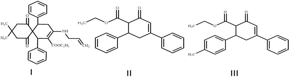
Due to perpetual increasing antimicrobial resistance, functionally substituted alicyclic compounds are envisioned as probable antimicrobial agents of future. From screening of in house library of compounds, here we report the antimicrobial properties of three novel functionally substituted monocyclic and spirocyclic cyclohexane derivatives involving ethyl-3 (allylamino)-9,9-dimethyl- 7,11-dioxo-1,5-diphenylspiro[5.5]undec-2-en-2-carboxylate (I), ethyl-4,6-diphenyl-2- dicyanomethylene cyclohex-3-ene 1-carboxylate (II) and ethyl-4-phenyl-6-(4-chlorophenyl)-2- dicyanomethylenecyclohex-3-ene 1-carboxylate (III).Initially, these compounds were screened for in vitro antimicrobial activity against Gram-positive bacteria, Gram-negative bacteria and fungi by agar well diffusion method. In second step, minimum inhibitory concentration of test compounds was determined against bacterial species by using resazurin microplate assay. All the tested compounds exhibited variable antimicrobial properties against various test cultures. All the compounds showed stronger antimicrobial activity against Gram-negative bacteria as compared to Gram-positive bacteria and fungi. Compound III was found to be the most effective compound. These results demonstrate the potential antimicrobial properties of mono and spiro cyclohexane derivatives and accentuate the need of in vivo trials for their application at clinical level.
Short Communication · Volume 52, Issue 1, 8 January 2020 · Open Access
Antimicrobial Activity of Novel Functionally Substituted Monocyclic and Spirocyclic Cyclohexane Derivatives
· · · ·
Affiliations & Notes ⌄
- 1Department of Parasitology and Microbiology, Faculty of Veterinary and Animal Sciences, PMAS Arid Agriculture University Rawalpindi, Pakistan 2Department of Organic Chemistry, Baku State University, Azerbaijan 3Department of Microbiology, Baku State University, Azerbaijan 4Department of Veterinary Pathology, Faculty of Veterinary and Animal Sciences, PMAS Arid Agriculture University Rawalpindi, Pakistan , * Corresponding author: shoaib1676@gmail.com
Article Info ⌄
- Journal:
- Pakistan Journal of Zoology
- Published:
- 8 January 2020

From the article
- Test compounds showed better antimicrobial activity against Gram-negative bacteria as compared to Gram-positive bacteria.
- Functionally substituted monocyclic and spirocyclic cyclohexane derivatives exhibited remarkable antimicrobial properties against various test cultures.
- Compound III showed strong antibacterial activity against Escherichia coli and Acinetobacter baumannii.
- Test compounds demonstratedweak to moderate antifungal activity.
Selected from the article's own text.
Abstract
Materials and methods
Functionally substituted monocyclic and spirocyclic cyclohexane derivatives; ethyl-3(allylamino)-9,9-dimethyl- 7,11-dioxo-1,5-diphenylspiro[5.5]undec-2-en-2-carboxylate (I), ethyl-4,6-diphenyl-2- dicyanomethylene cyclohex-3-ene 1-carboxylate (II) and ethyl-4-phenyl-6-(4-chlorophenyl)-2- dicyanomethylenecyclohex-3-ene 1-carboxylate (III) were obtained from Department of Organic Chemistry, Baku State University Azerbaijan. Structure of these compounds is shown in Figure 1. Test compounds were evaluated for in vitro antibacterial and antifungal activity against Gram-positive bacteria (Staphylococcus aureus BDU-23, Bacillus subtilis BDU-50, Bacillus mesentericus BDU-51 and Bacillus megaterium BDU-N2), Gram-negative bacteria (Escherichia coli BDU-12, Klebsiella pneumoniae BDU-44, Acinetobacter baumannii BDU-32 and Pseudomonas aeruginosa BDU-49) and fungi (Candida tropicalis BDU LK30, Candida pelliculosa BDU KT55 and Candida pseudotropicalis BDU MA88). All the test cultures were obtained from our own collection at Department of Microbiology, Baku State University Azerbaijan.
Test compounds were dissolved in Dimethyl sulphoxide (DMSO) and three different concentrations of test compounds were prepared; 0.3%, 0.1% and 0.05%. Standard agar well diffusion assay as described by was used to determine diameter of zone of inhibition. Mueller-Hinton agar was used for antibacterial activity and sabouraud dextrose agar was used for antifungal screening. Briefly, 100 µL of 24 hour fresh broth culture (0.5 McFarland) of each test culture was aseptically spread over agar surface. Wells with diameter 8 mm were punched aseptically in the agar plate and 150 µL of test compound was poured in each well. Agar plates were incubated at 37oC for 24 hours for bacteria and 30oC for 72 hours for fungi.Diametre of zone of inhibition was measured.All the experiments were performed in triplicates and DMSO was used as control.
Resazurin microplate assay was used to determine minimum inhibitory concentration (MIC) as described by Palomino et al. (2002). Briefly, bacterial inoculum was prepared from 24 hour fresh broth culture and density was adjusted to 1×10⁵ colony forming units per mL. The resazurin microtiter assay (REMA) plate method was performed in Mueller-Hinton broth. Serial twofold dilutions of each test compound in 100 μL of medium were prepared. The range of concentrations tested was 8–2000 mg/mL. Sterility controls and growth controls with no test compound were also included. Titer plate was covered and incubated at 37oC for 24 hours. After incubation, 30 µL of 0.01% resazurin solution was poured into each well, and the plate was again incubated for 3-4 hours. A change in color of resazurin from blue to pink indicates the growth of bacteria. Lowest concentration of compound that inhibited the change in color was defined as MIC.
Results and discussion
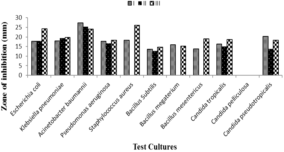
The antimicrobial activity of functionally substituted monocyclic and spirocyclic cyclohexane derivatives were determined against four Gram-negative bacteria, four Gram-positive bacteria and three fungi. Test compounds showed better antimicrobial activity against Gram-negative bacteria as compared to Gram-positive bacteria. These results contradict the findings of Urzua et al. (2008), who reported that benzofuran cyclohexane-5-carboxylate derivatives have better activity against Gram-positive bacteria. This difference in the findings can be attributed to different nature of functional group in cyclohexane. In agar well diffusion assay, all the tested compounds exhibited remarkable antimicrobial properties at concentration of 0.3% (Fig. 2). Moderate to weak antibacterial activity was observed against some of pathogen at concentration of 0.1% and 0.05% (Table I). Candida pelliculosa was most resistant strain as none of tested compound inhibited its growth. Candida tropicalis and Candida pseudotropicalis were found to be sensitive at 0.3% concentration, showed resistance at 0.1% and 0.05% concentration. These findings are supported by Sharma et al. (2011) who demonstrated antifungal activity of cyclohexane diamine derivatives against Candida sp. Among Gram-positive bacteria, Staphylococcus aureus was more sensitive as compared to Bacillus species which coincides with findings of Beena et al. (2014) who reported moderate antibacterial activity of adamantyl based cyclohexane derivatives against Staphylococcus aureus. Acinetobacter baumannii was most sensitive bacteria with zone of inhibition 27.3 mm, 25.3 mm and 24 mm for compounds I, II and III respectively.
Table I. Average diameter of zone of inhibition (mm) and Minimum inhibitory concentrations (µg/mL).
| Test Culture | Diameter of zone of inhibition (mm) | Minimum inhibitory concentrations (µg/mL) | |||||||||
| I | II | III | DM SO | I | II | III | |||||
| 0.1% | 0.05% | 0.1% | 0.05% | 0.1% | 0.05% | ||||||
| Escherichia coli | - | - | 16.5 ±0.2 | 13.4 ±0.3 | 16± 0.2 | 13± 0.4 | - | 1000 | 500 | 250 | |
| Klebsiella pneumoniae | - | - | 17.7 ±0.3 | 12.7 ±0.6 | 15± 0.5 | 12± 0.3 | - | 500 | 500 | 250 | |
| Acinetobacter baumannii | 25 ±0.8 | 14± 0.3 | 22.3 ±0.5 | - | 20.3± 0.4 | 16± 0.3 | - | 500 | 250 | 125 | |
| Pseudomonas aeruginosa | - | - | 15.7 ±0.3 | 12.7 ±0.2 | 18± 0.3 | 14.3± 0.6 | - | 1000 | 1000 | 500 | |
| Staphylococcus aureus | 15. 3±0.3 | 12.3 ±0.3 | - | - | 15.3 ±0.1 | 13.3± 0.4 | - | 2000 | - | 500 | |
| Bacillus subtilis | - | - | - | - | - | - | - | 2000 | 500 | 1000 | |
| Bacillus megaterium | 14.7 ±0.1 | 12.5 ±0.1 | - | - | 12± 0.2 | - | - | - | - | 1000 | |
| Bacillus mesentericus | - | - | - | - | 15± 0.2 | - | - | - | - | 1000 | |
| Candida tropicalis | - | - | - | - | - | - | - | NA | NA | NA | |
| Candida pelliculosa | - | - | - | - | - | - | - | NA | NA | NA | |
| Candida pseudotropicalis | 18.7 ±0.2 | 12.7 ±0.1 | - | - | - | - | - | NA | NA | NA | |
(-): Inactivity; NA: not available.
Monocyclic cyclohexane derivatives showed better antimicrobial activity as compared to spirocyclic derivative. Compound III was found to be most potent antimicrobial agent at all the three concentrations. This can be attributed to lipophilicity of the compounds. Antimicrobial activity increases with increase in number of hydrophobic groups (Urzua et al., 2008). Results of agar well diffusion assay were validated by resazurin microplate assay (Table I).
From Gram-negative bacteria, MIC values were found to be ≤1000 µg/mL. Compound III showed least MIC value (125µg/mL and 250µg/mL) against Acinetobacter baumannii and Escherichia coli respectively, which demonstrated maximum activity of this compound.Bogdanov et al. (2007) and Nief et al. (2017) also reported that Escherichia coli was most sensitive bacteria against spirobenzo pyran and phenyl cyclohexane derivatives. From Gram-positive bacteria, MIC values were found to be between 1000-2000 µg/mL. Compound III showed strong antimicrobial activity, while compound II and I exhibited moderate to weak antimicrobial activities. Keeping in view the antimicrobial potential of functionally substituted cyclohexane derivatives, further synthesis and exploration of these compounds or their hybrid analogues as broad spectrum antimicrobial drugs should be envisioned.
Conclusion
Functionally substituted monocyclic and spirocyclic cyclohexane derivatives exhibited remarkable antimicrobial properties against various test cultures. Gram-Negative bacteria were found to be more sensitive to these compounds as compared to Gram-Positive bacteria. Compound III showed strong antibacterial activity against Escherichia coli and Acinetobacter baumannii. Test compounds demonstratedweak to moderate antifungal activity. Findings depict that these substancescan act as potential future antimicrobial agents.
Statement of conflict of interests
The authors declare that there is no conflict of interests regarding the publication of this article.
Figures
Structure of compounds used in this study (Ismiev et al., 2018; Magerramov et al., 2014).
Average zone of inhibition (mm) at 0.3% concentration.
References
- Amin, K.M., Kamel, M.M., Anwar, M.M., Khedr, M. and Syam, Y.M., 2010. Eur. J. med. Chem., 45: 2117-2131. https://doi.org/10.1016/j.ejmech.2009.12.078[CrossRef]
- Bagdanov, M.G., Gocheva, B.T., Dimitrova, B.T. and Palamareva, M.D., 2007. Chem. Inform., 38: 1-7.
- Balouiri, M., Sadiki, M. and Ibnsouda, S.K., 2016. J. Pharm. Analys. 6: 71–79. https://doi.org/10.1016/j.jpha.2015.11.005[CrossRef]
- Beena, Kumar, D., Kumbukgolla, W., Jayaweera, S., Bailey, M., Alling, T. and Rawat, D.S., 2014. RSC Advan., 4: 11962-11966. https://doi.org/10.1039/c4ra00224e[CrossRef]
- Bielawski, K., Leszczyńska, K., Kałuża, Z., Bielawska, A., Michalak, O., Daniluk, T., Staszewska-Krajewska, O., Czajkowska, A., Pawłowska, N. and Gornowicz, A., 2017. Drug Des. Devel. Ther., 11: 2015-2028. https://doi.org/10.2147/DDDT.S133250[CrossRef]
- Flefel, E.M., Sayed, H.H., Hashem, A.I., Shalaby, E.A., El-sofany, W., Abdel-megeid, F.M.E., 2014. Med. Chem. Res., 23: 2515-2527. https://doi.org/10.1007/s00044-013-0830-y[CrossRef]
- Hussain, H.I., Iqbal, Z., Saleem, M.N., Huang, D., Sattar, A., Hao, H. and Yuan, Z., 2017. Sci. Rep., 7: 1-15. https://doi.org/10.1038/s41598-017-07798-1[CrossRef]
- Iqbal, Z., Saleem, M.N., Hussain, H.I., Huang, L., Hao, H. and Yuan, Z., 2016. Sci. Rep., 6: 1-12. https://doi.org/10.1038/srep35442[CrossRef]
- Ismiev, A.I., Potekhin, K.A., Maleev, A.V., Askerov, R.K. and Maharramov, M.K., 2018. J. struct. Chem., 59: 1911–1917. https://doi.org/10.1134/S0022476618080206[CrossRef]
- Kumar, D., Rohilla, R.K., Roy, N. and Rawat, D.S., 2011. Chem. Biol. Inter., 1: 263-278.
- Lallo, S., Lee, S., Dibwe, D.F., Tezuka, Y. and Morita, H., 2014. Nat. Prod. Res., 28: 1754-1759. https://doi.org/10.1080/14786419.2014.945175[CrossRef]
- Li, B. and Webster, T.J., 2018. J. Orthop. Res., 36: 22–32.
- Magerramov, A.M., Ismiev, A.I., Potekhin, K.A. and Askerov, R.K., 2014. J. Struct. Chem., 55: 1-4.
- Neif, O.A., Salman, H.N. and Ahamed, L.S., 2017. J. Sci., 58: 1998-2011.
- Palomino, J.C., Martin, A. and Camacho, M., 2002. Antimicrob. Agents Chemother., 46: 2720–2722. https://doi.org/10.1128/AAC.46.8.2720-2722.2002[CrossRef]
- Sharma, M., Joshi, P., Kumar, N., Joshi, S., Rohilla, R.K., Roy, N. and Rawat, D.S., 2011. Eur. J. med. Chem., 46: 480-487. https://doi.org/10.1016/j.ejmech.2010.11.027[CrossRef]
- Shoaib, M. and Ganbarov, G.K., 2019. J. Microbiol. Biotechnol., 4: 1-4.
- Song, L., Kang, H., Liu, D., Dai, Z., He, J. Wang, B., Zhao, W., Wang, X. and Xi, W., 2015. Trop. J. Pharm. Res.,14: 1719-1722.
- Tsemeugne, J., Fondjo, E.S., Tamokou, J., Rohand, T., Ngongang, A.D., Kuiate, J.R. and Sondengam, B.L., 2018. Int. J. med. Chem., 2: 1-8. https://doi.org/10.1155/2018/9197821[CrossRef]
- Urzúa, A., Echeverría, J., Rezende, M.C. and Wilkens, M., 2008. Molecules, 13: 2385-2393. https://doi.org/10.3390/molecules13102385[CrossRef]
- Useglio, M., Castellano, P.M., Operto, M.A., Torres, R. and Kaufman, T.S., 2006. Bioorg. med. Chem. Lett., 16: 5097-5101. https://doi.org/10.1016/j.bmcl.2006.07.029[CrossRef]
Cited by 1 article
In journals published by ResearchersLinks.
Services
Article usage statistics
Loading…