
In accordance with the global action plan on antimicrobial resistance established by the WHO, and with the aim of obtaining better therapeutic agents more effective than conventional antibiotics, we evaluated the antimicrobial activity of two synthetic organic compounds containing phosphate, i.e. (((cyanomethyl) (ethoxy) phosphoryl) oxy) zinc (II) chloride (compound I) and (Z)-(1-(3-(dichlorophosphoryl)-3-methyl-4-oxooxetan- 2-ylidene) ethyl) phosphonic dichloride (compound II), against ten pathogenic microorganisms including four Gram-negative bacteria (Acinetobacter baumannii, Escherichia coli, Klebsiella pneumoniae and Pseudomonas aeruginisa), three Gram-positive bacteria (Bacillus mesentericus, Bacillus subtilis and Staphylococcus aureus) and three yeasts (Candida albicans, Candida guilliermondii and Candida tropicalis). Agar well diffusion method was applied to estimate zone of inhibition, and the double dilution method of compounds was used to determine the minimum inhibitory concentration (MIC) of both test compound. The results obtained showed an excellent antimicrobial potential of compounds against test cultures with inhibition zones ranging from 34.2 mm to 39.3 mm for compound I, and from 35.5 mm to 41.2 mm for compound II against Gram-negative and Gram-positive bacteria. The diameters of inhibition zones for antifungal activity varied from 26.3 mm to 28.0 mm for compound I, and from 30.3 mm to 31.0 mm for compound II against the Candida species. The MIC values showed that the genus Candida is very sensitive to both test compounds as compared to Gram-negative and Gram-positive bacteria.
Short Communication · Volume 57, Issue 6, 14 November 2025 · Open Access
Two Synthetic Organic Compounds Containing Phosphorus against Pathogenic Microorganism
· · · · ·
Affiliations & Notes ⌄
- 1Research Laboratory of Microbiology and Virology, Baku State University, Azerbaijan 2PMAS Arid Agriculture University, Rawalpindi, Pakistan * Corresponding author: shoaib1676@gmail.com
Article Info ⌄
- Journal:
- Pakistan Journal of Zoology
- Published:
- 14 November 2025

From the article
- For Gram-positive bacteria, the diameters of inhibition zones ranged from 34.2 mm to 39.3 mm for compound I, and from 35.5 mm to 41.2 mm for compound II.
- Acinetobacter baumannii is more sensitive to compound II as compared to compound I having MIC value of 62.5 µg/mL.
- The MIC of fungi showed that Candida albicans and Candida guilliermondii are more susceptible to compound I compared to compound II.
- However, the MIC results showed that the genus Candida is the most sensitive to both compounds compared to gram-negative and gram-positive bacteria.
Selected from the article's own text.
Abstract
Materials and methods
The structures of test compounds used to evaluate the antimicrobial activity are illustrated in the Figure 1. These compounds were obtained from Department of Organic Chemistry, Baku State University (BSU), Azerbaijan.
The pathogenic microorganisms against which these compounds were tested were taken from our own repository at BSU Azerbaijan. Test cultures included Acinetobacter baumannii BDU-32, Escherichia coli BDU-12, Klebsiella pneumoniae BDU-44, Pseudomonas aeruginosa BDU-49, Bacillus mesentericus, Bacillus subtilis, Staphylococcus aureus BDU-23, Candida albicans, Candida guilliermondii and Candida tropicalis.
Agar well diffusion method was used to find out zone of inhibition for 0.3% concentration of test compounds . Test compounds were dissolved in dimethyl sulph oxide (DMSO), because it has no antimicrobial activity. We dissolved 0.3 µg of test compound in 1 mL of DMSO to prepare 0.3% concentration of each compound. Bacteria were grown on nutrient agar and fungi were grown on sabouraud dextrose agar. 150 μL of 24 h fresh broth culture of test microorganisms (0.5 McFarland) was aseptically spread on agar surface. Two wells were made aseptically in agar plate by sterile tips, and wells were labeled for test compounds. 150 μL of each compound was added in respective well. Agar plates were incubated at 37°C for bacteria and at 30°C for fungi for 24 h. After incubation, the diameter of inhibition zones was measured carefully. Procedure was performed four times to find mean values.
Minimum inhibitory concentration (MIC) of test compounds were determined using double dilution method as mentioned by Balouiri et al. (2016) for different dilutions of compounds. Different concentrations of test compounds ranging from 1000 µg/mL to 7.8 µg/mL were used to evaluate MIC. The smallest concentration of test compound that inhibited the growth of microorganisms was considered as MIC for that particular microorganism .
Table I. Antimicrobial activity of test compounds by agar well diffusion method.
| Test cultures | Diameter of inhibition zone (mm), M±m | |
| Compound I | Compound II | |
| Acinetobacter baumannii | 37.0±2.2 | 40.5±2.4 |
| Escherichia coli | 36.0±2.1 | 38.7±2.2 |
| Klebsiella pneumoniae | 36.8±2.1 | 38.2±2.2 |
| Pseudomonas aeruginosa | 34.2±2.0 | 38.0±2.2 |
| Bacillus mesentericus | 34.8±2.0 | 35.5±2.0 |
| Bacillus subtilis | 34.2±2.0 | 38.2±2.2 |
| Staphylococcus aureus | 39.3±2.3 | 41.2±2.4 |
| Candida albicans | 26.8±1.0 | 30.3±1.4 |
| Candida guilliermondii | 26.3±1.0 | 31.0±1.4 |
| Candida tropicalis | 28.0±1.2 | 30.8±1.4 |
Results and discussion
The diameters of zones of inhibition of two organophosphates are shown in the Table I. At 0.3% concentration, test compounds exhibited excellent antimicrobial activities against all tested microorganisms. For Gram-negative bacteria, the diameters of inhibition zones ranged from 34.2 mm to 37.0 mm for compound I, and from 38.0 mm to 40.5 mm for compound II. For Gram-positive bacteria, the diameters of inhibition zones ranged from 34.2 mm to 39.3 mm for compound I, and from 35.5 mm to 41.2 mm for compound II. For Candida species, the diameters of inhibition zones varied from 26.3 mm to 28.0 mm for compound I, and from 30.3 mm to 31.0 mm for compound II. These results are better than those mentioned by Kathiriya et al. (2015), Rim et al. (2022). Furthermore, Rim et al. (2022) evaluated the antifungal activity of two compounds i.e. diethylhydroxyphenyl α-aminophosphonate compounds (4a-k) and diethyl phosphate diethyl α-aminophosphate derivatives (6a-k) against Fusarium oxysporum and Botrytis cinerea. The latter was more effective for inhibiting mycelium growth with a value of 87% for Fusarium oxysporum and 92% for Botrytis cinerea.
Our results clearly show the capacity of these two organic compounds to inhibit the growth of all the ten tested microorganisms, unlike certain organic compounds, which have a selective action. Functionally substituted cyclohexane derivatives are more portent against Gram- negative bacteria as compared to Gram- positive bacteria and fungi . The findings of Urzua et al. (2008) are not in agreement with our results. They reported that benzofuran derivatives were more effective against Gram- positive bacteria. This is due to difference in structure of test compounds.
The different values of the MIC varying from 62.5 µg/mL to 125 µg/mL showed that Gram-negative and Gram-positive bacteria were more sensitive as compared to fungi for tested compounds. Our findings are similar to those reported by Ismiyev et al. (2020). They demonstrated that diethyl esters of dicarboxylic acids are batter antibacterial agents than antifungal agents. This is thought to be due to difference in cell wall of bacteria and fungi . Acinetobacter baumannii is more sensitive to compound II as compared to compound I having MIC value of 62.5 µg/mL. Pseudomonas aeruginosa and Bacillus subtilis are more sensitive to compound I compared to compound II. Escherichia coli, Klebsiella pneumoniae and Staphylococcus aureus have the same susceptibility to both tested compounds with MIC value of 62.5 µg/mL. Bacillus mesentericus is equally vulnerable to both tested compounds with 125 µg/mL as MIC value. Furthermore, the organophosphates used by have much lower MIC values compared to our compounds. Indeed, the amino phosphonates introduced into aloe emodin and sulfonamide hybrids by showed remarkable antibacterial potential. Aloe emodin derivative containing dimethyl phosphate moiety showed excellent antibacterial activity against Enterococcus faecalis, Escherichia coli and Acinetobacter baumannii at low concentrations, ranging from 0.25 to 2 μg/mL, higher than tested antibiotics.
The MIC of fungi showed that Candida albicans and Candida guilliermondii are more susceptible to compound I compared to compound II. Candida tropicalis has the same vulnerability to both compounds with 15.6 µg/mL as MIC value. For 50 μg/mL concentration, the organophosphate derivatives tested by against phytopathogenic fungi including Fusarium oxysporum and Cytospora mandshurica inhibited the mycelial growth of these pathogens, and all showed superiority compared to hymexazol, the commercial fungicide. Our results contradict the reports of Ismiyev et al. (2019), who demonstrated that toluenesulfonyl derivatives of pyrazoles were ineffective against Candida species and exhibited considerable antibacterial properties.
There are numerous factors which decide the antimicrobial potential of tested synthetic organic substances. This can be attributed to chemical structure of compounds, functionally substituted groups and positions of different functional groups in the compound. Numerous functional groups impart diverse range of biological properties to organic compounds. Differences in the susceptibility of different test cultures is due to their diverse genetic make up and variation sin the structures such as cell wall and cell membranes .
Conclusion
The two tested compounds, (((cyanomethyl) (ethoxy) phosphoryl) oxy) zinc (II) chloride and (Z)-(1-(3-(dichlorophosphoryl)- 3-methyl-4-oxooxetan-2-ylidene) ethyl) phosphonic dichloride, inhibited the growth of all pathogenic microorganisms, with very high zone of inhibitions. However, the MIC results showed that the genus Candida is the most sensitive to both compounds compared to gram-negative and gram-positive bacteria. Additional and more specific studies should be carried out on these two compounds to explore their mode of action and allow their use in clinical therapy.
Declarations
Acknowledgement
Authors acknowledge the contribution of Department of Organic Chemistry, Baku State University, Azerbaijan for providing synthetic compounds.
Funding
The research was funded by Research Laboratory of Microbiology and Virology, Baku State University, Azerbaijan.
IRB approval
The study was approved by the Ethical Committee of the Faculty of Biology, Baku State University, Azerbaijan.
Statement of conflict of interest
The authors have declared no conflict of interests.
Figures
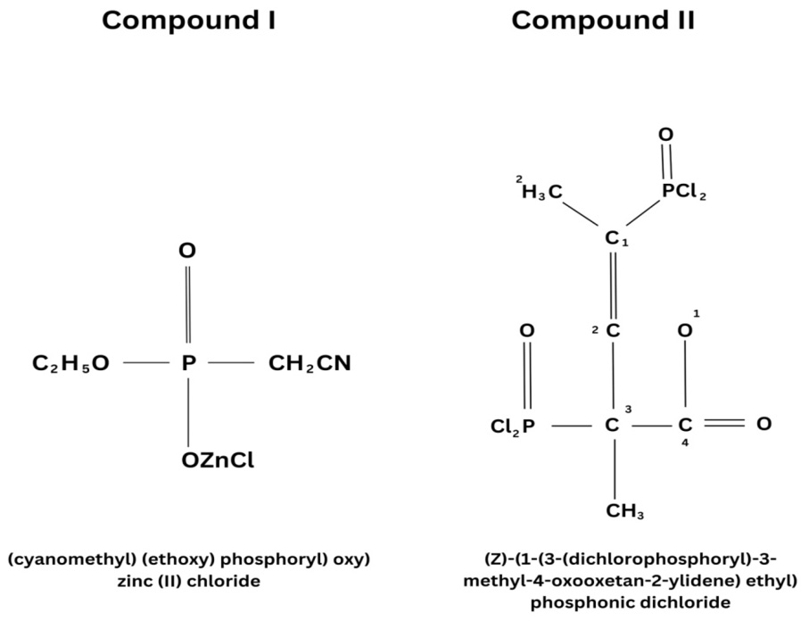
Structure of test compounds.
References
- Andrei, B., Olga, T., Alexandra, V., Anna, L., Syumbelya, A., Ekaterina, B., Sergey, B., Alexander, S., Valentin, P., 2022. ADMET DMPK., 10: 163-179.
- Balouiri, M., Sadiki, M. and Ibnsouda, S.K., 2016. J. Pharma. Anal., 6: 71–79. https://doi.org/10.1016/j.jpha.2015.11.005[CrossRef]
- Chandran, C.S., Thomas, S. and Unni, M.R., 2019. Org. Farm. Chem., pp. 71–87.
- Daisley, B.A., Trinder, M., McDowell, T.W., Collins, S.L., Sumarah, M.W. and Reid, G., 2018. App. environ. Microbiol., 9: 17.
- Fiore, M., 2018. Org. Biomol. Chem., 16: 3068–3086. https://doi.org/10.1039/C8OB00469B[CrossRef]
- Francesca, P., Patrizio, P. and Annalisa, P., 2015. Pathogen. Glob. Hlth., 109 : 309-3018.
- Gaoussou, B., Valeh, I., Niftali, Y. and Khudaverdi, G., 2023. Antimicrobial activity of (E)-2-(1-chloro-1-ethoxyprop-1-en-2-yl)-1, 3, 2-dioxaphospholane-2-oxide. The modern vector of the development of science, proceedings of the VIII international scientific and practical conference, Philadelphia, USA, 28-29 September; Tarmo Vesik; World of Conferences. pp. 17-19.
- Ismiyev, A.I., Shoaib, M., Dotsenkob, V.V., Ganbarova, K., Israilova, A.A. and Magerramov, M., 2020. Russ. J. Gen. Chem., 90: 1418-1425. https://doi.org/10.1134/S1070363220080071[CrossRef]
- Ismiyev, A.I., Shoaib, M., Ganbarova, K. and Agayeva, N., 2019. Adv. Biol. Earth Sci., 4: 88-92.
- Johnson, D.W. and Hils, J.E., 2013. Lubrifiants, 4: 132–148.
- Jolanta, B.Z., Jakub, W. and Agnieszka, B.O., 2013. Pharmacol. Rep., 65: 1–14.
- Kathiriya, P.J., Purohit, H.D. and Purohit, D.M., 2015. Int. Lett. Chem. Phys. Astron., 49: 143-148. https://doi.org/10.3390/lubricants1040132[CrossRef]
- Michele, C., Julia, L. and Luke, S., 2015. Antimicrobial resistance in G7 countries and beyond: Economic issues, policies and options for action. pp. 17.
- O’Neill, J., 2016. Tackling drug-resistant infections globally: Final report and recommendations. pp. 7-12. https://doi.org/10.4103/2045-080X.186181[CrossRef]
- Percy, Z., Vuong, A.M., Ospina, M., Calafat, A.M., La Guardia, J.M., Xu, Y., Hale, R.C., Dietrich, K.N., Xie, C., Lanphear, B.P., Braun, J.M., Joseph, M., Cecil, K.M., Yolton, K. and Chen, A., 2019. Environ. Res., 184: 109255. https://doi.org/10.1016/j.envres.2020.109255[CrossRef]
- Renzo, R. and Maurizio, C., 2020. Molecules, 25: 5133. https://doi.org/10.3390/molecules25215133[CrossRef]
- Rim, A., Samia, G.L., Lynda, G., Martial, T., Anna, I., Marta, A., Mounia, M.K., Regis, G., Louisa, A.Z., 2022. J. mol. Struct., 1247: 131336.
- Shikhaliyev, N.G., Ganbarov, K.G., Gaoussou, B., Qajar, A.M., Atakishiyeva, G.T., Ahmedova, N.E., Mukhtarova, S.H., Akkurt, M., Babayeva, G.V., Maharramov, A.M., 2023. Int. Sci. J. Theor. appl. Sci., 125: 132-152. https://doi.org/10.15863/TAS.2023.09.125.9[CrossRef]
- Shoaib, M., Arif, I., Ganbarov, K., Aygun, I. and Umar, S., 2020. Pakistan J. Zool., 52: 1-4. https://doi.org/10.17582/journal.pjz/2020.52.1.sc12[Full text][CrossRef]
- Shoaib, M. and Ganbarov, K., 2019. Open Access J. Microbiol. Biotechnol., 4: 000136.
- Shoaib, M., Israyilova, A. and Ganbarov, K., 2019. J. Microbiol. Biotechnol. Fd. Sci., 9: 84-87. https://doi.org/10.15414/jmbfs.2019.9.1.84-87[CrossRef]
- Shobhit, S. and Dharmendra, A., 2023. World J. Pharma. Res., 10: 734-755.
- Sujan, M., Anindita, P. and Satyapriya, S., 2022. Fd. Sci. Rep., 3: 10-14.
- Urzúa, A., Echeverría, J., Rezende, M.C. and Wilkens, M., 2008. Molecules, 13: 2385-2393. https://doi.org/10.3390/molecules13102385[CrossRef]
- World Health Organization, 2017. Prioritization of pathogens to guide discovery, research and development of new antibiotics for drug-resistant bacterial infections, including tuberculosis. pp. 5.
- Zhao, D., Hang, S., Rammohan, R.Y.B., Yan, L. and Cheng-He, Z., 2022. Bioorgan. Med. Chem. Lett., 64: 128695.
Services
Article usage statistics
Loading…