ResearchersLinks

Research Article · Volume 14, Issue 11, 29 September 2026 · Open Access

A Comparative Study of Serological Tests Used in The Diagnosis of Toxoplasma gondii Infection in Small Ruminants in South Sulawesi, Indonesia

· · · · · ·

Affiliations & Notes ⌄
  1. 1Doctoral Program of Veterinary Science, Faculty of Veterinary Medicine, Universitas Gadjah Mada, Jogjakarta, Indonesia
  2. 2Department of Animal Science, Faculty of Animal Husbandry and Fisheries, Sulawesi Barat University, Sulawesi Barat, Indonesia
  3. 3Department of Parasitology, Faculty of Veterinary Medicine, Universitas Gadjah Mada, Jogjakarta, Indonesia
  4. 4Veterinary Professional Education Study Program, Faculty of Medicine Hasanuddin University, Makassar, Indonesia. *Correspondence | Raden Wisnu Nurcahyo, Department of Parasitology, Faculty of Veterinary Medicine, Universitas Gadjah Mada, Jogjakarta, Indonesia
  5. 5Email: wisnu-nc@ugm.ac.id
Article Info ⌄
Journal:
Advances in Animal and Veterinary Sciences
Published:
29 September 2026

From the article

  • Among the 100 goat serum samples analyzed, 27 (27.0%) were seropositive by ELISA, whereas 19 (19.0%) were seropositive by CATT (Table 2).
  • Despite slight differences in the number of seropositive samples identified by each assay, ELISA and CATT showed a comparable distribution of serological findings across age, sex, and market categories (Table 2).
  • ELISA and CATT demonstrated substantial diagnostic agreement (κ = 0.664; p < 0.001), indicating that CATT provides diagnostic classifications largely consistent with those obtained by ELISA.
  • Diagnostic concordance remained consistent across sex and age groups, demonstrating that the performance of CATT was stable among different demographic categories of goats.

Selected from the article's own text.

Abstract

This study evaluated the diagnostic concordance between the Enzyme-Linked Immunosorbent Assay (ELISA) and the Card Agglutination Test for Toxoplasmosis (CATT) for detecting Toxoplasma gondii antibodies in goats traded at traditional markets in Makassar, South Sulawesi, Indonesia. A cross-sectional survey was conducted using 100 serum samples collected from clinically healthy native goats from five major traditional markets. All samples were analyzed using both serological methods, with ELISA serving as the reference assay. Diagnostic concordance was assessed using Cohen’s Kappa coefficient, while descriptive analyses compared agreement across sex and age groups. ELISA detected a higher seropositivity rate (27.0%) than CATT (19.0%), although both assays identified seropositive animals in all surveyed markets. Overall, ELISA and CATT demonstrated substantial diagnostic agreement (κ = 0.664; p < 0.001), indicating largely consistent classifications. Similar concordance was observed between male and female goats and across all age categories, suggesting stable diagnostic performance under field conditions. The widespread detection of seropositive animals across market locations indicates continuous environmental exposure to T. gondii within the urban livestock trade chain and highlights the public health significance of traditional market systems. These findings also demonstrate the potential value of CATT for supporting routine epidemiological surveillance and early detection programs in regions with limited diagnostic capacity. Although ELISA remains the preferred reference method, CATT provides a rapid, simple, and cost-effective alternative for preliminary screening and surveillance, particularly in resource-limited settings. The study provides baseline evidence supporting the integration of CATT into field-based surveillance programs for caprine toxoplasmosis in Indonesia.
 
Keywords | Card Agglutination Test (CATT), Cohen’s Kappa, Diagnostic agreement, ELISA, Goats, Toxoplasma gondii

INTRODUCTION

Toxoplasma gondii is an obligate intracellular protozoan parasite capable of infecting a wide range of warm-blooded animals, including goats, while members of the family Felidae serve as the definitive hosts. Infection in goats is of considerable veterinary and public health importance because of its adverse effects on animal productivity and its zoonotic potential. In livestock, caprine toxoplasmosis is a major cause of reproductive disorders, including abortion, stillbirth, fetal mummification, and the birth of weak offspring, resulting in substantial economic losses for smallholder production systems ; . Beyond its economic impact, infected goats contribute to the transmission of T. gondii to humans, as viable tissue cysts may persist in edible tissues and pose a risk of infection through the consumption of raw or undercooked goat meat . Consequently, toxoplasmosis is recognized not only as a significant reproductive disease of small ruminants but also as an important food-borne zoonosis that continues to represent a public health concern, particularly in developing countries where traditional livestock production and meat marketing systems remain predominant.

Detection of T. gondii infection in livestock is commonly based on serological assays that measure host antibody responses to the parasite. Among these, the enzyme-linked immunosorbent assay (ELISA) is widely recognized as a reliable diagnostic method due to its high sensitivity and specificity ; . However, ELISA requires well-equipped laboratories, trained personnel, and longer processing times, which limit its applicability for large-scale or field-based surveillance in resource-constrained settings. As a practical alternative, the Card Agglutination Test for Toxoplasmosis (CATT) also known as the latex agglutination test (LAT), offers a rapid, simple, and equipment-free approach suitable for on-site screening . Several studies have reported moderate to high diagnostic agreement between CATT and ELISA under different field conditions, although performance may vary depending on local epidemiological factors and antigen quality. These attributes make CATT an attractive option for preliminary surveillance in low-resource areas, provided that results are interpreted cautiously and confirmed by reference assays when necessary.

Serological testing is the most widely used approach for diagnosing Toxoplasma gondii infection in livestock because it detects the host immune response following parasite exposure. Among the available serological methods, the enzyme-linked immunosorbent assay (ELISA) is considered the reference technique owing to its high diagnostic sensitivity, specificity, and suitability for large-scale antibody detection ; . Despite these advantages, the routine application of ELISA in field settings is often constrained by the need for specialized laboratory facilities, trained personnel, and relatively long processing times, limiting its practicality for surveillance programs in resource-limited areas.

To overcome these limitations, the Card Agglutination Test for Toxoplasmosis (CATT), also referred to as the latex agglutination test (LAT), has been proposed as a rapid and user-friendly alternative for serological screening. Unlike ELISA, CATT requires minimal laboratory equipment and technical expertise, making it suitable for point-of-care or field-based applications . Previous studies have demonstrated moderate to substantial agreement between CATT and ELISA for the detection of T. gondii antibodies in small ruminants, although diagnostic performance may vary depending on factors such as antigen quality, assay characteristics, and the epidemiological background of the study population. Consequently, CATT represents a practical tool for preliminary surveillance and large-scale screening, particularly in low-resource settings, while confirmatory testing using ELISA or other reference diagnostic methods remains essential when definitive diagnosis is required.

In Indonesia, research comparing serological tests for T. gondii in goats has largely been concentrated in Java and other western regions, where diagnostic facilities and veterinary laboratories are more established . In contrast, comparable data from eastern Indonesia remain scarce, despite the region’s significant livestock population and active inter-island animal trade. The absence of systematic monitoring in these areas limits understanding of local transmission dynamics and diagnostic reliability under field conditions. Makassar, the capital of South Sulawesi, represents a major urban hub for small-ruminant trade in eastern Indonesia. Goats are commonly sold in traditional markets rather than specialized livestock markets, and veterinary inspection at these venues is minimal. Such conditions create practical challenges for disease surveillance and highlight the need for simple, rapid, and field-applicable diagnostic tools. Evaluating the agreement between ELISA and CATT under these circumstances can therefore provide valuable insights into the feasibility of integrating rapid serological methods into urban surveillance systems.

This study aimed to evaluate the diagnostic agreement between ELISA and CATT in detecting antibodies to Toxoplasma gondii among goats sold at traditional markets in Makassar, South Sulawesi, Indonesia. The analysis focused on both the overall level of concordance and descriptive comparisons across sex and age groups to determine whether agreement patterns remained consistent among different subsets of animals. By addressing a regional knowledge gap, this work provides baseline evidence for the potential application of CATT as a practical screening tool within urban livestock surveillance systems in eastern Indonesia.

Materials and Methods

Study area, and animal sample

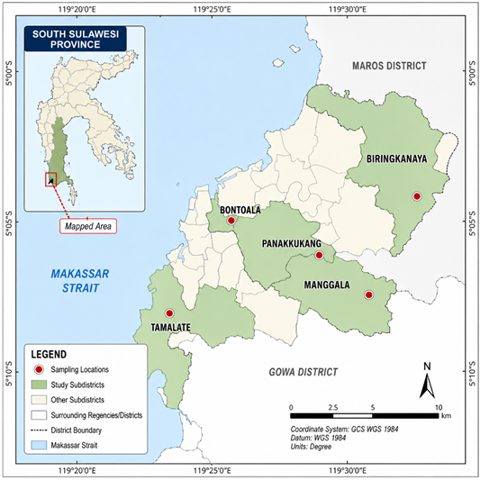
The study was conducted in five major traditional markets located within the administrative boundaries of Makassar City, South Sulawesi, Indonesia: Manggala, Bontoala, Panakkukang, Tamalate, and Biringkanaya (Figure 1). These markets serve as the primary trading centers for small ruminants in Makassar, as the city does not have a designated livestock market. Goats sold at these markets are predominantly sourced from smallholder farms in surrounding rural and peri-urban areas of South Sulawesi, representing diverse husbandry practices, environmental conditions, and management systems. Consequently, the sampled animals provide a representative overview of the goat population entering the urban livestock trade chain.

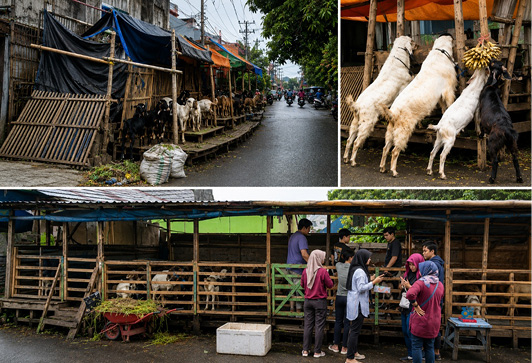
Figure 2 illustrates the typical marketing conditions of native goats (Capra aegagrus hircus, Kacang breed) at traditional markets in Makassar, South Sulawesi, Indonesia. The animals are commonly displayed in open roadside areas without designated livestock facilities or permanent holding pens. Most goats originate from smallholder production systems and are typically fed locally available agricultural by-products and household organic waste, including banana peels and vegetable residues. Commercial transactions are conducted directly between vendors and buyers under informal market conditions, reflecting the traditional livestock trading practices that remain prevalent in urban areas of Indonesia.

The study population consisted of clinically healthy, unvaccinated native goats (Capra aegagrus hircus, Kacang breed) aged six months or older that were offered for sale at five selected traditional markets in Makassar, South Sulawesi, Indonesia. Only apparently healthy animals showing no visible signs of acute illness at the time of sampling were included in the study. Because livestock vendors generally maintained limited animal records, goat age was estimated through dental examination based on the eruption and wear patterns of permanent incisors according to the criteria described by . Animals were subsequently classified into three age categories (1 year, >1–2 years, and >2–3 years) and by sex for descriptive analyses.

The minimum required sample size was determined using a 95% confidence level, an expected seroprevalence of 30%, and a desired precision of 10%, resulting in a minimum sample size of 81 animals. To improve statistical precision and ensure adequate representation across sampling locations, a total of 100 goats were enrolled using a stratified random sampling approach, with each traditional market considered an independent sampling stratum . Sampling was proportionally distributed among the five major traditional markets representing different subdistricts of Makassar, thereby capturing goats originating from diverse production systems and trading networks. The distribution of sampled animals according to market location, sex, and age group is presented in Table 1.

Blood collection and serum preparation

Approximately 5 mL of whole blood was aseptically collected from the jugular vein of each goat following standard veterinary blood collection procedures . Before sampling, the venipuncture site was shaved and disinfected with 70% ethanol, and the animals were gently restrained in a standing position to minimize stress and ensure safe handling. Blood was collected using a sterile vacuum blood collection system equipped with an 18–22 G needle. Immediately after collection, samples were placed in insulated cool boxes and transported to the Serology Laboratory of the Maros Veterinary Center for processing. Upon arrival, the blood samples were centrifuged at 3,000 rpm for 15 min to separate the serum. The recovered serum was carefully transferred into sterile, labeled cryovials and stored at −20°C until serological analysis.

Table 1: Distribution of goat samples by market location, sex, and age group in traditional markets of Makassar, South Sulawesi, Indonesia.

Traditional market

Male

Female

Total (n)

Age

Age

Age

Age

Age

Age

1 yr

2 yr

3+ yr

1 yr

2 yr

3+ yr

Biringkanaya

4

2

2

1

7

5

21

Bontoala

2

1

4

7

3

2

19

Manggala

3

0

2

5

0

0

10

Panakukang

0

13

2

0

0

0

15

Tamalate

1

6

2

8

17

1

35

Total

13

33

22

11

14

7

100

Serologi test

Serological examination was performed to detect antibodies against Toxoplasma gondii in goat serum samples using two serological assays: The Enzyme-Linked Immunosorbent Assay (ELISA) and the Card Agglutination Test for Toxoplasmosis (CATT). ELISA was employed as the reference serological method because of its well-established diagnostic sensitivity and specificity, whereas CATT was evaluated as a rapid and practical alternative suitable for field-based screening. All serum samples were tested using both methods under standardized laboratory conditions, and the results were subsequently compared to assess the level of diagnostic agreement between the two assays.

ELISA testing procedure

Serological detection of anti-Toxoplasma gondii IgG antibodies was performed using a commercial indirect ELISA kit (ID Screen® Toxoplasmosis Indirect Multi-species, IDvet, Grabels, France) following the manufacturer’s instructions. The assay reagents included wash buffer, anti-ruminant IgG conjugate, TMB substrate, stop solution, and the positive and negative control sera supplied with the kit.

Briefly, diluted serum samples, together with positive and negative controls, were dispensed into antigen-coated microplate wells at a volume of 100 µL per well. Following incubation at 37 °C, the plates were washed according to the manufacturer’s protocol to remove unbound components. Subsequently, 100 µL of conjugate solution was added to each well, followed by a second incubation and washing step. Thereafter, 100 µL of TMB substrate was added, and the plates were incubated in the dark to allow color development. The enzymatic reaction was terminated by adding 100 µL of stop solution to each well. Optical density (OD) values were measured at 450 nm using a Thermo Scientific Multiskan™ ELISA microplate reader.

The test results were interpreted according to the manufacturer’s recommendations using the sample-to-positive (S/P) ratio. Serum samples were classified as seropositive or seronegative based on the kit-specific cutoff values. All assays were performed under standardized laboratory conditions using calibrated micropipettes and appropriate laboratory equipment to ensure analytical reliability and reproducibility.

CATT testing procedure

Serological detection of anti-Toxoplasma gondii antibodies using the Card Agglutination Test for Toxoplasmosis (CATT) was performed in accordance with the manufacturer’s instructions. Before testing, all serum samples and reagents were allowed to equilibrate to room temperature. A measured volume of serum was dispensed onto the designated reaction area of the test card and mixed thoroughly with an equal volume of latex antigen reagent. The reaction mixture was gently spread over the entire test circle using a disposable mixing stick.

The test card was placed on a mechanical rotator and agitated for the time specified by the manufacturer to facilitate antigen antibody interaction. After completion of the reaction, the card was examined visually under adequate lighting conditions. Samples showing visible agglutination were interpreted as seropositive for T. gondii antibodies, whereas the absence of visible agglutination was considered seronegative. Positive and negative control sera supplied with the kit were included in each testing batch to verify assay performance. All samples were tested under identical laboratory conditions, and doubtful reactions were repeated to ensure result consistency.

Research parameters

The primary outcome of this study was the serological detection of Toxoplasma gondii antibodies in goat serum samples using two diagnostic assays: The Enzyme-Linked Immunosorbent Assay (ELISA) and the Card Agglutination Test for Toxoplasmosis (CATT). ELISA was designated as the reference method because of its well-established diagnostic sensitivity and specificity, whereas CATT was evaluated by comparing its diagnostic agreement with the ELISA results. Based on the interpretation criteria of each assay, serum samples were classified as either seropositive or seronegative for T. gondii antibodies. In addition to the serological findings, biological characteristics of the sampled goats, including sex (male or female) and age group (1 year, >1–2 years, and >2–3 years), were recorded. These variables were included solely for descriptive subgroup analyses to assess whether the level of diagnostic agreement between ELISA and CATT remained consistent across different animal categories and were not intended to identify biological risk factors associated with T. gondii infection.

Statistic analysis

All statistical analyses were performed using R software (version 4.4.0; R Foundation for Statistical Computing, Vienna, Austria). Descriptive statistics were used to summarize the distribution of goat samples according to traditional market, sex, and age group. The level of diagnostic agreement between the ELISA and CATT results was assessed using Cohen’s Kappa coefficient (κ), implemented through the irr package . Cohen’s Kappa measures the level of agreement between two categorical diagnostic methods after accounting for agreement expected by chance, with values ranging from −1 (complete disagreement) to 1 (perfect agreement).

The strength of agreement was interpreted according to the classification proposed by Landis and Koch (1977) as follows: κ < 0.00, poor agreement; κ = 0.00–0.20, slight agreement; κ = 0.21–0.40, fair agreement; κ = 0.41–0.60, moderate agreement; κ= 0.61–0.80, substantial agreement; and κ= 0.81–1.00, almost perfect agreement. To estimate the precision of the agreement analysis, 95% confidence intervals (95% CI) for Cohen’s Kappa were calculated using the epi.kappa function available in the epiR package . Diagnostic agreement was evaluated for the entire dataset (n= 100) and subsequently stratified by sex (male and female) and age group (1 year, >1–2 years, and >2–3 years) to assess the consistency of agreement across demographic subgroups. The results were summarized in tables and further illustrated using matrix plots to visualize the distribution of concordant and discordant serological results between ELISA and CATT.

Result and Discussion

Among the 100 goat serum samples analyzed, 27 (27.0%) were seropositive by ELISA, whereas 19 (19.0%) were seropositive by CATT (Table 2). Both serological assays detected Toxoplasma gondii antibodies across all age groups, sexes, and traditional market locations, although the proportion of seropositive animals varied among these categories.

Based on ELISA, seroprevalence increased with age, rising from 22.6% in one-year-old goats to 35.0% in goats aged ≥3 years. A comparable trend was observed with CATT, although the proportion of seropositive animals was generally lower, with positivity rates of 9.7%, 18.4%, and 35.0% for the respective age groups. Female goats exhibited a higher seropositivity rate than males in both assays, with ELISA identifying 27.3% of females and 12.5% of males as seropositive. Similar distributions were observed for CATT.

Table 2: Seroprevalence of Toxoplasma gondii antibodies in goats by age, sex, and market location as determined by ELISA and CATT in Makassar, Indonesia. Values represent the number of seronegative and seropositive samples, with percentages shown in parentheses.

Variable

Category

ELISA

CATT

Seronegative

Seropositive

Seronegative

Seropositive

n (%)

Age

1 yr

24 (77.4)

7 (22.6)

28 (90.3)

3 (9.7)

2 yr

36 (73.5)

13 (26.5)

40 (81.6)

9 (18.4)

≥3 yr

13 (65.0)

7 (35.0)

13 (65.0)

7 (35.0)

Sex

Male

49 (87.5)

7 (12.5)

49 (87.5)

7 (12.5)

Female

32 (72.7)

12 (27.3)

32 (72.7)

12 (27.3)

Trad. Market

Biringkanaya

20 (95.2)

1 (4.8)

18 (85.7)

3 (14.3)

Bontoala

13 (68.4)

6 (31.6)

14 (73.7)

5 (26.3)

Manggala

5 (50.0)

5 (50.0)

7 (70.0)

3 (30.0)

Panakukang

12 (80.0)

3 (20.0)

14 (93.3)

1 (6.7)

Tamalate

23 (65.7)

12 (34.3)

28 (80.0)

7 (20.0)

Total N=100

73 (73.0)

27 (27.0)

81 (81.0)

19 (19.0)

Differences in seroprevalence were also evident among the five traditional markets. ELISA recorded the highest seropositivity in Manggala (50.0%), followed by Tamalate (34.3%), whereas the lowest prevalence was observed in Biringkanaya (4.8%). The distribution of CATT-positive samples generally followed the same pattern, although lower seropositivity rates were recorded in several markets, particularly Biringkanaya and Panakukang.

Overall, the findings demonstrate the presence of T. gondii antibodies in goats traded across all surveyed traditional markets in Makassar. Despite slight differences in the number of seropositive samples identified by each assay, ELISA and CATT showed a comparable distribution of serological findings across age, sex, and market categories (Table 2).

The diagnostic agreement between ELISA and CATT was evaluated using Cohen’s Kappa coefficient, and the corresponding results are summarized in Table 3. Overall, the two serological assays demonstrated substantial agreement across all 100 goat serum samples (κ= 0.664; p < 0.001). Similar levels of agreement were observed in the subgroup analyses by sex, with Kappa values of 0.687 for male goats and 0.628 for female goats, both indicating substantial agreement.

Table 3: Cohen’s Kappa coefficients showing diagnostic agreement between ELISA and CATT results for Toxoplasma gondii in goats by sex and age group.

Group

n

Kappa

p-value

Interpretasi

Male

56

0.687

<0.001

Substantial

Female

44

0.628

<0.001

Substantial

Age = 1yr

31

0.537

<0.001

Moderate

Age = >1–2yr

49

0.652

<0.001

Substantial

Age = >2–3yr

20

0.780

<0.001

Substantial

Total (all samples)

100

0.664

<0.001

Substantial

Table 4: Cross-tabulation of ELISA and card agglutination test for toxoplasmosis (CATT) results for the detection of Toxoplasma gondii antibodies in goat serum samples (n = 100).

CATT

ELISA Positive

ELISA Negative

Total

Positive

17

2

19

Negative

10

71

81

Total

27

73

100

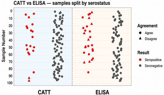
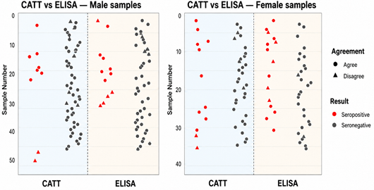
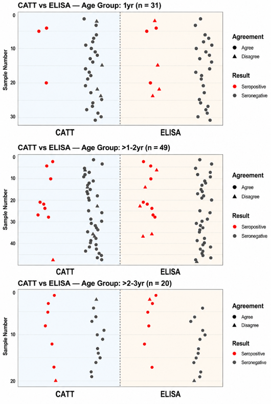
The cross-tabulation of ELISA and CATT results is presented in Table 4. Of the 100 serum samples examined, 88 (88.0%) showed concordant results between the two assays, comprising 17 samples that were seropositive by both ELISA and CATT and 71 samples that were seronegative by both methods. In contrast, 12 (12.0%) samples showed discordant results, including 10 samples that were positive by ELISA but negative by CATT and two samples that were negative by ELISA but positive by CATT. These findings indicate a high proportion of agreement between the two serological methods, although ELISA detected a greater number of seropositive samples than CATT. When stratified by age, the level of agreement showed a gradual increase with advancing age. The youngest age group (1 year) exhibited moderate agreement (κ = 0.537), whereas goats aged >1–2 years and >2–3 years demonstrated substantial agreement, with Kappa values of 0.652 and 0.780, respectively. Overall, the pattern of agreement between ELISA and CATT remained consistent across all demographic subgroups despite slight differences in seropositivity rates. The distribution of concordant and discordant classifications between the two diagnostic methods is further illustrated in the matrix plots presented in Figures 3, 4 and 5.

Figure 3 presents the overall distribution of ELISA and CATT results for all 100 goat serum samples. Each point represents the serological status of an individual goat as determined by both assays, with red symbols indicating seropositive samples and gray symbols representing seronegative samples. Triangular symbols denote discordant classifications between ELISA and CATT. Most samples were classified as seronegative by both assays, whereas only a limited number of discordant results were observed.

The subgroup analysis by sex (Figure 4) showed a comparable distribution of concordant and discordant results between male and female goats. Although slight differences in the number of seropositive samples were observed, the overall pattern of agreement between ELISA and CATT remained similar for both sexes.

Similarly, the age-stratified analysis (Figure 5) demonstrated comparable agreement patterns across all age groups. Older goats (>2–3 years) exhibited a greater number of seropositive samples than younger animals; however, the distribution of concordant and discordant classifications remained consistent among the three age categories. Overall, the matrix plots visually illustrate the substantial level of agreement between ELISA and CATT observed across the entire dataset and within each demographic subgroup, consistent with the Cohen’s Kappa results presented in Table 3.


Discussion

Diagnostic and technical

The present study demonstrated a substantial level of diagnostic agreement between ELISA and CATT (κ= 0.664; p < 0.001), indicating that both assays produced highly comparable classifications of Toxoplasma gondii serostatus in goat serum samples. Although ELISA remains the reference method for serological diagnosis because of its superior diagnostic sensitivity and specificity, the observed level of concordance indicates that CATT can provide reliable results for preliminary screening under field conditions. The relatively small number of discordant samples observed in the matrix plots, together with the consistent Kappa values across sex and age groups, suggests that disagreement between the two assays was limited and not associated with specific demographic characteristics.

These findings are consistent with previous studies reporting good diagnostic concordance between agglutination-based assays and ELISA for the detection of T. gondii antibodies in small ruminants. Toaleb et al. (2025), for example, reported a high level of agreement between a commercial latex agglutination test and ELISA when evaluating sera from farm animals. Similarly, Kim et al. (2024) highlighted the excellent analytical performance of ELISA while emphasizing its dependence on laboratory infrastructure, specialized equipment, and trained personnel. Dong (2024) likewise identified ELISA as a highly accurate serological assay but noted that its operational requirements may limit its routine application in large-scale surveillance programs.

Collectively, these findings indicate that agglutination-based assays such as CATT represent a practical alternative for rapid field screening, particularly in resource-limited settings where access to laboratory facilities is restricted. Nevertheless, because ELISA provides greater analytical sensitivity, positive CATT results should ideally be confirmed using ELISA or molecular diagnostic methods when accurate case confirmation is required for epidemiological investigations or disease control programs.

Minor differences between ELISA and CATT results in this study could partly reflect technical variability intrinsic to serological assays rather than limitations of either method. As noted in the systematic review by , ELISA performance parameters such as sensitivity, specificity, and Kappa agreement can vary substantially depending on antigen type, antibody-binding reagents, and sample matrices used across studies. Variability in antigen sources may influence test reactivity, particularly in field samples collected under non-standardized conditions. These methodological factors may therefore explain the small number of discordant samples observed between ELISA and CATT in the present analysis. Comparable outcomes have also been documented in Indonesia, where a Modified Agglutination Test (ToMAT) demonstrated good diagnostic accuracy and substantial agreement with both commercial ELISA and immunoblotting assays . Together with the present results, these findings support the feasibility of agglutination-based techniques for routine screening of T. gondii infection in field conditions. Nevertheless, given the known variability among serological assays, it remains advisable that CATT results be interpreted as preliminary indicators, with confirmatory testing using ELISA or molecular methods when feasible.

Compared with laboratory-based serological methods, CATT offers several practical advantages for field application, particularly in resource-constrained settings. The assay is rapid, easy to perform, and requires only minimal equipment and technical expertise, making it well suited for preliminary screening before confirmatory testing by ELISA. Previous studies have likewise demonstrated that agglutination-based assays provide satisfactory diagnostic performance, repeatability, and operational feasibility under field conditions where laboratory infrastructure is limited ; ; .

In the present study, the substantial diagnostic agreement observed across different sex and age groups further supports the robustness of CATT under routine field conditions. The consistency of agreement among demographic subgroups indicates that the assay maintains stable diagnostic performance despite potential variations in animal characteristics, sample handling, and environmental conditions commonly encountered in smallholder production systems. Similar observations have been reported for other agglutination-based serological methods, which have shown reliable performance across heterogeneous livestock populations ; .

These operational advantages make CATT a valuable complementary tool for routine surveillance and epidemiological monitoring of T. gondii infection in areas where access to laboratory facilities is limited. Rather than replacing ELISA, CATT can serve as an efficient first-line screening assay within tiered diagnostic strategies, thereby improving surveillance coverage while reducing the logistical and financial constraints associated with large-scale serological monitoring .

Despite its operational advantages, CATT should be considered a complementary rather than a replacement diagnostic method for ELISA. Although the assay demonstrated substantial agreement with ELISA in the present study, its relatively lower analytical sensitivity, particularly in samples with low antibody titers or borderline serological responses, may result in occasional false-negative classifications. Consequently, confirmation of positive or inconclusive CATT results using ELISA or molecular diagnostic techniques remains advisable, especially in epidemiological investigations, disease surveillance programs, and animal health certification. Therefore, the findings of this study support the application of CATT as a rapid and cost-effective first-line screening tool that complements, rather than substitutes, laboratory-based diagnostic methods, particularly in resource-limited settings where logistical, technical, or financial constraints restrict routine ELISA testing.

Epidemiological and public-health

The detection of Toxoplasma gondii antibodies in goats traded at traditional markets in Makassar confirms the continued circulation of this zoonotic parasite within the local small-ruminant population. Although the present study was not designed to estimate the regional seroprevalence of toxoplasmosis, the identification of seropositive animals in all surveyed markets indicates widespread environmental exposure and the potential for parasite transmission at the livestock human interface. Similar observations have been reported in other geographic regions, including Indonesia , Malaysia , Brazil , and Ethiopia , where moderate-to-high seroprevalence has frequently been associated with smallholder production systems and traditional livestock marketing practices.

The epidemiological context of Makassar may further contribute to the persistence and dissemination of T. gondii. Goats are commonly traded in open-market environments with limited veterinary supervision and biosecurity measures, conditions that may facilitate the unnoticed circulation of the parasite within the livestock value chain ; . In addition, close contact between animals, traders, and consumers, together with inadequate sanitation and meat-handling practices, may increase the risk of environmental contamination and food-borne transmission . These findings underscore the importance of strengthening surveillance at traditional livestock markets, not only to improve animal health management but also to support public health protection through a One Health approach, particularly in densely populated urban areas where human animal interactions are frequent.

From a public-health perspective, these findings underscore the importance of traditional markets as potential nodes of zoonotic risk, particularly where slaughtering or meat handling occurs close to residential areas. In Indonesia, most goat transactions take place in smallholder or informal market settings where veterinary supervision is limited and slaughtering often occurs directly at or near the point of sale. In a review highlighted that over 99% of goats and sheep are managed by smallholders and marketed through traditional outlets, especially during religious festivals such as Eid al-Adha and Aqiqah, when meat processing and hygiene controls are minimal. Similarly, documented that goats traded from South Sulawesi to East Kalimantan are commonly slaughtered in roadside or market facilities with inadequate biosecurity and sanitation, reinforcing the potential for pathogen transmission along the meat value chain.

At the epidemiological level, the relatively uniform pattern of infection across age and sex groups suggests environmental exposure rather than management-specific factors as the likely source of infection pressure. Similar age- and sex-related tendencies have been reported in other regions, such as in Espírito Santo, Brazil, where T. gondii seroprevalence was higher among female and older goats . Comparable observations have also been described in Malaysia and Indonesia, where infection probability increased with age and reproductive activity in smallholder goats ; . These consistent patterns across diverse settings suggest that host age and sex reflect cumulative exposure rather than intrinsic susceptibility, reinforcing the dominant role of environmental contamination by oocysts shed from cats as the main driver of infection risk in goat populations ; .

Policy and research perspectives

The findings of the present study provide practical evidence supporting the incorporation of rapid serological assays such as CATT into existing surveillance frameworks for Toxoplasma gondii in Indonesia. Although ELISA remains the preferred reference method because of its superior diagnostic accuracy, the substantial agreement observed between ELISA and CATT indicates that CATT can serve as a reliable first-line screening tool, particularly in urban and peri-urban settings where access to laboratory infrastructure is limited. Integrating CATT into a tiered diagnostic algorithm would enable broader surveillance coverage while maintaining the accuracy of disease confirmation through subsequent ELISA testing.

The detection of seropositive goats across all surveyed traditional markets further highlights the need to strengthen routine veterinary surveillance and animal traceability within informal livestock marketing systems. Implementing basic veterinary inspection services, standardized health record systems, or mobile diagnostic units at major trading points could improve the early detection of zoonotic pathogens and facilitate monitoring of animal movements throughout the livestock value chain. Such interventions would not only enhance disease surveillance but also reduce the potential risk of zoonotic transmission associated with traditional livestock markets.

These recommendations are consistent with Indonesia’s One Health strategy for zoonotic disease prevention, which promotes coordinated collaboration among the veterinary, environmental, and public health sectors . Strengthening surveillance through accessible diagnostic tools and integrated monitoring systems would contribute to more effective control of T. gondii and other food-borne zoonotic pathogens while supporting evidence-based animal and public health policies.

Conclusion

As the conclusion of this study, three key conclusions can be drawn that collectively address the study objectives.

  • ELISA and CATT demonstrated substantial diagnostic agreement (κ = 0.664; p < 0.001), indicating that CATT provides diagnostic classifications largely consistent with those obtained by ELISA. These findings support the use of CATT as a reliable and operationally feasible first-line screening method for detecting Toxoplasma gondii antibodies in goats under field conditions.
  • Diagnostic concordance remained consistent across sex and age groups, demonstrating that the performance of CATT was stable among different demographic categories of goats. This consistency supports its application as a practical screening tool for heterogeneous goat populations managed under smallholder production systems.
  • The detection of seropositive goats in all surveyed traditional markets confirms the circulation of T. gondii within the urban livestock trade chain and underscores the importance of strengthening veterinary surveillance, routine inspection, and animal traceability to reduce the risk of zoonotic transmission.
  • Overall, this study provides baseline evidence supporting the integration of CATT into Indonesia’s veterinary surveillance programs as a complementary diagnostic method within a tiered testing strategy, whereby positive screening results are confirmed by ELISA. Future studies incorporating molecular diagnostics, environmental sampling, and broader geographic coverage are recommended to improve understanding of transmission dynamics and to support the development of effective One Health based control strategies for toxoplasmosis and other food-borne zoonotic diseases.

Acknowledgements

The author expresses his gratitude and appreciation to the Indonesia Endowment Funds for Education and the Center for Higher Education Funding for Publication.

Novelty Statement

Although several studies have compared ELISA and latex agglutination based assays (LAT/CATT) for the serological detection of Toxoplasma gondii in small ruminants, evidence regarding their diagnostic agreement in goats traded through traditional urban markets remains unavailable. These markets are characterized by informal livestock trading, limited veterinary supervision, and frequent human animal interactions, creating unique epidemiological conditions for zoonotic disease transmission. This study is the first to evaluate the diagnostic concordance between ELISA and CATT in goats from traditional markets in Makassar, Indonesia, while also assessing the consistency of diagnostic agreement across different age and sex groups under real field conditions.

Authors Contribution

AR: Collecting data, animal maintenance, and data analysis. RWN: Assembled research design and review manuscript. DP: Assembled research design, data analysis, and manuscript writing. JP: Assembled research design and reviewed the manuscript. R: Reviewed and advised on manuscript improvement and data analysis. S: Advised on fine-tuning the manuscript. M: Contributed to manuscript refinement and provided suggestions for manuscript enhancement.

Study design and ethical approval

A cross-sectional study was conducted from November to December 2024 to determine the seroprevalence of Toxoplasma gondii infection in native goats sold at traditional markets in Makassar, South Sulawesi, Indonesia. A stratified random sampling approach was applied to ensure representative coverage from five main urban districts: Manggala, Bontoala, Panakkukang, Tamalate, and Biringkanaya. Each market was considered a stratum, and individual goats were selected randomly within each stratum. All sampling and handling procedures followed established animal welfare standards and biosafety guidelines. Ethical approval for animal handling, restraint, and blood collection was granted by the Institutional Animal Care and Use Committee of Hasanuddin University, under protocol number 010/UN4.1.1.RSHUHB/PP36/2025.

Funding

Scholarships were provided by the Indonesia Endowment Funds for Education and the Center for Higher Education Funding.

Data availability

All data supporting the findings of this study are available within the manuscript, and no additional data sources are required.

Generative AI and AI-assisted technology statement

AI-assisted technologies were employed exclusively for non-scientific purposes, including grammar improvement, reference formatting, and minor figure editing (e.g., resolution enhancement and label alignment). No AI tools were used for data analysis, interpretation of results, or scientific writing. All findings and conclusions presented in this manuscript are the sole responsibility of the authors.

Conflict of interest

The authors have declared no conflict of interest.

Figures

Figure 1:
Figure 1:.

Map of the study area showing the sampling locations in three urban subdistricts of Makassar City, South Sulawesi, Indonesia: Biringkanaya, Manggala, and Tamalate (Map vector retrieved from: https://tanahair. indonesia.go.id).

Figure 2:
Figure 2:.

Native goats (Kacang breed) sold under typical market conditions at a traditional livestock section of Makassar, Indonesia.

Figure 3:
Figure 3:.

Matrix plot showing overall diagnostic agreement between ELISA and CATT for Toxoplasma gondii in goat serum samples (n = 100).

Figure 4:
Figure 4:.

Matrix plots comparing diagnostic agreement between ELISA and CATT according to sex (male and female goat samples).

Figure 5:
Figure 5:.

Matrix plots comparing diagnostic agreement between ELISA and CATT across age groups (1 year, >1–2 years, and >2–3 years).

References

  1. Adnyana IM, Utomo B, Eljatin DS, Sudaryati NL (2023). One Health approach and zoonotic diseases in Indonesia: Urgency of implementation and challenges. Narra J., 3(3). https://doi.org/10.52225/narra.v3i3.257[CrossRef]
  2. Asja MA, Natsir A, Murray-Prior R, Murray P, Nurhayu A (2010). Goat supply from Enrekang, South Sulawesi to East Kalimantan: A long and winding road. Proceedings of the 5th International Seminar on Tropical Animal Production. 5th International Seminar on Tropical Animal Production, Yogyakartya.
  3. Dong H, Zhang J, Wang Q, Shen Y, Zhou B, Dai L, Zhu W, Sun H, Xie X, Xie H, Xu C, Zhao G, Yin K (2024). Development of an indirect ELISA for detecting Toxoplasma gondii IgG antibodies based on a recombinant TgIMP1 protein. PLoS Negl. Trop. Dis., 18(8): e0012421. https://doi.org/10.1371/journal.pntd.0012421[CrossRef]
  4. Dubey JP (2021). Toxoplasmosis of animals and humans (3rd ed.). CRC Press. https://doi.org/10.1201/9781003199373[CrossRef]
  5. Fatmawati,M, Suwanti LT, Mufasirin M, Fong S, Valinata S, Subekti DT, Ekawasti F, Awad HAA (2023). Serological survey of immunoglobulin G from Toxoplasma gondii infection in dairy goats in East Java, Indonesia. Vet. World, pp. 1926–1932. https://doi.org/10.14202/vetworld.2023.1926-1932[CrossRef]
  6. Greenfield HJ, Arnold ER (2008). Absolute age and tooth eruption and wear sequences in sheep and goat: Determining age-at-death in zooarchaeology using a modern control sample. J. Archaeol. Sci., 35(4): 836–849. https://doi.org/10.1016/j.jas.2007.06.003[CrossRef]
  7. Hung PHS, Duong LD, Thao TN, Dung HT, Hoa NX, Hien BT, Hoa NT, Na TT, Vui TQ, Nhung NPN, Thuy NT (2023). Toxoplasma gondii contamination in fresh vegetables destined for human and livestock consumption in Thua Thien Hue, Vietnam. Vet. Integr. Sci., 22(3): 847–856. https://doi.org/10.12982/VIS.2024.057[CrossRef]
  8. Kim M, Park SJ, Park H (2024). Trend in serological and molecular diagnostic methods for Toxoplasma gondii infection. Euro. J. Med. Res., 29(1): 520. https://doi.org/10.1186/s40001-024-02055-4[CrossRef]
  9. Klun I, Djurković-Djaković O, Thulliez P (2007). Comparison of a commercial ELISA with the modified agglutination test for the detection of Toxoplasma gondii infection in naturally exposed sheep. Zoonoses Publ. Health, 54(3–4): 165–168. https://doi.org/10.1111/j.1863-2378.2007.01032.x[CrossRef]
  10. Landis JR, Koch GG (1977). The measurement of observer agreement for categorical data. Biometrics, 33(1): 159–174. https://doi.org/10.2307/2529310[CrossRef]
  11. Li M, Gao Q, Yu T (2023). Kappa statistic considerations in evaluating inter-rater reliability between two raters: which, when and context matters. BMC Cancer, 23: 799. https://doi.org/10.1186/s12885-023-11325-z[CrossRef]
  12. Liyanage KLDTD, Wiethoelter A, Hufschmid J, Jabbar A (2021). Descriptive comparison of ELISAs for the detection of Toxoplasma gondii antibodies in animals: A systematic review. Pathogens, 10(5): 605. https://doi.org/10.3390/pathogens10050605[CrossRef]
  13. Martin SW, Meek AH, Willeberg P (1987). Veterinary epidemiology: Principles and methods. Iowa State University Press.
  14. Mishra V, Mitra P, Barbuddhe S, Thorat Y, Chavan K, Shinde S, Chaudhari S, Khan W, Deshmukh AS (2024). Serological and molecular detection of Toxoplasma gondii and Neospora caninum in free-ranging rats from Nagpur, India. Parasitol. Res., 123(1): 63. https://doi.org/10.1007/s00436-023-08095-9[CrossRef]
  15. Ragavan GK, Selvaraj P, Murugesan D, Gopalan TK, Chinnaiyan S, Gopal DR, Parthiban ABR, Kumaragurubaran K, Palavesam A (2023). Management practices and technologies for efficient biological sample collection from domestic animals with special reference to Indian field conditions. Animal Diseases, 3: 34. https://doi.org/10.1186/s44149-023-00096-6[CrossRef]
  16. Rossi F, Santonicola S, Amadoro C, Marino L, Colavita G (2024). Food and drinking water as sources of pathogenic protozoans: An update. Appl. Sci., 14(12): 5339. https://doi.org/10.3390/app14125339[CrossRef]
  17. Sadiq MB, Muhamad AS, Hamdan SA, Ramanoon SZ, Zakaria Z, Aziz NAA, Mansor R, Arshad SS, Khalid N, Abdul Hamid N, Kamaludeen J, Syed-Hussain SS (2023). Seroprevalence and factors associated with Toxoplasma gondii, Neospora caninum, and Besnoitia besnoiti infections in cattle and goats in Selangor, Malaysia. Animals, 13(5): 948.. https://doi.org/10.3390/ani13050948[CrossRef]
  18. Shapiro K, Bahia-Oliveira L, Dixon B, Dumètre A, De Wit LA, VanWormer E, Villena I (2019). Environmental transmission of Toxoplasma gondii: Oocysts in water, soil and food. Food Waterb. Parasitol., 15: e00049. https://doi.org/10.1016/j.fawpar.2019.e00049[CrossRef]
  19. Stelzer S, Basso W, Benavides Silván J, Ortega-Mora LM, Maksimov P, Gethmann J, Conraths FJ, Schares G (2019). Toxoplasma gondii infection and toxoplasmosis in farm animals: Risk factors and economic impact. Food Waterb. Parasitol., 15: e00037. https://doi.org/10.1016/j.fawpar.2019.e00037[CrossRef]
  20. Stevenson M., Nunes T., Heuer C., Marshall J., Sanchez J., Thornton R., Reiczigel J., Robison-Cox J., Sebastiani P., Solymos P., Yoshida K., Jones G., Pirikahu S., Firestone S., Kyle R., Popp J., Jay M., Reynard C., Cheung A., Singanallur N., Szabo A., Rabiee A. 2022. epiR: Tools for the Analysis of Epidemiological Data. 2022. https://cran.r-project.org/package=epiR
  21. Sujarwanta RO, Afidah U, Suryanto E, Rusman, Triyannanto E, Hoffman LC (2024). Review: Goat and Sheep Meat Production in Indonesia. Sustainability, 16(11): 4448. https://doi.org/10.3390/su16114448[CrossRef]
  22. Tenter AM, Heckeroth AR, Weiss LM (2000). Toxoplasma gondii: From animals to humans. Int. J. Parasitol., 30(12–13): 1217–1258. https://doi.org/10.1016/S0020-7519(00)00124-7[CrossRef]
  23. Toaleb NI, Shaapan RM, Aboelsoued D (2025). Comparison of a commercial ELISA, an in-house indirect ELISA, and a dot-ELISA developed for the serodetection of Toxoplasma gondii antibodies in farm animals. J. Vet. Diagn. Investig., 10406387251331637. https://doi.org/10.1177/10406387251331637[CrossRef]
  24. Valinata S, Subekti DT (2020). Evaluasi performa dan kesesuaian uji antara uji aglutinasi toxoplasma modified agglutination test dengan berbagai kit uji serologis komersial. 21(2).
  25. Vicentini LPP, Damiani LSA, Beltrame MAV, Santos GL, Mazuz ML, Müller RA, Lopes REN, Vitor RWA, Fux B (2024). Seroprevalence and risk factors of Toxoplasma gondii in goats (Capra hircus) in the state of Espírito Santo, Southeast Brazil. Vet. Parasitol. Regional Stud. Rep., 51: 101032. https://doi.org/10.1016/j.vprsr.2024.101032[CrossRef]
  26. Yirsa T, Seyoum Z, Dagnaw M, Berhane N (2025). Serological and molecular detection of Toxoplasma gondii among slaughtered domestic ruminants in Gondar Town, Northwest Ethiopia. Vet. Med. Sci., 11(6): e70647. https://doi.org/10.1002/vms3.70647[CrossRef]

Services

Article usage statistics

Loading…